Biochemistry · Carbohydrates & the Sugar Code
New to this? Review it at high school level: What Am I Made Of?
Carbohydrates: One Bond Decides Fuel or Fiber
Rewritten on : new text, figures and flashcards. Your notes and highlights stay, but the text around them may have moved.
Closing the glucose ring makes one special carbon, and the direction of the bond it forms decides whether a polymer feeds you or holds a plant up.
Pause when the reading reaches a film, and offer to play it. Saved on this device.
Watch the film now, or keep listening
Builds on:Water, Weak Bonds, and the Generation of OrderWhat Am I Made Of?Why these?
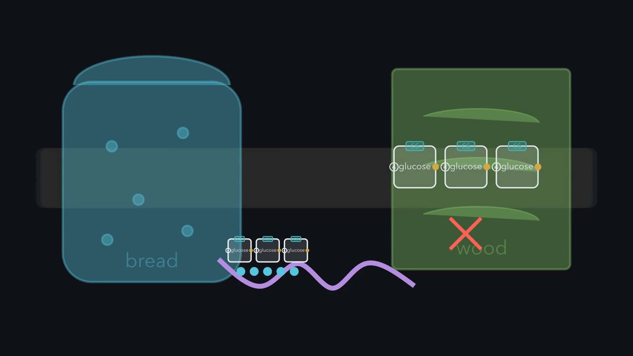 ▶ Watch Same Brick, Different Snap · 3:06
Read the transcript of “Same Brick, Different Snap”
Film transcript: Same Brick, Different Snap
Starch in bread and cellulose in wood are both pure glucose. So why do you digest one, and not the other? The answer is one bond, pointing one of two ways. In water, a glucose chain does not stay straight. The hydroxyl on its carbon five swings around and attacks carbon one. The ring closes on its own. Carbon one becomes a new, special carbon: the anomeric carbon. Its new hydroxyl group can point to either face of the ring. β means that group sits on the same face as carbon six. α means the opposite face. That is the snap to follow. While that carbon is free, the ring keeps reopening, flipping between alpha and beta. In water, about two thirds settle as beta.
Now bond that carbon to carbon four of the next glucose. Once used, the ring cannot open. The face is locked in. Starch and cellulose both use this 1,4 bond. Starch makes it from the α face. Cellulose makes it from the β face. Put simply, the real ring is puckered. In beta, the carbon one bond points straight across from carbon four. In alpha, it leaves at a sharp angle. Now chain glucoses together each way. Which chain runs straight? Which one bends at every glucose?
Picture it first. Beta runs straight, like a rod through beads. Each glucose sits flipped from its neighbors, so the chain lies flat, a ribbon. Alpha bends at every joint, always the same way. So the chain winds into a helix, about six glucoses per turn in amylose, a starch. Flat beta ribbons lie side by side and hydrogen bond into rigid fibers. That is cellulose, which holds plants up. Alpha helices cannot lie flat together that way. They pack into compact granules instead: starch in plants, glycogen in animals.
Stored fuel. Shape is only part of the story. Digestion needs an enzyme that fits the bond. Your amylases fit α-1,4. You make no cellulase to cut the β bonds of cellulose. A cow lives on grass because its gut microbes make cellulase for it. The brick never changes. Only the face of one bond does. For glucose joined 1 to 4, α coils into fuel and β runs straight into fiber.
From the film
- Starch and cellulose are both glucose polymers; their 1,4 bonds use opposite faces.
- Closing the glucose ring turns carbon one into the anomeric carbon, whose new group can point to either face.
- β puts that group on the same face as carbon six; α puts it on the opposite face.
- For glucose joined 1 to 4, β chains run straight into cellulose fibers; α chains coil into starch and glycogen.
- You digest starch because your amylases fit α-1,4; you make no cellulase to cut cellulose's β bonds.
Starch and cellulose are both made of nothing but glucose. You digest one every day, and you could not digest the other if you ate a tree.
What differs is the direction of one bond: a small difference in structure, an enormous difference in outcome.
Carbohydrates store energy, build structure and carry information. Plants fix carbon from air and sunlight into glucose; the rest of us eat plants, or things that ate plants. Everything downstream is built from that one borrowed monomer.
Meet glucose: just enough to number it
A monosaccharide is a single sugar unit: a polyhydroxy aldehyde or ketone. Two units make a disaccharide, a handful an oligosaccharide, many a polysaccharide.
Classify the single unit by its carbonyl. An aldehyde at the end (C1) makes it an aldose. A ketone tucked inside (usually C2) makes it a ketose. Add the carbon count and the name assembles itself: glucose is an aldohexose (six carbons), fructose a ketohexose. Glucose and fructose have the same formula but the carbonyl on a different carbon, which makes them constitutional isomers.
Number the carbons from the carbonyl end, C1 to C6; linkage names like “1,4” use these numbers.
Almost every internal carbon carries four different groups, making it a chiral center: swap any two of its groups and you get a different molecule, a stereoisomer. Glyceraldehyde, the simplest aldose, has one chiral center and two mirror-image forms. Dihydroxyacetone, its ketose cousin, has none and is achiral. Whether a sugar is D or L is set by the chiral carbon farthest from the carbonyl (see the technique box), and biology runs almost entirely on D-sugars.
Six sugars carry most of the biological traffic: ribose and deoxyribose in your nucleic acids, and glucose, fructose, mannose and galactose in your metabolism.
One spelling trap returns on every exam: “-ose” marks a sugar, “-ase” an enzyme. Sucrose is the sugar; sucrase is the enzyme that cleaves it.
Closing the ring makes one special carbon
The flat Fischer drawing is a useful mugshot, but it lies about behavior. The film above shows the C5 hydroxyl closing onto the C1 aldehyde in water; the ring it makes has six members.
An alcohol adding to an aldehyde makes a hemiacetal. A ketose such as fructose, whose carbonyl is at C2, makes a hemiketal. Either way, the old carbonyl carbon now carries four different groups and becomes a new chiral center, the anomeric carbon.
The two forms the film names, one for each face of the new hydroxyl, are the anomers: α on the opposite face from the CH₂OH group (C6) on C5, β on the same face.
In the standard flat ring drawing of a D-sugar (the Haworth projection), the CH₂OH points up, so α has its C1 hydroxyl below the ring and β above. Flip the drawing and “below” flips too; the CH₂OH reference does not.
The real ring is not flat. It puckers into a chair. Each ring carbon has two bonds that are not part of the ring. One is axial: it points straight up or down, parallel to the ring’s axis. The other is equatorial: it points outward around the rim. Groups on equatorial bonds crowd each other least. In β-glucose every bulky group, including the C1 hydroxyl, sits equatorial. In α-glucose the C1 hydroxyl is axial.
Predict. The two anomers interconvert in water. Which one should be the majority, and why?
Check your prediction
β. A glucose solution settles near one-third α and two-thirds β (about 36:64), with well under one percent open-chain at any instant. β wins mostly because every bulky group, the C1 hydroxyl included, sits equatorial. An electronic effect at the ring oxygen favors axial α a little, which keeps the win modest (Going deeper, “Why the split is not more lopsided”).
A six-membered sugar ring is a pyranose, a five-membered one a furanose. Glucose favors the pyranose, so its stable form, spelled out, is α- or β-D-glucopyranose. Fructose forms the five-membered furanose when it is locked into sucrose or a polysaccharide, though free in solution it is mostly the pyranose.
The one idea to carry through this lesson. Much of what a sugar does turns on one carbon. Closing the ring turns the old carbonyl carbon (C1 in glucose, C2 in fructose) into the anomeric carbon, a new center whose group can point two ways, α or β. While that carbon is free, the ring can reopen into a reactive carbonyl (in glucose, an aldehyde): the sugar flips between α and β, reduces copper, and slowly tags proteins. Once it is used in a bond to another molecule, it is locked, and two things about that bond, the face it leaves from (α or β) and the carbon it joins (4 or 6 in the glucose polymers), set the shape of the chain.
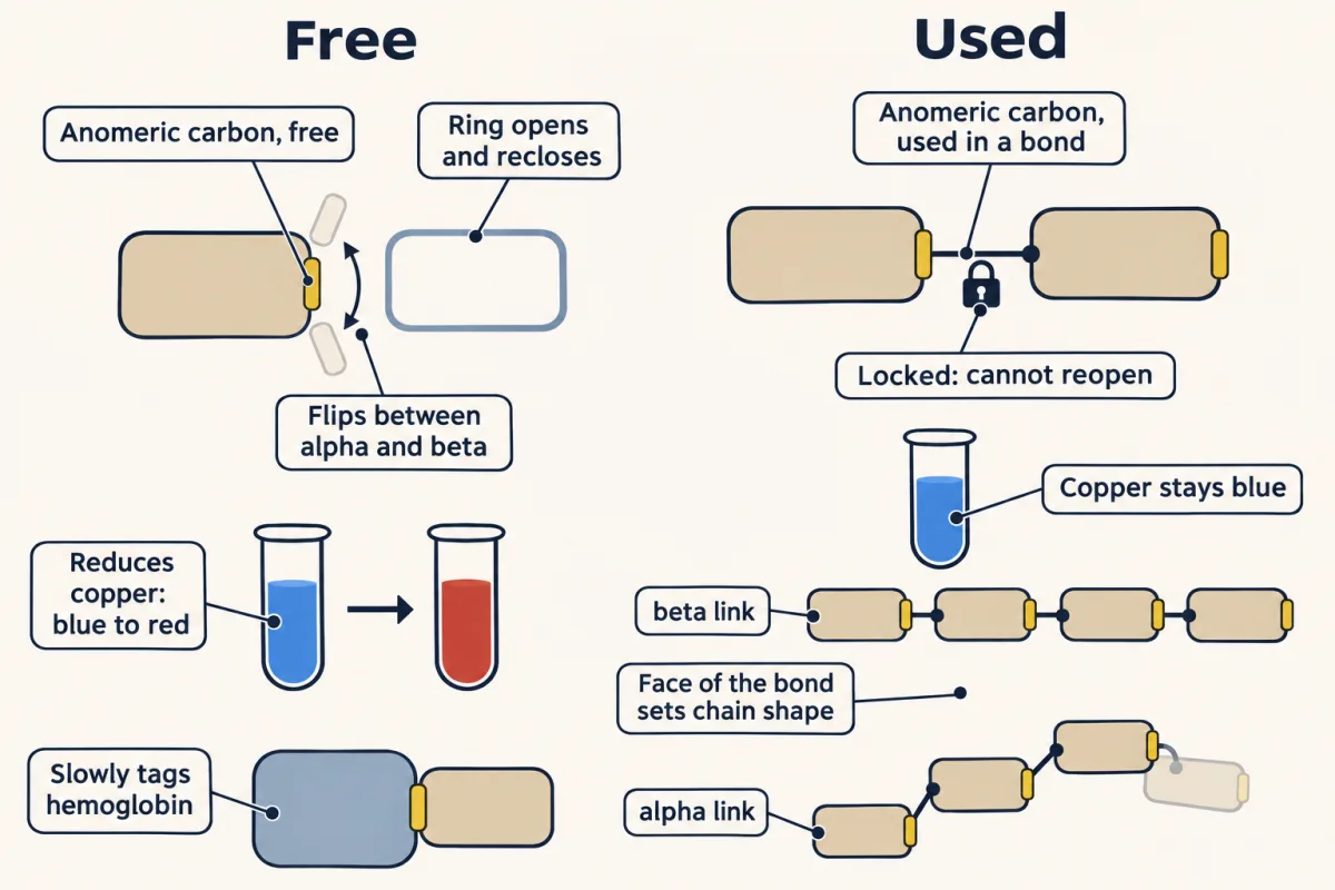
This rule explains the α–β flip (mutarotation), the reducing-sugar test, HbA1c, which disaccharides are reducing, why starch and glycogen coil while cellulose runs straight, and why branching speeds fuel release. It does not tell you which sugar sits in a chain, which matters most on the cell surface; that needs a second rule, near the end.
Naming the relationships
With the anomers in hand, every isomer term has an example you know. All of these are stereoisomers: same formula and connectivity, different arrangement in space.
| Relationship | What differs | A pair you know |
|---|---|---|
| Enantiomers | Mirror images that cannot be superimposed; every chiral center is flipped | D- and L-glucose |
| Diastereomers | Stereoisomers that are not mirror images (the umbrella for the next two rows) | Glucose and galactose |
| Epimers | Exactly one chiral center | Glucose and mannose (C2); glucose and galactose (C4) |
| Anomers | Only the anomeric carbon (a special kind of epimer) | α- and β-glucose |
Hold the C4 fact: with lactose’s β bond, it is why milk sugar needs its own enzyme.
A free anomeric carbon can reopen
A ring that formed on its own can open on its own: while the anomeric carbon is free (not bonded to another molecule), the ring keeps opening and reclosing.
Predict. You dissolve crystals of pure α-glucose in water. What is in the beaker the next day?
Check your prediction
The usual mix: about one-third α, two-thirds β and a trace of open chain. Each time the ring reopens, it can reclose onto either face. This drift is called mutarotation, after the change in how the solution rotates polarized light.
The open chain is rare but always there, and it carries an aldehyde. That makes glucose a reducing sugar. A reducing sugar is one that reduces something else: its aldehyde gives up electrons and is oxidized. In Benedict’s test, blue Cu(II) takes those electrons and becomes brick-red Cu₂O. Glucose is reducing not because it is usually open-chain, but because it can become open-chain.
The explorer below walks the same path in three steps: flip C1 between α and β, watch the equilibrium and its tiny open-chain fraction, then snap glucose units into a polymer with either bond.
1 · Anomer
α: C1–OH on the opposite face from CH₂OH (below, in this standard drawing). The flip goes through the open chain.
2 · Equilibrium & reducing power
β wins: its bulky groups sit equatorial, lower strain.
Benedict's test: positive. The open-chain aldehyde reduces blue Cu²⁺ to brick-red Cu₂O in minutes.
HbA1c: —
The same open-chain trace glycates hemoglobin, slowly. HbA1c rises for a few months, then levels off as old red cells (about a 120-day lifespan) are replaced, so it reports your average glucose over the past 2–3 months. Model: a normal 5% start; half the remaining change arrives each month.
3 · Build a polymer
—
—
Free, C1 reopens: the sugar flips α⇄β, reduces copper and slowly tags hemoglobin. Used in a bond, C1 is locked, and the face of that bond sets the shape of the chain.
The clinic hiding in “under one percent”
Here is one chemical reason high glucose harms a diabetic. The open-chain aldehyde reacts slowly and non-enzymatically with free amino groups on proteins. Glucose attached to the N-terminal valine of hemoglobin’s β chain is what is measured as HbA1c. Red cells live about 120 days, so HbA1c is a molecular diary of your average blood glucose over the prior two to three months, the standard for long-term diabetic monitoring. The same chemistry, run on other proteins, builds advanced glycation end products (AGEs), which accumulate in tissues and contribute to diabetic complications. A vanishingly small open-chain fraction, multiplied by months of exposure, becomes a clinical number.
A used anomeric carbon: the glycosidic bond
The anomeric carbon is also the sugar’s handle for joining to something else, through a glycosidic bond: O-glycosidic through an oxygen, N-glycosidic through a nitrogen (the linkage that holds the base onto the sugar in the standard nucleotides). Once the anomeric carbon is used this way, its ring can no longer open.
The name of a linkage says which carbons it joins. In α-1,4, the first number is the anomeric carbon of the first sugar (C1), the Greek letter is the face that bond leaves from, and the second number is the carbon it joins on the next sugar (C4).
Disaccharides are the simplest test: a sugar is reducing if at least one anomeric carbon is still free to open.
- Maltose is glucose-glucose, α-1,4. The glucose on the receiving end keeps its anomeric carbon free, so maltose is reducing.
- Lactose is galactose-glucose, β-1,4, also with one free anomeric carbon, so it is reducing too. Lactase, an enzyme on the lining of the small intestine (the brush border), cleaves it. Lose that enzyme in adulthood, as most adults worldwide do, and you can become lactose intolerant.
- Sucrose is glucose-fructose, joined α-1,β-2 (also written α1→β2). Here both numbers are anomeric carbons: C1 of glucose and C2 of fructose, the ketose.
Predict. Is sucrose a reducing sugar?
Check your prediction
No. Neither ring can open, so sucrose is the exception worth remembering: it leaves Benedict’s reagent blue, while maltose and lactose turn it brick-red.
Bond geometry sets polymer shape
α-1,4 and β-1,4 join the same two carbons, C1 of one glucose and C4 of the next, through an oxygen. Only the face at C1 changes.
Now look at both in the chair. In β-glucose the bond leaving C1 is equatorial, and so is the bond at C4. C1 and C4 sit at opposite ends of the ring, and their equatorial bonds point in exactly opposite directions. In α-glucose the bond leaving C1 is axial instead, at a sharp angle to the C4 bond rather than straight across from it.
Predict. A chain enters each glucose at C4 and leaves at C1. Which linkage gives a straight chain, and which gives a chain that bends at every glucose?
Check your prediction
β-1,4 runs straight. The chain enters and leaves each glucose along nearly one line, like a rod through a bead. Each glucose also sits flipped 180° from its neighbors, so the chain lies flat, as a ribbon. α-1,4 bends at every junction, and every bend turns the same way, so the chain winds into a helix, about six glucoses per turn in amylose.
The film shows what the shapes do together: β ribbons hydrogen-bond into cellulose fibrils, the structural form, and α helices pack into starch granules and glycogen particles, the storage forms. (This straight-versus-helix result is for 1,4-linked glucose, not a general law: β-1,3 glucans, such as callose in plants and the curdlan some bacteria make, form helices.)
Shape is only part of the story, because an enzyme’s active site fits one bond geometry. Your amylases cut α-1,4 in the middle of chains, not end by end. You make no cellulase for the β-1,4 bonds of cellulose. (Lactase splits lactose’s β-1,4 bond, but it does not attack cellulose.) A cow gets its cellulases from a large community of microbes in its rumen; your gut carries far fewer. Packing adds to the problem: tightly bonded cellulose fibrils break down slowly even for microbes that have the enzyme.
To see the storage-versus-structure split in one place, pick a linkage in the lab below and compare what the same glucose becomes.
- Shape
- Job
- Humans digest it?
One choice at the anomeric carbon decides food or fiber: α (C1 bond on the opposite face from CH₂OH) or β (same face). Every other anomeric carbon is used in a bond; only the reducing end's is free. Branching adds a second lever: each α-1,6 branch adds one more non-reducing end for phosphorylase.
Branching: more ends, faster fuel
Storage polymers add a second variable. They run α-1,4 chains and tack on α-1,6 branch points, where the first glucose of one chain bonds through its C1 to C6 of a glucose in another chain. Glycogen branches every 8 to 12 residues (often rounded to about ten), amylopectin about every thirty, and amylose stays linear.
Every chain has two kinds of end, and the anomeric carbon tells them apart.
- The reducing end is the one residue whose anomeric carbon is not bonded to another glucose. A whole branched particle has just one. (In cells this end is attached to the protein glycogenin, so it is a reducing end by name more than in practice.)
- A non-reducing end is every other chain tip: a glucose whose anomeric carbon is used, bonded inward, with its C4 hydroxyl free.
Each branch point uses one more anomeric carbon and starts one more chain, so it adds one more non-reducing end. A glucan with b branch points has b + 1 non-reducing ends and one reducing end.
Predict. A single unbranched α-1,4 chain and a branched glycogen particle hold the same number of glucoses. The enzyme that releases glucose, glycogen phosphorylase, works only from non-reducing ends. Which releases glucose faster?
Check your prediction
The branched particle. The single chain offers one non-reducing end; the particle offers b + 1, so many enzyme molecules work at once. That is parallel processing for energy: when you sprint, branching is what lets your muscles pull glucose out of their own glycogen fast.
Phosphorylase cannot do the whole job. It stops a few residues short of each branch, and a separate debranching enzyme handles the α-1,6 bond. Building the polymer runs the same logic in reverse: glucose is activated as UDP-glucose first, glycogen synthase adds the next glucose to a non-reducing end, and a separate branching enzyme installs the α-1,6 links.
How many ends? One theoretical model of a full-size particle holds about 55,000 glucose units with about 2,000 chains in its outer layer, the ends enzymes reach first (Going deeper, “Glycogen particle size: a model”).
Why store sugar at all, when fat packs more energy per gram? Because glucose mobilizes faster, which is why a sprinter runs on sugar and not on body fat (Going deeper, “Sugar or fat for a sprint”).
The sugar coat: a second rule
On the cell surface a second rule takes over: what a reading protein recognizes is which sugar, joined by which bond, at which position. Every sugar in the coat is still attached through its anomeric carbon, so the first rule tells you where each link is, and the second tells you what is being read.
Proteins rarely work undressed. Most secreted and cell-surface proteins are glycoproteins, decorated with sugars the cell and its neighbors can read, along a spectrum. A typical glycoprotein is mostly protein with a few sugar chains. A proteoglycan is mostly carbohydrate, with glycosaminoglycan chains (long, unbranched, negatively charged sugar polymers) making up most of its weight. A mucin is so heavily O-glycosylated it works as a lubricant.
Two attachment routes dominate.
- N-linked glycans attach to asparagine, almost always within a consensus sequence: asparagine, then any residue except proline, then serine or threonine. They are built on a common core of three mannoses and two N-acetylglucosamines.
- O-linked glycans attach to serine or threonine and follow no single consensus sequence.
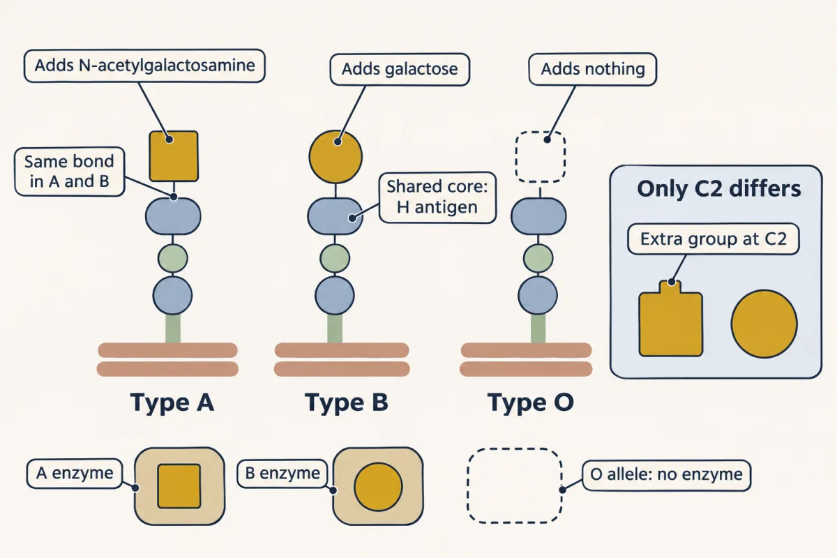
Your ABO blood type comes down to which glycosyltransferase (a sugar-adding enzyme) you inherit, acting on a shared core antigen called the H antigen. The A enzyme adds N-acetylgalactosamine, the B enzyme adds galactose, and the O allele adds nothing. A and B add their sugar by the same α1-3 bond, and the two sugars differ only at C2, where N-acetylgalactosamine carries an acetylated amino group in place of galactose’s hydroxyl. One sugar’s worth of difference, and a small one, sets your ABO type, a key match for which blood you can receive.
Sugar coats do physical work too. Cartilage cushions your knees because the proteoglycan aggrecan, about 90 percent glycosaminoglycan by weight, traps water in its sugar chains and releases it under load, like a molecular sponge.
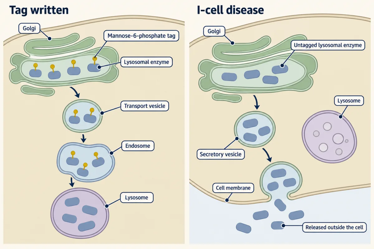
And the cell labels its own deliveries with sugar. A mannose-6-phosphate tag is the address that routes enzymes to the lysosome. Lose the ability to write that tag and the enzymes are dumped into the blood instead, the molecular lost-mail problem behind I-cell disease.
Lectins read what the sugar code wrote
A code is only useful if something reads it, and the readers are lectins, proteins that bind specific oligosaccharides. Any one sugar-protein grip is weak, typically with a dissociation constant (Kd) in the micromolar to millimolar range. Lectins solve that with multivalency: many weak handholds at once add up to a strong, selective grip, the way a strip of hook-and-loop holds though no single hook would. Selectins, a lectin family, use exactly this trick to slow circulating immune cells at a site of injury, and in a proposed role they also help an embryo attach to the uterine wall.
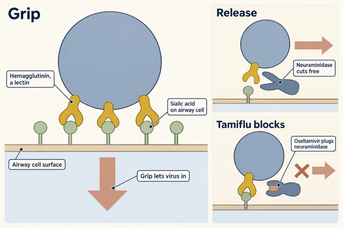
The flu exploits the same chemistry against you. Influenza’s hemagglutinin is a lectin that binds sialic acid, the negatively charged terminal sugar on your airway cells, and that grip is how the virus latches on before it gets in. To leave, new virions must cut themselves free, which is the job of the viral enzyme neuraminidase. Oseltamivir, sold as Tamiflu, inhibits neuraminidase, so new virus has trouble releasing and spreading: a drug aimed at a sugar-cutting enzyme, designed because a virus reads your sugar code.
The idea, with its scope
Free, the anomeric carbon runs mutarotation, reducing power and HbA1c; used, its bond sets the shape of the chain. The sugar coat adds which sugar, on which bond, read by which protein.
Going deeper
Why the split is not more lopsided
The 36:64 α:β ratio is a small energy difference, about 1.4 kJ/mol in favor of β at 25 °C. Crowding alone predicts more: illustrative costs for an axial hydroxyl (2.5 to 3.8 kJ/mol) would give about 73 to 82% β. The observed 64% is lower because of the anomeric effect: an electronegative group on C1, next to the ring oxygen, is stabilized when axial by the ring oxygen’s lone pairs. “Everything equatorial” is the right first answer; the anomeric effect is the correction.
Glycogen particle size: a model
Where do the model’s numbers come from? Build 12 tiers of 13-residue chains, each inner chain carrying two branches: that gives 4,095 chains, 53,235 glucoses and 2,048 outer chains. Its spacing, one branch per 13 residues, sits just above the textbook 8 to 12. By the b + 1 rule it has 4,094 branch points and so 4,095 non-reducing ends, one per chain; the 2,048 on the outer chains are the ones phosphorylase reaches first, which is the “about 2,000” quoted above.
Sugar or fat for a sprint
Strictly, a sprint draws on phosphocreatine and on glycogen through glycolysis; fat oxidation is too slow and needs oxygen. Over a longer time, the energy of both sugar and fat comes from oxidation. Sugar carbon is partly reduced, and oxidizing it to CO₂ and water releases energy because the products are held together by stronger bonds than the starting sugar and oxygen.
A hormone with a sugar fingerprint
Erythropoietin, the hormone that drives red-cell production, is about 40 percent carbohydrate by weight. Its glycosylation both stabilizes it in the blood and gives anti-doping labs a glycan fingerprint to catch synthetic EPO.
Your notes
Select any text in the lesson to highlight it or add a note to it. Your highlights and notes are saved in this browser and collected in your notebook.
This browser is not letting the site save anything (a private window or strict privacy settings), so highlights and notes will be lost when you leave the page.
How we measure it
Reading the isomer decision tree
Same formula but different connectivity → constitutional isomers. Same connectivity, different 3-D arrangement → stereoisomers; mirror images → enantiomers, non-mirror → diastereomers; differ at one chiral center → epimers (glucose/galactose at C4); differ at the anomeric carbon → anomers. Walk the tree and any two sugars get a name.
Drawing and decoding a Fischer projection
Vertical bonds point back, horizontal bonds point toward you. Look at the chiral carbon farthest from the carbonyl (the lowest one in the drawing): its hydroxyl on the right means D, on the left means L. Biology runs almost entirely on D-sugars, so the convention does real work.
The reducing-sugar test (Benedict's / Fehling's)
A free anomeric carbon can open to a free carbonyl (an aldehyde in glucose) and reduce blue Cu(II) to brick-red Cu₂O. Maltose and lactose pass; sucrose fails because its bond ties up both anomeric carbons. The same open-chain reactivity is what glycates your proteins.
Practice questions on this lesson (3) → MCAT-style, original, each with an explanation
Where this lesson fits
Before thisReview first
- Water, Weak Bonds, and the Generation of OrderThis course · Lesson 3
You need hydrogen bonding and weak interactions from there to see how cellulose chains pack and why glucose dissolves.
- What Am I Made Of?Biology · High school
It introduces carbohydrates as polymers built from sugar monomers, which this lesson then explains in detail.
After thisStudy next
- Glycolysis: The Investment-and-Payoff PathwayThis course · Lesson 16
The free glucose ring you met here is the molecule this pathway breaks down.
- Digestion and Absorption: Turning a Meal into Molecules Your Cells Can UseThis course · Lesson 14
Amylases cutting α-1,4 bonds and lactase cutting β-1,4 bonds are followed here through the gut.
- Integration of Metabolism: How the Body Switches FuelsThis course · Lesson 30
Glycogen branching and fast mobilization from non-reducing ends are used here when the body switches fuels.
Suggested by an AI model from the lessons themselves, then checked. How we use AI
Finish line
Test yourself
8 questions
Practice and MCAT-style, with explained answers.
Play it · about 4 min
Metabolic Autopsy: Glycogen
A glycogen storage disease case. Find the missing enzyme.
Play →Next · Lesson 11
Lipids: The Building Blocks That Refuse to Polymerize
Lipids make the barriers around every cell and the densest fuel in your body. One packing rule, how well the tails lie together, explains why a fat is solid or liquid and why a membrane is stiff or fluid.
Continue →← Lesson 9 · Protein Binding and Allostery: How Hemoglobin Loads and Delivers Oxygen