Cell Biology · Unit 3: Traffic and movement
New to this? Review it at high school level: Organelles as a System
The Secretory Pathway
Rewritten on : new text, flashcards and film. Your notes and highlights stay, but the text around them may have moved.
Label a pancreas cell's new proteins for a few minutes, then watch them move: what route do they take, and how do you know?
Pause when the reading reaches a film, and offer to play it. Saved on this device.
Watch the film now, or keep listening
Builds on:Protein Sorting into CompartmentsVesicle Budding, Targeting and FusionExperiments That Distinguish MechanismsWhy these?
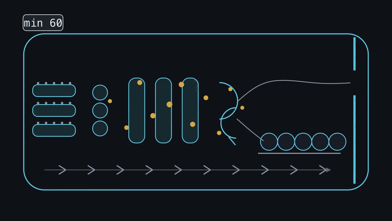 ▶ Watch Forward Is the Default · 3:37
Read the transcript of “Forward Is the Default”
Film transcript: Forward Is the Default
Label the new proteins a pancreas cell makes in a few minutes. An hour later, where are they? Could the proteins themselves tell you how far they have come? A pulse-chase gives a brief pulse of radioactive amino acid, then a flood of the unlabelled one. Only the batch made during the pulse is labelled: the cohort. Caro and Palade did this in the guinea-pig pancreas.
Silver grains in a photographic layer marked the label. At about 5 minutes they lay over the rough ER. At about 20, over the Golgi. At about an hour, over the storage granules. So the cohort's peak moves forward through a fixed order of stations, though it spreads out. The ER, a sorting dock, the Golgi stack, and the trans-Golgi network, where routes split. Now follow one protein. In the ER it gets a ready-made tree of 14 sugars, trimmed as it folds. In the Golgi, enzymes in successive sacs cut and rebuild it, roughly in order. One, mannosidase II, marks a red line. But these enzymes overlap from sac to sac, so the tree cannot say which sac the protein is in. It records the enzymes it has met: a rough odometer, not an address.
To read the record, sample at each chase time and treat each sample with an enzyme, Endo H. It cuts chains still mostly mannose, but not those rebuilt past mannosidase II. Cut protein loses sugar and runs faster on a gel. Over the chase, the cut band gives way to the uncut one. Some proteins should stay.
ER chaperones, which help others fold, leak forward too. Many end in a tag, KDEL. Past the ER, in slightly acidic compartments, a receptor grabs the tag. A coated vesicle carries both back, and in the near-neutral ER the receptor lets go. ER chaperones, which help others fold, A coded vesicle carries both back, and a coded vesicle carries both back, too. A coded vesicle carries both back, Now snip the tag off. Sean Munro and Hugh Pelham did this to the chaperone grp78, in cultured monkey cells. Where does it end up?
It is secreted with the cargo, because its KDEL tag is gone. For soluble cargo in animal cells, forward is the default unless a protein is held, fetched back, or sorted out. For soluble cargo, the trans-Golgi network can split the routes. Some carriers go straight to the surface: constitutive secretion, the default door. In this cell, digestive enzymes are sorted into granules instead: regulated secretion. They wait on a shelf until a stimulus releases them. Does that stimulus also speed up the line? So where are the labelled proteins an hour later? Mostly in storage granules. A glycoprotein among them can carry a sugar record of the enzymes it met.
For soluble cargo, going on is the default. Staying means being held or fetched back. Storing means being sorted, in ways still argued.
From the film
- A pulse-chase labels one batch of new protein, the cohort: in the guinea-pig pancreas it lay over the rough ER at about 5 minutes, the Golgi at about 20 and the storage granules at about an hour.
- The sugar chain goes on whole in the ER and is cut and rebuilt step by step by overlapping Golgi enzymes, so it records which enzymes a protein has met: a rough odometer, not an address.
- Endo H, added to samples taken over a chase, cuts chains not yet rebuilt past mannosidase II; as the cohort is processed, the faster cut band gives way to the uncut one.
- For soluble cargo in animal cells, forward is the default destination: in COS cells, the ER chaperone grp78 lost its KDEL tag and was secreted, while tagged proteins are fetched back.
- For the soluble cargo compared here, the trans-Golgi network can release it at once by constitutive secretion (the door) or store it in granules of regulated secretion (the shelf) until a stimulus.
The first film ended on its rule; here it is with its scope.
The one idea to carry through this lesson. Forward is the default. It sets a protein’s destination, not its speed, and a few proteins leave by other routes (Going deeper).
The route, station by station
A secretory protein enters the ER as it is made, guided by its signal sequence and SRP (the signal recognition particle, which brings the ribosome to the ER); protein sorting covers the ribosome and channel, and translation covers protein synthesis. In the lumen, the space inside the ER, the enzyme signal peptidase cuts the signal off. Then come the stations, each with characteristic roles that overlap at the edges.
| Station | Its main roles |
|---|---|
| Rough ER | Makes the protein, adds the core sugar chain, checks folding |
| ERGIC (ER-Golgi intermediate compartment) | The sorting dock between ER and Golgi, marked by the protein ERGIC-53 |
| cis Golgi | The entry face of the stack; Golgi sugar processing begins |
| Medial and trans Golgi | Flattened sacs (cisternae) that continue and elaborate the sugar processing, overlapping into the TGN |
| Trans-Golgi network (TGN) | A tubular exit hall on the far side, where the routes split |
| Door or shelf | Release at once, or storage in a granule until a stimulus |
Folded cargo leaves at ER exit sites in carriers coated with the protein coat COPII, long drawn as small vesicles, though some may be tubules. A second coat, COPI, makes carriers that travel backward. Both coats, and the SNAREs that fuse carriers, are in vesicle traffic.
The first film’s pulse-chase is Lucien Caro and George Palade’s 1964 experiment in living guinea pigs, read by electron-microscope autoradiography. James Jamieson and Palade then found the same order by fractionating pancreatic slices. Going deeper covers both methods and how one fraction misled them; distinguishing experiments sets out pulse-chase in general.
Keep the scope exact. These experiments, in one tissue of one species, showed the order of compartments and roughly when the labelled cohort reached each one; later work placed the ERGIC and the TGN within that coarse order. Jamieson and Palade argued that the kinetics were direct evidence for passage through small Golgi vesicles, but no one watched cargo ride inside a vesicle. Order and timing are what pulse-chase gives you.
The odometer: what the sugar chain records
The first film drew the sugar tree as a cartoon; here is the fuller chemistry.
In the ER the chain goes on whole. Very soon after the protein enters the lumen, the enzyme oligosaccharyltransferase attaches a preformed core of 14 sugars (Glc3Man9GlcNAc2: three glucoses, nine mannoses, two GlcNAc) to an asparagine in the sequence Asn-X-Ser/Thr (asparagine, almost any amino acid, then serine or threonine). So N-glycosylation begins in the ER, not the Golgi; the sugar chemistry is in the carbohydrates lesson. Glucoses are trimmed while the protein folds, and the folding check in quality control holds back protein that has not folded. What leaves carries a high-mannose glycan, made mostly of mannose.
In the Golgi the chain is rebuilt, step by step. Early enzymes cut mannoses off and add GlcNAc; one of them, mannosidase II, trims two more mannoses on the way to the complex type. Trimming by mannosidase II and then GlcNAc branching lead to a complex glycan, which many cells go on to decorate with galactose and often sialic acid. A hybrid glycan is part way: one branch rebuilt, the other still high-mannose. The enzymes are enriched in different cisternae but overlap (Going deeper).
Endo H reads the record at one mark. Endoglycosidase H (Endo H) cuts high-mannose and most hybrid glycans, but not the complex glycans made after mannosidase II has acted. Cut protein is smaller, so it runs faster on an SDS gel, which sorts proteins by size. Resistance is a threshold in a sequence, not a switch between organelles: it shows that a glycan has been processed past mannosidase II. Sensitivity alone does not locate the protein, which could be in the ER, the ERGIC or the first cisterna, or carry a glycan that is never processed. The record branches, and some glycans stay high-mannose even after their protein has crossed the Golgi.
Worked problem: reading an Endo H time course
The first film’s gel, as numbers. The data are illustrative. A glycoprotein in a cultured cell line is pulse-labelled for 5 minutes and chased. At each time you immunoprecipitate it, digest with Endo H, and measure the two bands. Assume nothing is secreted or degraded in 60 minutes, so each pair sums to 1,000.
| Chase (min) | Resistant | Sensitive |
|---|---|---|
| 0 | 0 | 1,000 |
| 15 | 330 | 670 |
| 30 | 550 | 450 |
| 60 | 800 | 200 |
Step 1, fractions resistant: 0, 33%, 55% and 80%.
Step 2, test for first order. If the fraction still sensitive, S, falls as S = e−kt, then k = -ln S / t at every time.
k = -ln(0.67) / 15 = 0.0267 per min; -ln(0.45) / 30 = 0.0266 per min; -ln(0.20) / 60 = 0.0268 per min
The three agree, so the data fit a single first-order process with an apparent k = 0.0267 per minute. That does not mean only one physical step is involved.
- Resistant : sensitive
- Fraction resistant
- Fraction still sensitive, S
- k = −ln S / t
- Half-time, t½ = ln 2 / k
Endo H cuts high-mannose glycans but not the complex glycans made after GlcNAc transferase I and mannosidase II have acted, so the resistant band times arrival at those Golgi enzymes. These data are illustrative, each pair sums to 1,000 if nothing is secreted or degraded, and the picture is a sketch of the logic.
Step 3, half-time.
t½ = ln 2 / k = 0.693 / 0.0267 = 26 min
Step 4, interpret. Resistance appears only after ER exit and the Golgi processing that follows, so 26 minutes covers every step up to resistance. It is an upper bound for ER exit alone; telling which step is slow needs a separate measure of ER exit. Real cargoes leave the ER at clearly different rates (Going deeper).
Predict. One snapshot can be read too. In isolated rat hepatocytes under normal conditions, only about 10% of the haptoglobin inside the cells was Endo H resistant. What does that say about where the protein spends its time in the cell?
Check your prediction
Most of the haptoglobin in the cells had not yet been processed past mannosidase II. If flow is steady and little protein is lost, each pool’s share of the protein matches its share of the time spent in it, and if essentially every molecule becomes resistant before secretion, haptoglobin spent about nine times as long in the cell before becoming resistant as after (90 / 10). A sensitive bypass population would break this inference. The snapshot does not say which step before resistance is slow.
Fetched back: the KDEL signal
The chaperone that lost its tag in the first film was grp78 (also called BiP), in Sean Munro and Hugh Pelham’s 1987 work in COS cells, a line of cultured monkey cells. They had noticed that many soluble ER residents end in KDEL (lysine, aspartate, glutamate, leucine). The converse test settled it: adding grp78’s last six residues to chicken lysozyme, a secreted protein, kept lysozyme in the ER. In that system the tag was both needed and enough. (Yeast uses HDEL in place of KDEL.)
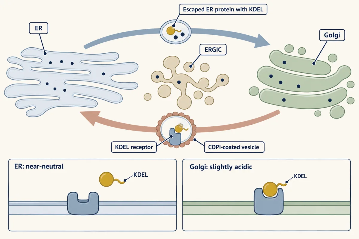
Predict. A tag could keep a protein in the ER by holding it there, or by fetching it back after it leaves. In 1988 Pelham put the tag on cathepsin D, a lysosomal enzyme whose sugar normally receives the first step of the mannose 6-phosphate mark (its lysosome address) from an early-Golgi enzyme. The tagged enzyme stayed in the ER. Under each model, would its sugar carry that Golgi step?
Check your prediction
Held in place, it would never meet the Golgi enzyme, so its sugar would lack the step. Fetched back, it would carry the step while sitting in the ER. Pelham found the Golgi-linked modification on enzyme in the ER: it had gone forward and been brought back. That is retrieval, read from the sugar record; later work on the receptor that binds the signal supported it.
Retrieval keeps ER residents home, but Golgi enzymes face the same problem one step on. The second film picks up here: how does cargo cross the stack while those enzymes stay at their level?
Read the transcript of “Crossing the Golgi Stack”
Film transcript: Crossing the Golgi Stack
Cargo moves through the Golgi, yet its enzymes stay at their level. Chapter 1 showed the same problem. An ER chaperone drifted forward. A receptor used its KDEL tag to bring it back. Each flattened sac of the stack is a cisterna. The cis face receives cargo arriving from the ER. The trans face passes it on to the trans-Golgi network. Early sugar enzymes are enriched toward cis, later ones toward trans, and they overlap. Picture one is the stable-compartment model. Each cisterna is long-lived and keeps a characteristic set of enzymes.
Small vesicles bud from one sac and carry cargo forward to the next. The compartments keep their cis, medial, or trans identities as cargo passes through. Rothman's lab rebuilt Golgi transport without intact cells. VSV G began in donor Golgi from one cell population.
Acceptor Golgi from another population added a sugar after transfer. That sugar was the assay's readout. Transport needed ATP and cytosol. Now weigh what the assay showed. Does it prove vesicles carry cargo forward, sac by sac, inside one stack? Separate what the experiment measured from the mechanism its inventors proposed.
Not on its own. The assay inferred transfer between separate Golgi populations from the sugar readout. It did not watch cargo move within one stack. Nor did it identify the carrier that crossed between populations. And transfer between populations fits the second picture too. Picture two is the cisternal maturation model. Here the cisterna itself carries the cargo. It forms at the cis face with cargo inside. Then it moves through the stack and ends as trans Golgi and trans-Golgi network. The compartment changes identity while its cargo remains inside. So how do the enzymes stay at their level? They keep being carried backward, in COPI vesicles, to the younger cisterna behind.
Backward recycling replaces early enzymes with later ones, as each cisterna matures. The sacs flow forward, while the enzymes keep going back. That is the KDEL retrieval logic: recycling against the flow. The comparison is only the logic. Golgi enzymes use different retention and retrieval machinery, not the KDEL receptor. Put the two pictures side by side. In one, the sacs stay put and the cargo hops between them. In the other, the cargo stays inside a sac that changes character from cis to trans. Which is right? In the text, predict how fast one filmed cisterna should change. For stacked mammalian Golgi, the question remains open. Glick and Luini judged that no single model explains every observation.
They proposed maturation as the best candidate for a shared core. So how does cargo cross the stack while its enzymes stay put? Fixed sacs and moving vesicles, or moving sacs and recycled enzymes. Either way, the cargo goes forward.
From the film
- Golgi enzyme populations stay enriched at different levels—early enzymes toward cis and later ones toward trans—even as cargo moves through.
- In the stable-compartment model, each cisterna keeps its enzymes and small vesicles carry cargo from one to the next.
- Rothman's cell-free assay inferred VSV G transfer between donor and acceptor Golgi from a sugar readout and needed ATP and cytosol; it did not observe transport within one stack.
- In the cisternal maturation model, the cisterna carries the cargo from cis to trans, and its enzymes are carried back in COPI vesicles to the younger cisterna behind.
- For the stacked mammalian Golgi the question is open; maturation has been proposed as the best candidate for a shared core.
Crossing the Golgi: residents carried backward
The second film set out the stable-compartment model and the cisternal maturation model; the figure keeps them side by side. What it left out is the test that tells them apart. Rothman’s cell-free system used Golgi from Chinese hamster ovary (CHO) cells (Going deeper).
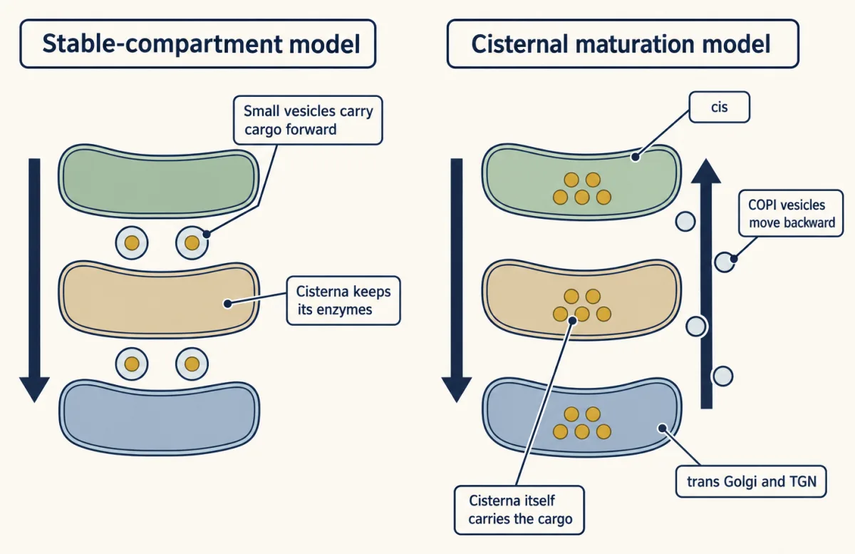
In the maturation picture, a cisterna forms at the cis face, progresses through trans Golgi to the trans-Golgi network (TGN), and keeps cargo inside while resident enzymes recycle backward in COPI vesicles. That recycling resembles KDEL retrieval—movement against the flow—but Golgi enzymes use their own retention and retrieval machinery.
Predict. Budding yeast has scattered Golgi cisternae rather than a stack, so one cisterna can be filmed alone. Label an early Golgi protein green and a late one red. If maturation carries cargo forward, how should the time for green-to-red conversion compare with the independently measured transit time of secretory cargo?
Check your prediction
The times should be similar: if cisternal maturation carries cargo forward, marker conversion should keep pace with cargo transit. A stable compartment, by contrast, should retain one identity while cargo moves onward. In 2006 the laboratories of Benjamin Glick and Akihiko Nakano each filmed single cisternae carrying an early and a late fluorescent marker. A cisterna lost the early marker and gained the late one, and Glick’s group found the rate matched cargo transit. Later imaging showed cargo staying within a cisterna as it matured: in yeast, maturation is seen directly.
For the stacked mammalian Golgi, as the second film said, the question is open: Glick and Alberto Luini proposed maturation as the best candidate for a shared core.
Two exits: the door and the shelf
In the first film’s terms, constitutive secretion is the door. Regulated secretion is the shelf, with protein stored at high concentration in granules.
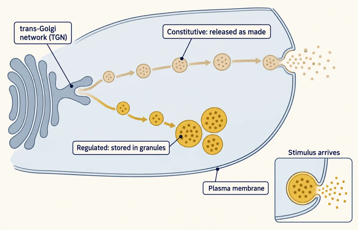
Getting onto the shelf takes sorting, and how the selection is made is still argued; clumping of regulated cargo, such as granins, may help (Going deeper). In pancreatic beta cells, entry from the trans-Golgi network (TGN) into immature granules is not very selective: regulated proteins condense inside, and lysosomal enzymes that entered with them are removed later. There too, in human and rat, proinsulin is cut to insulin mainly in clathrin-coated immature secretory granules, not in the Golgi stack.
Predict. In the first film a stimulus empties the shelf. Does it also speed the line? James Jamieson and Palade stimulated guinea-pig pancreatic slices with carbamylcholine, a secretagogue (a drug that triggers secretion). Would you expect faster protein synthesis, faster transit through the cell, or neither?
Check your prediction
Neither. The secretagogue did not speed protein synthesis, drainage of the rough ER, or overall transit through the cell; the results suggested that it mainly acted on discharge. The line kept its own pace, and the stimulus emptied the shelf.
Design problem
AtT-20 cells, a mouse pituitary tumour line, make the hormone adrenocorticotropic hormone (ACTH) from a larger precursor. They also carry an endogenous leukaemia virus and make its viral envelope glycoprotein. Design an experiment to show that one cell has two exit routes.
Saved in this browser only.
One way to do it
Pulse-label the cells, then chase, collecting the medium at intervals. Split the chase into two arms, with and without 8-bromo-cyclic AMP, a cyclic AMP analogue that stimulates these cells. Immunoprecipitate the viral glycoprotein, precursor ACTH and mature ACTH from medium and cells.
Expected result, as Gumbiner and Kelly reported in 1982: the viral glycoprotein and some precursor ACTH leave steadily, with little change from the stimulus, while mature ACTH is held in the cells and released four to five times more when stimulated. As a further test, Moore, Gumbiner and Kelly found that 200 micromolar chloroquine diverted new ACTH into the steady route: cargo that fails to be stored falls through to the default door.
Controls: equal synthesis in both arms (labelled protein in cells at the end of the pulse), viability under the stimulus, and a cell-associated count to show stored hormone falls when medium hormone rises. Limits: this is a tumour line, and the experiment does not identify the sorting signal.
Going deeper
Autoradiography, microsomes and the granules that filled too early
Caro and Palade injected labelled leucine and, in most animals, followed it with a large excess of unlabelled leucine. In autoradiography, photographic emulsion laid over thin sections forms silver grains where radiation from the label strikes it. The grains moved from the rough ER to Golgi elements to zymogen granules, stores of digestive enzymes made as inactive precursors (zymogens). Caro and Palade read their data as suggesting two steps in the Golgi region, small peripheral vesicles first and then large central condensing vacuoles, sacs in which secretory protein is concentrated before it becomes a granule.
Jamieson and Palade then moved to slices of pancreas kept alive in a dish, which allowed a precise three-minute pulse and chases of 7, 17 and 57 minutes. They broke the cells open and separated microsomes, small vesicles that form from broken membranes, not compartments of the living cell. Label was in rough microsomes (from the ER) at the end of the pulse, peaked in smooth microsomes (from the Golgi periphery) after 7 minutes of chase, and arrived in the zymogen granule fraction later. That is a precursor-product relation: each compartment fills as the one before it empties.
The two methods disagreed at one point. The granule fraction was half-maximally labelled at 17 minutes, yet in intact cells the granules carried label no earlier than 57 minutes. The cause was contamination: labelled condensing vacuoles were sedimenting with the granules. The principle is that a fraction is only as good as its purity. A year later the same slices showed that the step between the ER exit region and the peripheral Golgi vesicles needs energy, and is blocked by inhibitors of respiration but not of glycolysis.
Inside the Golgi: enzyme order and the cell-free assay
Mannosidase I, GlcNAc transferase I, mannosidase II and GlcNAc transferase II act early; galactose and sialic acid are added later. In Chinese hamster ovary (CHO) cell membranes the early enzymes partly separated from the late ones on density gradients, and the authors suggested that they work in some or all of the cis-side cisternae.
Rothman’s cell-free assay measured transfer of VSV G between Golgi stacks from donor and acceptor CHO cells, and showed that transport needs ATP and cytosol. It did not show that vesicles carry cargo forward within one stack, and transfer between stacks fits maturation too.
Export rates of real cargoes
Four secretory proteins in isolated rat hepatocytes left the ER with half-times from 14 to 137 minutes, and in human HepG2 cells five secreted proteins matured at rates differing at least threefold. That spread was first read as evidence against pure bulk flow, and Lodish and colleagues suggested a receptor that selects cargo, though folding and retention in the ER also contribute. For comparison, VSV G protein fused to green fluorescent protein moved from ER to Golgi in living COS cells at about 2.8% per minute (a first-order rate constant), a half-time of 0.693 / 0.028 = 24.8 minutes.
Leaving the TGN, and other ways out
In a cell-free system from PC12 neuroendocrine cells, a regulated protein and a constitutive one were sorted into different vesicles as they left the trans-Golgi network (TGN). Granins, typical regulated cargo, stay aggregated at pH 6.4 with 1 to 10 mM calcium, conditions then believed to hold in the TGN. Later calcium probes found less free calcium in the trans-Golgi than in the medial Golgi or the ER, so how closely those test conditions match the living TGN is unclear.
Not every secreted protein follows this whole path: some bypass the Golgi after entering the ER, and some proteins without a signal sequence leave without entering the ER at all. Lysosomal enzymes leave the flow at the TGN by a different signal, mannose 6-phosphate.
Your notes
Select any text in the lesson to highlight it or add a note to it. Your highlights and notes are saved in this browser and collected in your notebook.
This browser is not letting the site save anything (a private window or strict privacy settings), so highlights and notes will be lost when you leave the page.
How we measure it
Pulse-chase with autoradiography
Cells or tissue get a radioactive amino acid for a few minutes, then a large excess of the unlabelled one, so only the protein made during the pulse carries label. Fixing samples at chase times and laying photographic emulsion over thin sections shows, by the silver grains, which compartment holds the labelled cohort at each time.
Subcellular fractionation
Homogenized tissue is separated by centrifugation into fractions such as rough microsomes, smooth microsomes and secretory granules. Measuring label in each fraction over a chase gives a precursor-product time course, as long as each fraction is pure; contamination by a neighbouring compartment can make label appear early.
Endo H digestion and SDS gel electrophoresis
An immunoprecipitated glycoprotein is treated with endoglycosidase H, which removes high-mannose and most hybrid N-glycans but not complex ones, and run on a gel. Sensitive protein runs faster after digestion; resistant protein does not. The rise of the resistant band over a chase times arrival at the Golgi enzymes that make glycans resistant.
Live imaging of a fluorescent cargo
A cargo such as the temperature-sensitive VSV G protein, fused to a fluorescent protein, is held in the ER and then released all at once by a temperature shift (or a chemical switch in the RUSH system). Fitting a kinetic model to fluorescence in the ER, Golgi and surface over time gives effective rate constants for each stage in living cells.
Method films · the methods library
- Binding assays: immunoprecipitation, pull-down and gel shift 3:43
- Cell-free extracts and reconstitution 4:01
- Centrifugation and subcellular fractionation 2:50
- Engineered mutants: site-directed, dominant-negative and constitutively active 4:25
- Live-cell time-lapse imaging 4:01
- Pulse-chase labelling 2:40
Practice questions on this lesson (3) → MCAT-style, original, each with an explanation
Where this lesson fits
Before thisReview first
- Protein Sorting into CompartmentsThis course · Lesson 7
You need signal sequences and SRP entry into the ER from there before following the protein onward.
- Vesicle Budding, Targeting and FusionThis course · Lesson 11
COPII and COPI carriers and SNARE fusion are used here without being re-taught.
- Experiments That Distinguish MechanismsThis course · Lesson 3
Pulse-chase logic and fractionation are used here to read order and timing.
After thisStudy next
- Endocytosis, Recycling and DegradationThis course · Lesson 13
Cargo that reached the surface by this route is taken back in and sorted to recycling or degradation.
- Cell Identity, Cell State and DiseaseThis course · Lesson 25
Secretory capacity and secretion routes feed into how cell identity and cell state shape what a cell exports.
Elsewhere on the siteFilms, simulations and method films in other courses
- Build a big molecule from small blocksSimulation · Biology
Building large molecules from blocks shows how sugar chains are put onto and trimmed from a protein.
- One Cell, One Shipping LineFilm · Biology
It follows insulin from nucleus through rough ER and Golgi to secretion, the same route traced here.
- A Room With No Way OutFilm · Biochemistry
It shows how membrane-bound compartments keep a lysosomal enzyme's cargo in its own room, as the Golgi stack does.
Suggested by an AI model from the lessons themselves, then checked. How we use AI
Finish line
Test yourself
8 questions
Practice and MCAT-style, with explained answers.
Next · Lesson 13
Endocytosis, Recycling and Degradation
Why can a patient's cells grip cholesterol-carrying LDL normally and still leave most of it on the surface?
Continue →