Cell Biology · Skim version
Adhesion and the Extracellular Matrix
Read the full lesson → Skim the whole course →
The guiding idea
Take the calcium away and a sheet of epithelial cells comes apart. What was the calcium holding together?
Cells in a tissue hold on to each other through cadherins and to the matrix around them through integrins.
From the film: One Chain, Four Links
- In its simplest form, each anchoring grip is one chain: partner outside, receptor, adaptors and a cable inside.
- In one simple chain, the pull passes through every link, so one missing link cuts that path.
- At adherens junctions, calcium keeps the cadherin arms stiff; without it, the outer link lets go.
- In epithelia, desmosomes and hemidesmosomes build the same kind of chain but end on keratin cables, not actin.
- Collagen ropes resist pulling far better than squeezing; water-holding proteoglycan gels resist the squeeze.
From the film: Links That Feel the Pull
- Myosin II can pull on actin cables, so a cell can load its adhesion chains from inside.
- Stretching a purified talin rod exposes hidden sites to a vinculin-head test piece; in cells, activated vinculin can add a tie to actin.
- α-Catenin has a hidden vinculin site that tension opens, so the cadherin chain can reinforce itself too.
- Normal talin and vinculin did not bind at mitochondria; releasing either protein's autoinhibition enabled force-independent binding.
- Active forms supported disorganized adhesions with myosin II blocked, but how normal signals bypass force in a tissue is unknown.
-
The chain, link by link
-
Where the chain breaks: blisters
-
The far end: what the matrix is made of
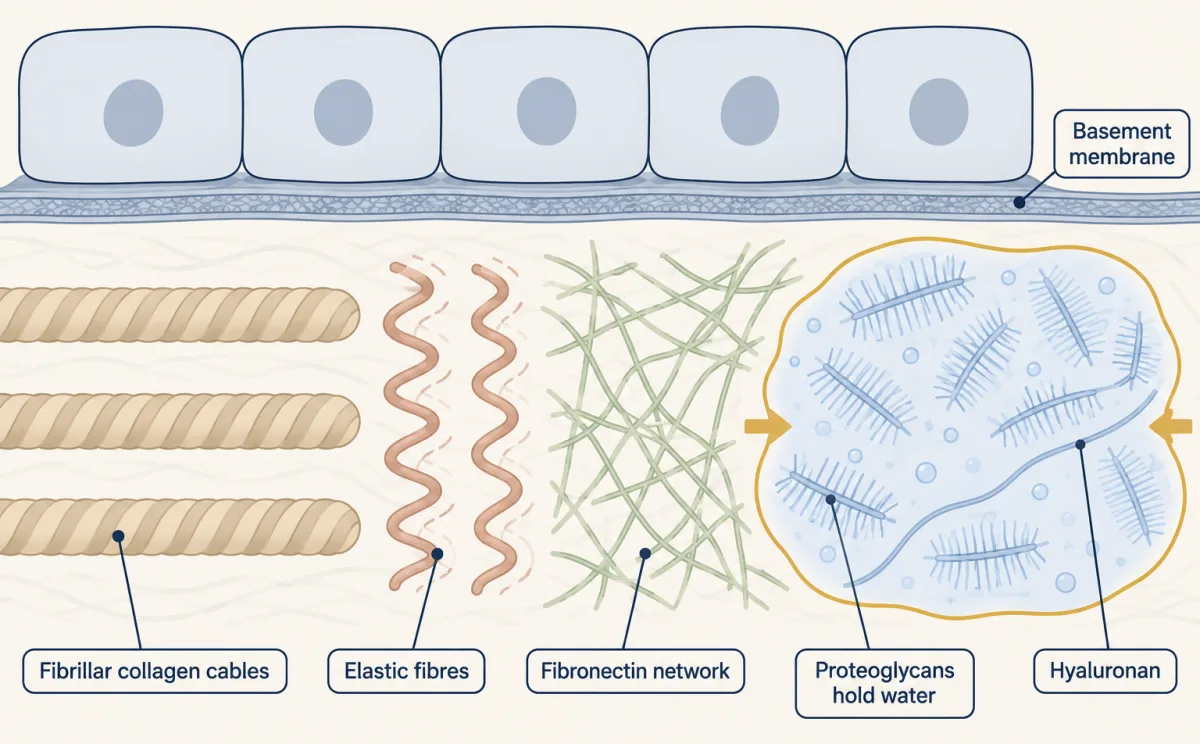
The matrix has parts matched to jobs: collagen ropes resist pulling, elastic fibres recoil, fibronectin is a mesh cells grip, basement membranes are thin sheets of laminin and type IV collagen, and a proteoglycan gel resists compression. AI-generated illustration -
Which links feel the pull?
-
Reading adhesion assays
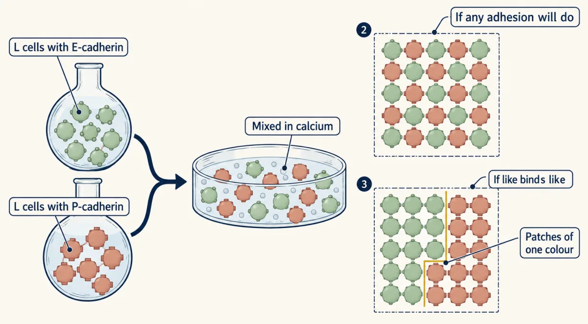
Two L-cell lines carry different cadherins and are mixed in calcium. If contacts are equally good whatever the cadherin, the colours intermix; if cells favour partners with the same cadherin, each aggregate shows patches of one colour. AI-generated illustration -
Going deeper
Where this lesson fits
Before thisReview first
- Cytoskeletal Polymers and DynamicsThis course · Lesson 14
The cable at the end of every chain is actin or keratin, so you need how these filaments are built.
- Membrane Organization and TopologyThis course · Lesson 4
Cadherins and integrins are single-pass membrane proteins whose tails face the cytosol, which this lesson assumes.
- The Secretory PathwayThis course · Lesson 12
Collagens and other matrix proteins are made and trimmed through this route before they reach the matrix.
After thisStudy next
- Epithelia and Tissue OrganizationThis course · Lesson 23
Sheets of cells held by adherens junctions, desmosomes and a basement membrane are put to work in tissue organization here.
- Stem Cells and Tissue RenewalThis course · Lesson 24
Niche attachment and matrix signals use these adhesion chains to hold stem cells and steer their fate.
- Designing a Cell Biology ExperimentThis course · Lesson 26
The blocking-antibody and control-well logic from the adhesion assays carries into designing a full experiment.
Elsewhere on the siteFilms, simulations and method films in other courses
- A Path Is Not a PumpFilm · Biochemistry
It shows how the membrane holds receptors like cadherins and integrins.
Suggested by an AI model from the lessons themselves, then checked. How we use AI