Physiology · Skim version
Thyroid Hormone: Tissues Decide What T4 Becomes
Read the full lesson → Skim the whole course →
The guiding idea
A person with no thyroid who takes only T4 still has T3 in the blood. Where does it come from?
The thyroid secretes mostly T4, yet T3 is the hormone that acts, and tissues help decide how much T3 a cell gets: deiodinases switch T4 on or off, and transporters let it in.
-
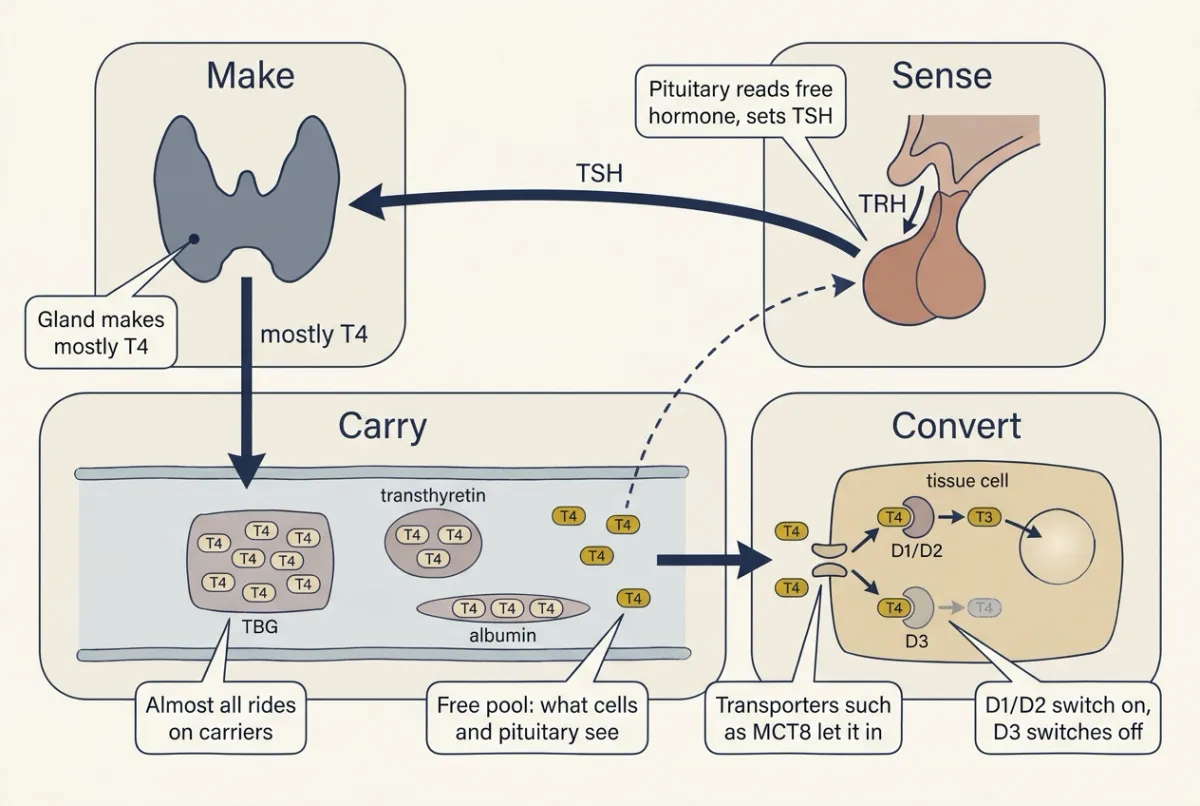
Thyroid hormone runs around one loop: the gland makes mostly T4, the blood carries almost all of it on proteins, each tissue switches T4 on or off, and the pituitary senses the small free pool and sets TSH. To read a test, ask which station failed. AI-generated illustration -
The gland sends mostly T4; tissues decide how much becomes T3
-
What T3 does: development first, then heat and the heart
-
Sense: one loop holds free hormone steady
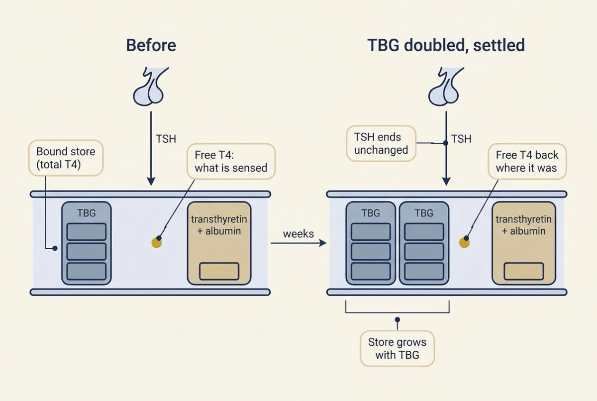
With the loop intact, doubling TBG enlarges the bound store, so total T4 rises (80 to 140 nmol per L in the made-up example), but once things settle free T4 and TSH end where they started: binding proteins change the total, not the free. AI-generated illustration -
Make: how the follicle builds hormone, and what the scan sees
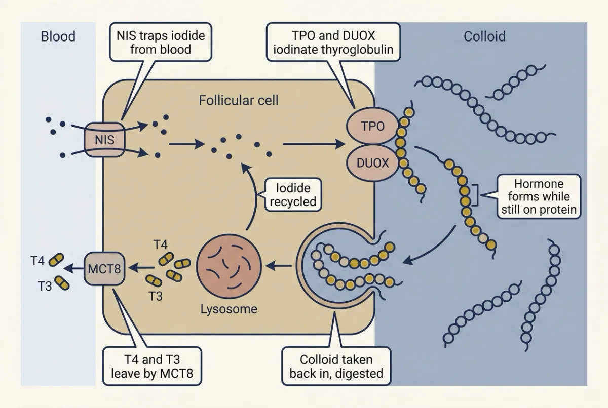
The follicle makes hormone across one cell: iodide is trapped on the blood side, fixed onto thyroglobulin on the colloid side, stored as hormone on that protein, then taken back in and digested so mostly T4 leaves into the blood. AI-generated illustration -
Locating a fault: read the pair, then the scan
-
Going deeper
Where this lesson fits
Before thisReview first
- Endocrine Axes and How to Test ThemThis course · Lesson 2
You need its paired hormone levels and feedback testing here to read TSH with free T4.
- Body Water and Its CompartmentsThis course · Lesson 1
Free versus bound hormone in plasma builds on how substances distribute between body compartments.
After thisStudy next
- The Adrenal Cortex and MedullaThis course · Lesson 20
Another hormone axis where tissue enzymes and binding proteins shape what reaches the cell, with a similar feedback loop.
- Exercise: The Whole Body at WorkThis course · Lesson 30
Thyroid hormone's effects on metabolic rate and cardiac output matter when the whole body works hard.
Suggested by an AI model from the lessons themselves, then checked. How we use AI