Biochemistry · Cellular Respiration
New to this? Review it at high school level: Respiration and ATP
Oxidative Phosphorylation: The Electron-Transport Chain
Rewritten on : new text, figures, flashcards and film. Your notes and highlights stay, but the text around them may have moved.
Electron flow and ATP synthesis are two separate machines linked by one thing: the proton gradient.
Pause when the reading reaches a film, and offer to play it. Saved on this device.
Watch the film now, or keep listening
Builds on:The Citric Acid Cycle: Harvesting ElectronsMetabolism: Basic Concepts and DesignMembranes: The Impermeable Sheet That Builds ItselfWhy these?
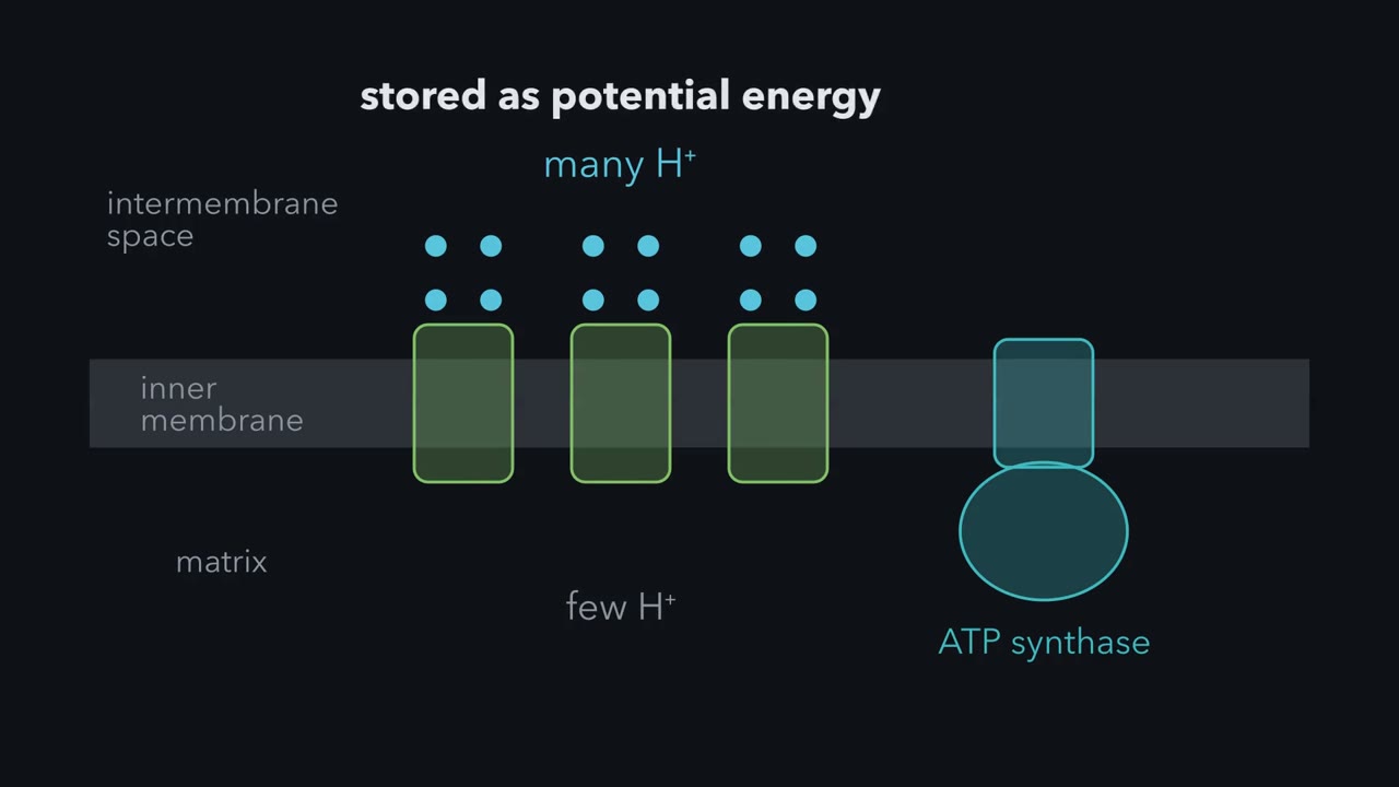 ▶ Watch Electrons build a proton battery · 3:54
Read the transcript of “Electrons build a proton battery”
Film transcript: Electrons build a proton battery
This is the single most important idea in the chapter, so hold on to it. Electron flow and ATP synthesis are two separate events linked only by a proton gradient. On the left, electrons hop downhill through carriers in the inner membrane, and the big exergonic steps do work by pushing protons from the matrix out into the intermembrane space. That accumulation of protons is potential energy, like water behind a dam. On the right, ATP synthase lets those protons flood back down their gradient, and that flow drives the phosphorylation of ADP. Peter Mitchell proposed this in 1961, was met with deep skepticism and won the Nobel Prize for being right.
Let me show you why electrons move at all. Every carrier has a reduction potential, plotted here on the vertical axis. NADH sits high, at a very negative potential, meaning it holds electrons loosely and donates them readily. Oxygen sits at the bottom, strongly positive, meaning it grabs electrons hungrily. Left to themselves, electrons flow from low potential to high potential, from generous donor to greedy acceptor, and that is why they cascade from NADH down to oxygen. The total drop is about 1.14 volts, which translates to roughly minus 220 kilojoules per mole. That released energy is exactly what gets spent pumping protons across the membrane.
Now, we assemble the chain from named parts. Electrons from NADH enter at Complex I, NADH-Q oxidoreductase, and are handed to ubiquinone. Complex III, Q-cytochrome c oxidoreductase, passes them to cytochrome c. Complex IV, cytochrome c oxidase, delivers them to oxygen. Those three are the proton pumps. Notice the side door. Complex II, succinate-Q reductase, feeds electrons from succinate-derived FADH2 into the Q pool, but it does not pump protons. That single fact, that Complex II is a feeder and not a pump, explains why FADH2 yields less ATP than NADH. The reduction potentials on the left confirm the path runs downhill overall, even though a few hops inside the complexes are slightly uphill.
This is a beautiful piece of experimental logic, and it is a favorite exam scenario, so reason through it with me. If you block the chain at one point, everything upstream of the block piles up in the reduced state because electrons cannot leave, and everything downstream becomes oxidized because no new electrons arrive. So with an NADH-linked fuel only, rotenone, blocking Complex I, leaves NADH reduced and the rest oxidized. Antimycin A, blocking Complex III, leaves carriers up to cytochrome b reduced. Cyanide, blocking Complex IV, leaves the entire chain reduced and only oxygen unreduced. By reading which carriers turn reduced versus oxidized, you can deduce the exact sequence of electron flow.
Tie the whole chapter together with one image, the mitochondrion as a rechargeable battery. The electron-transport chain is the charging cable, using the energy of electron flow to pump protons and build up charge, the proton-motive force, across the inner membrane. ATP synthase is the discharge circuit, letting protons flow back down to do useful work and make ATP. Now the diseases and drugs make sense. Cut the charging cable as cyanide does by blocking Complex IV and the battery never charges. Add an uncoupler, which pokes holes in the membrane, and the battery short-circuits, draining its charge as heat instead of ATP.
Cyanide kills in minutes, and the reason is one enzyme at the very bottom of the electron-transport chain. Block that last step and electrons have nowhere to fall, the chain backs up, the proton gradient runs down, and a cell suffocates while surrounded by air.
Oxidative phosphorylation is two machines, not one. The first drops electrons down a ladder and uses the energy to pump protons out of the matrix, the mitochondrion’s innermost compartment, into the intermembrane space, like pumping water up behind a dam. The second, ATP synthase, lets them flow back like water through a turbine and uses that to make ATP. The two are linked by exactly one thing: the proton gradient. Treat the gradient as the currency and the famous misconceptions dissolve.
The one idea to carry through this lesson. Count protons per pair of electrons. Each pair pushes a fixed number of protons out of the matrix, set by which of the three pumps (Complexes I, III and IV) it passes: from NADH, 4 + 4 + 2 = 10; from FADH₂, which joins below Complex I, 4 + 2 = 6. Each ATP spends about 4 protons coming back in. So the yield is protons ÷ 4: 2.5 ATP per NADH and 1.5 per FADH₂. And anything that changes the gradient changes both rates: electrons can push protons out only as fast as ATP synthase, or a leak, lets them back in.
The yield arithmetic assumes an intact, tightly coupled membrane and uses rounded textbook values (Going deeper, “Why 2.5 and not 2.7”). With a leaky membrane (an uncoupler, below) it fails, though the rate half still holds. Neither half covers electrons pushed backward through Complex I (reperfusion).
The energy ladder
Electrons fall the way water falls. The height is the reduction potential, E°′: a more negative E°′ means a molecule donates electrons eagerly; a more positive one means it grabs them hard. Electrons flow spontaneously from negative toward positive, downstairs (what the explainer film calls “from low to high potential”).
Picture the chain as a five-story building, its landmarks from top to bottom: NADH in the penthouse at −0.32 V, succinate at about +0.03 V, coenzyme Q just below it near +0.04 V, cytochrome c at +0.25 V, and oxygen in the basement at +0.82 V. Succinate and Q sit almost level. Succinate’s electrons reach Q as FADH₂ inside Complex II, whose FAD sits close to that level too: a relay on that short hop, not a floor of its own. The complexes are the stairways between floors (table below).
In words Energy released equals electrons moved, times Faraday's constant, times the drop in potential; the minus sign means a downhill fall releases energy.
- ΔG°′“delta G naught prime”
- the standard free-energy change. Negative means energy is released.kJ/mol
- n“n”
- electrons transferred: 2 for one NADH or FADH₂
- F“F”
- Faraday's constant, about 96.5 kJ per mole per voltkJ/(mol·V)
- ΔE°′“delta E naught prime”
- the acceptor's E°′ minus the donor'sV
Sanity check If the two potentials are equal, nothing is released.
Try one number
NADH to oxygen: ΔE°′ = +0.82 − (−0.32) = 1.14 V, so ΔG°′ = −2 × 96.5 × 1.14 ≈ −220 kJ/mol per electron pair.
Need a refresher? units and prefixes
The cell spends that 220 kJ in stages, not one crash:
| Step | Drop in E°′ | Energy released per pair |
|---|---|---|
| Complex I: NADH → Q | 0.36 V | about 70 kJ/mol |
| Complex II: succinate → Q | 0.01 V | under 3 kJ/mol |
| Complex III: Q → cytochrome c | 0.21 V | about 40 kJ/mol |
| Complex IV: cytochrome c → O₂ | 0.57 V | about 110 kJ/mol |
The three steep drops make up the whole fall (70 + 40 + 110 = 220), and their energy pumps protons uphill, out of the matrix.
Predict. Pushing one proton out against a typical gradient (about 0.18 V) costs about 17 kJ/mol. Can Complex II pump?
Check your prediction
No. Under 3 kJ/mol cannot pay for one proton, and Complex II has no proton-pumping machinery anyway: it pumps zero protons, the structural reason FADH₂ yields less ATP than NADH.
The chain, in order
Everything happens on the inner mitochondrial membrane, folded into cristae and impermeable to ions. That is the whole foundation: a gradient is only useful if it cannot leak away. The matrix inside runs the citric acid cycle and fatty-acid oxidation.
Two mobile carriers link the complexes: lipid-soluble coenzyme Q, moving within the membrane (reduced, it is QH₂), and water-soluble cytochrome c, on the inner membrane’s intermembrane-space face, carrying one electron at a time.
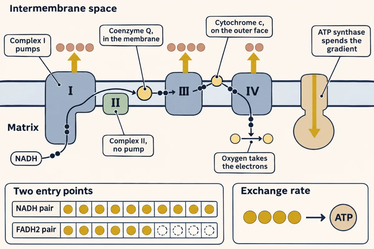
Complex I (NADH-Q oxidoreductase) is an L-shaped giant. NADH hands two electrons to FMN (a flavin, like FAD), which passes them through a relay of Fe-S clusters (small iron-sulfur groups that carry one electron at a time) to Q. It moves about 4 protons out per NADH.
Complex II (succinate dehydrogenase), also a citric acid cycle enzyme, oxidizes succinate to fumarate and passes the electrons through its FAD and Fe-S clusters to Q. It is small, only four subunits, and, as you predicted, pumps nothing.
Complex III (cytochrome bc1) passes electrons from QH₂ to cytochrome c. QH₂ carries two electrons but cytochrome c takes one, so the complex splits each pair by the Q cycle. Net per pair: one QH₂ in, two cytochrome c reduced, four protons out. The cost is a short-lived Q radical (a Q holding a single unpaired electron) that makes superoxide, O₂ with one extra electron, if it meets O₂: elegant and dangerous.
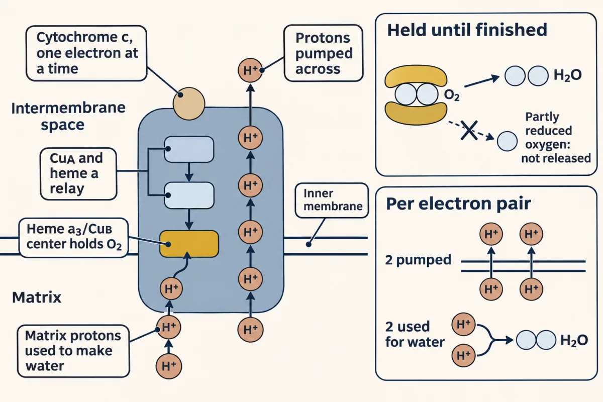
Complex IV (cytochrome c oxidase) is the terminal step, and it deals with oxygen, whose partly reduced forms are among the most damaging molecules a cell can make. Electrons pass through copper and heme centers to the binuclear center (a heme iron and a copper side by side), where O₂ binds. It holds oxygen and every partly reduced form of it until all of it is water, so partly reduced oxygen almost never escapes. Four electrons plus four protons plus O₂ give two waters. Cyanide and CO jam this site, and the chain dies here.
The proton ledger
One NADH, FADH₂ or QH₂ carries one pair of electrons; one oxygen atom (half an O₂) accepts one. Count per O₂ or per Q-cycle turn instead and the numbers will not add up.
| Step | Protons moved out per pair |
|---|---|
| Complex I: NADH → Q | 4 |
| Complex II: succinate → Q | 0 |
| Complex III: QH₂ → cytochrome c | 4 |
| Complex IV: cytochrome c → ½ O₂ | 2 |
- Complex IV pumps 2 protons out per pair and uses 2 more from the matrix to make water. Only the pumped ones go in this tally, but the water-making ones also move charge across the membrane, and that work takes part of the biggest drop. Per whole O₂ that is 4 pumped plus 4 used, the “8 per O₂” you may see elsewhere.
- Complex III releases 4, but two of them came from the matrix earlier: when Complex I or II loaded electrons onto Q, Q picked up two matrix protons with them, and QH₂ carried them across. That is how it can release 4 when its own 40 kJ pays to move only about 2.
The column gives the rule’s totals: 10 from NADH, 6 from FADH₂, whose pair joins at Q and never passes the first pump. Those four protons are the entire difference: both carriers bring two electrons, and every proton they move is identical.
The exchange rate: about 4 protons per ATP
The pumped protons store energy as the proton-motive force, the gradient’s push on protons to flow back in, which ATP synthase discharges. Each ATP made in the matrix and delivered to the cytosol costs about 4 protons back in:
- about 3 through ATP synthase itself (in mammals more exactly 2.7, set by the size of its rotor, which differs between species);
- about 1 more to move the goods: the carriers that bring ADP and phosphate in and send ATP out run on the same gradient.
So one NADH buys 10 ÷ 4 = 2.5 ATP and one FADH₂ 6 ÷ 4 = 1.5. Neither has to be a whole number, because no chemical step ties one electron pair to a fixed number of ATP: the protons go into one shared gradient, and ATP synthase draws from it.
Now one glucose. Glycolysis, pyruvate dehydrogenase and the citric acid cycle make 10 NADH and 2 FADH₂, plus 4 ATP or GTP made directly: 10 × 2.5 + 2 × 1.5 + 4 = 32 ATP. That counts the 2 cytosolic NADH fully, but they cannot cross the inner membrane, so a shuttle passes on their electrons. The malate-aspartate shuttle (heart, liver) delivers them into the matrix as NADH, giving the 32. The glycerol 3-phosphate shuttle (skeletal muscle, brain) hands them straight to Q from the intermembrane-space side, as if they were FADH₂, worth 1.5 ATP each instead of 2.5, which gives 30. So roughly 30 to 32 ATP per glucose, an order of magnitude beyond the 2 from glycolysis alone.
Switch the ledger below between the modern (2.5 / 1.5) and classic (3 / 2) factors, and between the two shuttles, to see where the old textbook figure of 38 came from.
| Stage | Direct ATP/GTP | NADH | FADH₂ | ATP from this stage |
|---|---|---|---|---|
| Glycolysis (cytosol) | 2 | 2* | 0 | — |
| Pyruvate → acetyl-CoA (×2) | 0 | 2 | 0 | — |
| Citric acid cycle (×2 turns) | 2 | 6 | 2 | — |
| Grand total per glucose | 4 | 10 | 2 | — |
— · —. * The 2 cytosolic NADH from glycolysis can't cross the inner mitochondrial membrane directly — a shuttle moves their electrons in, and which shuttle decides whether they're cashed as NADH or as FADH₂.
The famous "~38 ATP per glucose" is the old textbook number, built on round P/O ratios of 3 and 2. Measured proton stoichiometry gives the modern estimate of roughly 30–32, and the exact figure isn't a constant of nature — it depends on which shuttle a tissue uses and on the leakiness of the membrane. The lesson of this ledger isn't a number to memorize; it's that the cell does accounting, and the answer shifts with the assumptions you bring to the books.
Two machines that keep each other in step
The proton-motive force has two parts: a voltage, with the intermembrane side positive, and a pH difference, with that side more acidic. In mitochondria the voltage does most of the work, and both parts push protons back toward the matrix.
That push also works against the pumps. As the gradient builds, each proton costs more to push out. When a pair’s protons would cost nearly as much as its fall releases, electron flow stalls. That is the rule’s second half, and the return route is ATP synthase or a small leak. With little ADP to make into ATP, the synthase slows and the chain burns less oxygen.
Predict. Oligomycin blocks ATP synthase. What happens to oxygen use? Now add instead an uncoupler, a molecule that makes the membrane leaky to protons. What happens to oxygen use, ATP and heat?
Check your prediction
Oligomycin: protons cannot return through the synthase, the gradient fills, and oxygen use nearly stops, down to the leak rate.
Uncoupler: protons rush back without passing through ATP synthase. The gradient stays low, nothing holds the pumps back, and oxygen use speeds up. ATP output falls, and the energy comes out as heat.
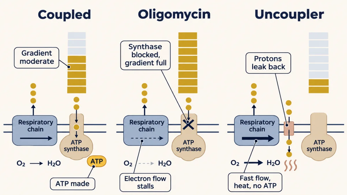
Brown fat uncouples on purpose, for warmth, through a regulated proton channel, UCP1. The diet drug DNP (dinitrophenol) did it with no control, and lethally.
Now cyanide makes sense. It stops the chain, so the pumps stop, while protons keep leaking back in. The gradient runs down and ATP synthase can no longer make ATP; it may even run backward, spending glycolytic ATP to prop the gradient up. Either way, the cell’s ATP supply falls fast.
Try it below: send NADH or FADH₂ pairs one at a time, switch on the DNP proton leak, then try an inhibitor.
Press Send one electron pair and follow it: down the chain, down the redox ladder, H⁺ pumped up, then ATP synthase spending the gradient. Then try an inhibitor, block ATP synthase with oligomycin, or switch on the uncoupler.
Here is the whole chapter in one sentence: the respiratory chain and ATP synthase are two different machines that never touch. The chain is a pump that charges a battery (the H⁺ gradient); ATP synthase is a motor that spends it. Cut the wire between them — add an uncoupler like 2,4-dinitrophenol — and electrons still flow, O₂ is still burned, but the protons leak back as heat and almost no ATP gets made. That is how brown fat keeps a newborn warm, through a regulated proton channel, UCP1. It is also why DNP, sold as a 1930s diet pill, melted fat by turning the battery into a heater, and killed people through uncontrolled overheating. Same physics, two destinies, decided entirely by whether the protons go through the motor.
Reading the order of the chain
Inhibitors freeze the chain at one point, and the freeze is diagnostic. Rotenone blocks Complex I, antimycin A Complex III, cyanide Complex IV. Every carrier upstream of the block piles up reduced, because electrons cannot leave; everything downstream goes oxidized, because none arrive.
Predict. Add rotenone to mitochondria fed an NADH-linked fuel. Which carriers end up reduced? Now feed them succinate instead. Does the chain still run?
Check your prediction
With NADH-linked fuel, NADH stays reduced, everything past Complex I goes oxidized, and the chain stops. With succinate it runs, because those electrons enter at Q, below the block, and each pair still moves 6 protons.
That crossover pattern, read by spectroscopy, is one of the main ways the order of the chain was worked out before anyone had a structure.
The price of running on oxygen
Reactive oxygen species are that price. Mitochondria leak a small fraction of the O₂ they handle as superoxide, and the leak is lower when they are busy making ATP. Complex I and the Complex III Qo site are among the main sources, and which sites dominate depends on the fuels present.
In Friedreich’s ataxia, a GAA-repeat expansion lowers levels of frataxin, a protein that helps build Fe-S clusters; cluster assembly is impaired, and Fe-S enzymes of the chain and the citric acid cycle lose activity, with sensory neurons and the heart hit hardest.
In ischemia-reperfusion injury, succinate builds up while the heart or brain is starved of oxygen. When blood returns, Complex II oxidizes it fast and over-reduces the Q pool. Complex I’s drop only just covers its four protons on standard values (about 70 kJ against 4 × 17), so it runs near balance, and shifts in the NADH and Q pool ratios easily tip it. With the gradient high and Q crammed with electrons, it tips and electrons are pushed backward through it, from QH₂ toward NADH, making a burst of superoxide. With calcium overload and opening of the mitochondrial permeability transition pore, that burst adds damage on top of the ischemia itself.
The next chapter follows the gradient into the second machine, ATP synthase, the rotary motor that spends the battery this chapter charged.
Where the oxygen, and the mitochondria, came from
The chain ends at oxygen, but oxygen was not always there to receive electrons. Early Earth had almost none; photosynthesis filled the air with it, and oxygen’s reactive forms became a hazard. Somewhere in the same long stretch of history a host cell came to keep a bacterium inside it, the ancestor of the mitochondrion; how oxygen figured in that event is still debated. Two short films tell that story, one for each half.
Read the transcript of “How Earth Got Its Oxygen”
Film transcript: How Earth Got Its Oxygen
The oxygen in your next breath is, in part, made of leftovers. What it was left over from is the story. Rewind about 3 billion years. Watch the gauge. The air holds almost no oxygen. Something had to fill this sky. And something held it back.
Nobody saw this world, so we rebuild it from ancient rocks. The sea held dissolved iron. That iron had electrons to give. But with little oxygen to take them, much of that iron stayed dissolved. Life was already here, and all of it was microbial.
Many of these cells likely made energy without oxygen, by fermenting or by making methane. Remember them. They come back near the end. Then came a new trick. Water holds its electrons tightly. Cyanobacteria learned to pull them out, powered by sunlight. They kept the electrons to build their own food. The waste was oxygen gas, and they let it go. In shallow seas, these cells spread as sticky mats. So here is a puzzle. The mats were releasing oxygen. Why did the air not fill with it right away?
Because oxygen had many other places to go. Oxygen strongly pulls in electrons. Each oxygen molecule can take four. And the sea held iron with electrons to give. So iron handed its electrons to oxygen, formed rust-like minerals, and sank. Layer by layer, iron and silica built striped rock on the sea floor. These are banded iron formations, and much of the iron we mine today comes from them. Some layers may be the work of microbes that rust iron without any oxygen. Iron was not the only taker. Volcanic gases, methane, and minerals on land all consumed oxygen too. As an analogy, picture a bathtub. The mats are the tap. Every one of those takers is an open drain.
Here is the idea to hold on to. Making oxygen is only half of the story. The air keeps only the oxygen that nothing else takes first. So when did the tap turn on? Cyanobacteria most likely arose well before oxygen built up. By how long is debated. Some rocks hint at brief whiffs of oxygen before the lasting rise. Then came the change. Between about 2.4 and 2.1 billion years ago, oxygen rose, wavered, and finally stayed. In bathtub terms, more was now arriving than the drains could take. Why the balance tipped then is still debated. Geologists call this the Great Oxygenation Event. But the tub was nowhere near full. Oxygen stayed far below today's level for a very long time.
Now remember the old microbes. An oxygen molecule that is only partly filled with electrons can damage DNA and proteins. For many of those microbes, oxygen was toxic. So what happened to them? Was there a mass die-off? A mass die-off is often claimed, but the evidence is thin.
Microbes that live without oxygen are called anaerobes. They clearly survived, in mud, deep water and other airless places. They thrive there still, including in your own gut. The oxygen that was left over did not end the old world of life. It pushed it into the shadows. Now run the clock forward. Oxygen stayed low for well over a billion years. Then, much later, it rose again, toward the level you breathe today. Take that breath again. It began as waste. And what reaches your lungs is what the rest of the planet left behind.
Read the transcript of “How Your Cells Got Mitochondria”
Film transcript: How Your Cells Got Mitochondria
Inside your cells, something acts like a separate living thing. Your mitochondria keep their own DNA, apart from the DNA in your nucleus. And they multiply by pinching in two. Why would a part of you do that? The answer is a very old story. Eukaryotes are living things with cells like ours. Their oldest widely accepted fossils are more than 1.5 billion years old. Exactly when this story began is uncertain. But rewind far enough, and the cell comes apart into two strangers. Meet the first stranger. It was most likely an archaeon, a kind of single-celled microbe. Whether it already had a nucleus is still debated. The second stranger was a bacterium, related to today's alphaproteobacteria.
It could probably respire. That means a chain in its membrane pulled energy out of food. And it may have used oxygen to do it. Those are long names. Just call them the host and the guest. Somehow, the host came to enclose the guest. What do you expect will happen next?
You might expect a meal. But instead of being digested, the guest stayed. Two strangers now lived as one. Here is the claim. Your mitochondria descend from that guest. But how could anyone know, so long afterwards? If a bacterium moved in, what would it leave behind?
Here they are. Most mitochondria still have two membranes. They have their own DNA. Their ribosomes, which build proteins, are bacteria-like. And they divide by pinching in two. One more clue comes from genes. On the family tree, mitochondria from animals, plants and fungi all sit on one branch. That suggests this happened only once. Every mitochondrion today descends from that single guest. So here is the idea to keep. A mitochondrion is not something your cells invented. It is a guest that stayed. But why did the partnership begin? Go back to the two strangers. The honest answer is that it is still debated. Here are two of the ideas. The hydrogen hypothesis says the host lived on hydrogen.
The guest gave off hydrogen as waste. So the host came to depend on the guest. The second idea is about protection. Oxygen can damage cells, and this idea says it harmed the host. The guest consumed oxygen, and shielded its partner. Both ideas fit some evidence, and neither is proven. One hint is real, though. A close relative of the host, grown in a lab, lives by trading hydrogen with partner microbes. So partnerships like this do exist. Whatever began it, the guest changed. Over time it became an organelle, a working part of the cell. Many of its genes were lost. Others moved to the nucleus. But it kept a small genome of its own. Human mitochondrial DNA encodes just 13 proteins.
All 13 are for respiration and for making ATP, the molecule that powers your cells. What did the host gain? Mitochondria make most of your ATP. One influential idea, still argued over, goes further. It says the energy from many mitochondria let cells like ours afford larger genomes and more complex lives. So look again. Why would a part of you act like a separate living thing? Because its ancestor once was one. Every breath you take delivers oxygen to a chain inherited from that ancient guest. Your mitochondria began as a guest, and never left.
The longer film below covers both halves in one sitting and adds the chemistry of reactive oxygen.
Read the transcript of “The Oxygen Bargain”
Film transcript: The Oxygen Bargain
Picture Earth about three billion years ago. There are oceans, volcanoes and sunlight. But there is almost no oxygen in the air. The sky may have been hazy, perhaps tinted orange by a methane haze. Nobody saw it, so we rebuild it from ancient rocks.
The oceans held dissolved iron, in its reduced, ferrous form. With little oxygen to take its electrons, much of that iron stayed dissolved. Life was already here, and all of it was microbial. Cells likely made energy by fermenting or by making methane. Some may have reduced sulfate.
Every one of those lifestyles moves electrons from a donor to an acceptor. Oxygen, a far more powerful acceptor, was scarce. The land had no plants and no animals, though microbes may have crusted damp patches. Plants would not arrive for over two billion years.
Then came a new trick. Cyanobacteria learned to pull electrons out of water, powered by sunlight. The waste product was oxygen gas. Water holds its electrons tightly. Photosystem two splits it, and photosystem one then boosts the electrons higher. This machinery evolved once, in the ancestors of cyanobacteria. Plants later gained it by taking in a cyanobacterium.
In shallow seas, these cells spread as glowing, sticky mats. Similar mats still build layered mounds, called stromatolites, in some bays and lakes today. Predict. If cyanobacteria were releasing oxygen, why did the air not fill with it right away?
Because oxygen had many places to go. Dissolved iron handed its electrons to oxygen, formed rust-like minerals, and sank. Layer by layer, iron and silica built banded iron formations. Some layers may record microbes rusting iron without oxygen. Much of the iron we mine today comes from them.
Their biggest pulse came just before and as oxygen began to rise. Iron was not the only sink. Volcanic gases, methane and minerals on land all consumed oxygen too. Cyanobacteria most likely arose well before oxygen built up, though by how long is debated. Some rocks hint at brief whiffs of oxygen before that lasting rise.
Then, between about two point four and two point one billion years ago, oxygen rose, wavered, and finally stayed. Geologists call this the Great Oxygenation Event. The methane haze may have thinned, leaving a clearer sky. Even so, oxygen stayed far below today's level for a very long time.
Was there a mass die-off of anaerobes? It is often said, but the evidence is thin. Anaerobes clearly survived, in mud, deep water and other airless places. They thrive there still, including in your own gut. Oxygen did not end the old world of life. It pushed it into the shadows.
Why should oxygen be dangerous at all? Oxygen strongly pulls in four electrons, to become water. But its two unpaired electrons make it slow to react with most molecules. It tends to take electrons one at a time. Predict. If oxygen picks up electrons one at a time, what forms along the way?
Partly reduced oxygen. One electron makes superoxide. Two make hydrogen peroxide. Three make a hydroxyl radical, plus water. That radical attacks almost anything it touches. Together these are called reactive oxygen species. They break DNA, damage proteins, and set off chain reactions in membrane lipids.
Iron makes it worse. Ferrous iron hands an electron to hydrogen peroxide, releasing the hydroxyl radical. This is one reason cells keep most of their iron tightly bound, much of it stored inside proteins. Here is the idea that ties this film together. Every way of living with oxygen is a way of handling its electrons.
Cells evolved and spread defenses. Superoxide dismutase turns superoxide into hydrogen peroxide. Catalase turns hydrogen peroxide into water and oxygen. Some bacteria went further and put oxygen to work. They placed it at the end of an electron-transport chain.
Oxygen is the strongest common acceptor. So an electron that falls all the way to oxygen releases much more energy. Fermenting glucose to lactate or ethanol yields two A T P. Respiring it fully with oxygen, as our cells do, yields around thirty.
But the chain is not perfect. A small fraction of electrons slip off early, often at complexes one and three, making superoxide. Now move forward, to some time after oxygen rose. The oldest widely accepted eukaryote fossils are more than one and a half billion years old.
The host was most likely an archaeon, related to today's Asgard archaea. Whether it already had a nucleus is still debated. Asgard archaea were first found as DNA in deep sea sediments. Their genomes carry many genes once thought unique to eukaryotes.
The guest was a bacterium related to today's alphaproteobacteria. It could probably respire, and may have used oxygen. Somehow, the archaeon came to enclose the bacterium. And instead of being digested, the guest stayed.
Gene trees suggest that this happened only once. Every mitochondrion today descends from that single guest. Most mitochondria still show their bacterial past. They have two membranes, their own DNA, bacteria-like ribosomes, and they divide by pinching in two.
Why did the partnership begin? The honest answer is that it is still debated. The hydrogen hypothesis says the host lived on hydrogen. The bacterium gave off hydrogen as waste, so the two fed each other. The oxygen detoxification hypothesis says oxygen harmed the host. The bacterium consumed oxygen and shielded its partner.
Both ideas fit some evidence, and neither is proven. The first Asgard archaeon grown in a lab gives hydrogen to partner microbes. That is the reverse of the proposed host, but it shows such partnerships are real. Oxygen changed the world the partners lived in. But eukaryote fossils appear long after the oxygen rise. How much oxygen drove their merger is still debated.
Over time, the guest became an organelle. Many of its genes were lost, and others moved to the nucleus. But it kept a small genome of its own. Human mitochondrial DNA encodes just thirteen proteins, all for respiration and A T P synthesis.
One influential idea is that many mitochondria let eukaryotic cells afford larger genomes and more complex lives. So think back to the idea. Rust, poison and power are all ways of handling oxygen's electrons. In this lesson, follow electrons from N A D H down the chain to oxygen. Watch complex four hold oxygen until it is fully reduced to water.
That careful grip is the bargain. Every breath you take delivers oxygen to a chain inherited from that ancient guest.
Going deeper
The full Q cycle
At the Qo site, facing the intermembrane space, QH₂ sends one electron through the Rieske Fe-S cluster and cytochrome c1 to cytochrome c. The other loops back through the b hemes to reduce a Q at the Qi site, facing the matrix, which antimycin A blocks. Two rounds oxidize two QH₂ and regenerate one, giving the net per pair above. The semiquinone radical (Q•⁻) forms fleetingly at Qo.
How Complex IV reduces oxygen
Electrons go from cytochrome c to CuA, heme a and the heme a3/CuB center, which binds O₂ once it holds two electrons, then breaks the O–O bond in one step, drawing four electrons from its own metals and a nearby tyrosine (the heme a3 iron goes to the ferryl state). Electrons from cytochrome c then reset those groups and complete the two waters.
Protons released versus charges moved
The ledger counts protons released, not charges each complex moves across the membrane. Complex III moves only 2 charges itself, and Complex IV moves 4 (its 2 pumped protons plus 2 matrix protons consumed with electrons arriving from the intermembrane-space side); with Complex I’s 4, both tallies total 10. Check energy on the whole chain: 10 protons at about 17 kJ is about 174 of the 220 kJ an NADH pair releases, and 6 is about 104 of the roughly 150 kJ from succinate.
Why 2.5 and not 2.7
The mammalian ATP synthase rotor has 8 c-subunits and makes 3 ATP per turn, about 2.7 protons per ATP. Add about 1 for the carriers and the cost is about 3.7, not 4, giving theoretical yields of about 2.7 and 1.6. Proton leak and slip bring measured values down to about 2.5 and 1.5.
Supercomplexes
Cryo-EM shows the complexes assembling into supercomplexes (the respirasome, I + III₂ + IV), which may shorten the diffusion path for the mobile carriers and reduce leak. Whether that matters for normal function is still debated.
Carriers between the landmark floors
The potentials here are standard values, E°′. Other carriers sit between the landmark floors: FMN at −0.30 V, the iron-sulfur clusters across a range (Complex I’s mostly negative, Complex III’s Rieske cluster strongly positive), and cytochrome a at +0.29 V. In Complex I, a machine of about 45 subunits, the relay from FMN to coenzyme Q passes through about seven Fe-S clusters.
The porous outer membrane
The gradient depends on the inner membrane being impermeable to ions; the outer membrane is the opposite. Its VDAC channels let most small molecules and ions, up to roughly 5000 Da, through.
Reactive oxygen species: numbers and cleanup
The old estimate that 1 to 2 percent of the O₂ mitochondria handle leaks as superoxide is now thought to be too high. Eleven leak sites have been mapped; Complex I’s main ones are its flavin and its quinone site. Superoxide dismutase converts two superoxides to H₂O₂ and O₂, then peroxiredoxins and glutathione peroxidases (and catalase, mostly in peroxisomes) finish the cleanup.
Low oxygen at ecosystem scale
Every aerobic animal needs O₂ waiting at the bottom of the ladder; without it the chain backs up just as it does under cyanide. The Gulf of Mexico dead zone is this chemistry at ecosystem scale: nutrient runoff fuels an algal bloom, bacteria decomposing the dead algae use up the dissolved oxygen, and the terminal electron acceptor that fish and other aerobic animals depend on runs too low to support them.
Your notes
Select any text in the lesson to highlight it or add a note to it. Your highlights and notes are saved in this browser and collected in your notebook.
This browser is not letting the site save anything (a private window or strict privacy settings), so highlights and notes will be lost when you leave the page.
How we measure it
Difference spectroscopy of cytochromes
Keilin's microspectroscope read respiration in real time: oxidized and reduced cytochromes absorb light at different wavelengths, so watching absorption bands appear and vanish as oxygen is added or consumed tells you which carrier is reduced and which is oxidized. Add an inhibitor and the chain freezes in a 'crossover' pattern (reduced upstream, oxidized downstream), and that pattern is one of the main ways the order of the carriers was deduced before any structure existed.
Inhibitor crossover analysis
Specific blockers cut the chain at known points: rotenone at Complex I, antimycin A at the Complex III Qi site, cyanide and CO at Complex IV. Apply one, and every carrier upstream of the block piles up reduced while everything downstream goes oxidized. Mapping that boundary for each inhibitor pins down where each component sits in the sequence and is the classic logic for ordering an electron-transfer pathway.
Submitochondrial particles and the pH-electrode pulse
Tear mitochondria apart by sonication and the inner membrane reseals into closed, inside-out vesicles that still pump and still make ATP, proving OxPhos lives on the membrane while the matrix runs the soluble reactions. Mitchell and Moyle put a fast glass pH electrode in a mitochondrial suspension, pulsed in oxygen, and watched the medium acidify, measuring the protons ejected per oxygen atom consumed (and, in separate ATP-hydrolysis pulses, per ATP). That turned chemiosmosis from hypothesis into numbers; the early ratios have since been revised upward.
Cryo-EM of supercomplexes
Single-particle cryo-electron microscopy resolved respiratory supercomplexes, including the 'respirasome' (Complex I + III2 + IV), which may shorten the diffusion path for CoQ and cytochrome c and reduce electron leak. It also solved mammalian Complex I, the L-shaped 45-subunit machine, at near-atomic resolution. The first structures of the bc1 complex and cytochrome c oxidase came from X-ray crystallography in the 1990s.
Practice questions on this lesson (5) → MCAT-style, original, each with an explanation
Where this lesson fits
Before thisReview first
- The Citric Acid Cycle: Harvesting ElectronsThis course · Lesson 19
It makes the NADH and FADH2 whose electrons start the chain, and Complex II is also a cycle enzyme.
- Metabolism: Basic Concepts and DesignThis course · Lesson 15
You need its energy coupling and free-energy ideas to follow how electron drops pay for pumping protons.
- Membranes: The Impermeable Sheet That Builds ItselfThis course · Lesson 12
The gradient only works because the inner membrane is impermeable to ions, which this lesson explains.
After thisStudy next
- The Proton-Motive Force and ATP SynthaseThis course · Lesson 21
It follows the gradient into ATP synthase, the machine that spends the proton battery built here.
- Integration of Metabolism: How the Body Switches FuelsThis course · Lesson 30
It shows how the body tunes fuel use around the oxygen-dependent ATP supply this chain provides.
- Photosynthesis: The Light ReactionsThis course · Lesson 22
Photosynthetic electron transport uses the same redox ladder and proton gradient, pointing the opposite way.
Elsewhere on the siteFilms, simulations and method films in other courses
- A cell’s energy budgetSimulation · Biology
It lets you budget a cell's ATP spending and rebuilding, the demand that electron transport and ATP synthase meet.
- Two Jobs in One Green CellFilm · Life Science
It shows mitochondria using oxygen day and night in the same cell where chloroplasts make it.
- Spending ATP, Rebuilding ATPFilm · Biology
It shows how a cell keeps rebuilding ATP from ADP, the job the proton gradient does here.
- A Tap, a Hole, a LevelFilm · Cell Biology
It shows that a membrane leaks, so a pump must keep paying to hold a gradient, as with protons here.
Suggested by an AI model from the lessons themselves, then checked. How we use AI
Finish line
Test yourself
8 questions
Practice and MCAT-style, with explained answers.
Play it · about 3 min
Ledger Rush: Electron transport
Complex by complex: which carriers are spent, how many protons are pumped, and what ATP synthase pays out.
Play →Next · Lesson 21
The Proton-Motive Force and ATP Synthase
Electron transport and ATP synthesis are one coupled machine: the chain pumps protons into a gradient, and protons falling back through ATP synthase turn a rotary motor that makes ATP.
Continue →Also in the arcade for this lesson: Wire the Cell: Burn glucose