Developmental Neuroscience · Unit 1: The problem and the tools
Seeing Neurons: Golgi, Cajal and the Neuron Doctrine
Rewritten on : new text, figures, flashcards and film. Your notes and highlights stay, but the text around them may have moved.
Are neurons separate cells that touch, or one fused network? The neuron doctrine says separate, and it took a new stain, a lot of embryos and the electron microscope to show it. Two men looked at the same stained brain and saw two different organs. Who was right, and how could anyone tell?
Pause when the reading reaches a film, and offer to play it. Saved on this device.
Watch the film now, or keep listening
Builds on:The Wiring Problem: How a Brain Gets BuiltWhy these?
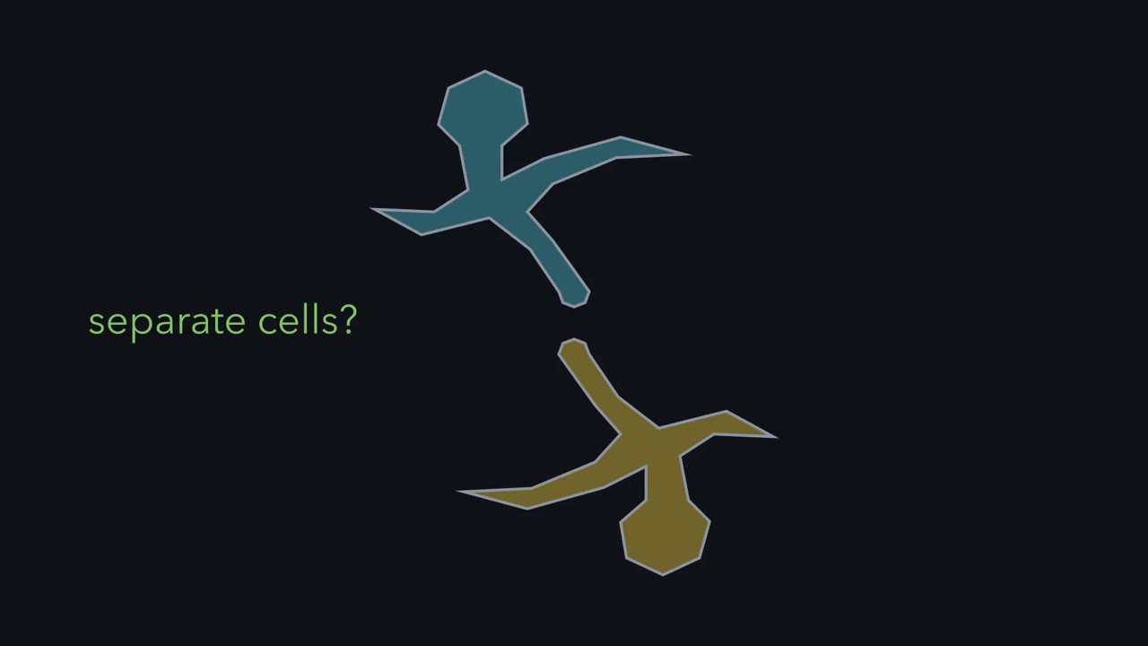 ▶ Watch A Touch Is Not a Fusion · 2:09
Read the transcript of “A Touch Is Not a Fusion”
Film transcript: A Touch Is Not a Fusion
Is your nervous system built of separate cells, like liver or skin? Or is it one continuous network of fused fibres? There were two answers. The neuron theory said each nerve cell is a separate cell, bounded by its own membrane. The reticular theory said the fine branches fuse into one continuous net. It comes down to the point where two branches meet. Does one membrane run on, or do two face each other across a gap? Here is the one idea to carry through. A touch is not a fusion. Whether two touching branches are one cell or two depends on the membrane where they meet. Settling it takes evidence that bears directly on that membrane: seeing it, or testing what crosses it.
Under the best light microscope, two points closer than about 200 nanometres blur into one. The gap at a chemical synapse is about 20 nanometres. That is about ten times below the limit. Depth is worse. Two branches close together, one above the other, can look like one line as you focus through. Picture one contact under the best light microscope of the 1890s. What does each theory predict you will see?
The same thing. Two facing membranes and a fused junction give the same smudge. So the evidence had to come from elsewhere: from whole cells, from embryos, and from the direction signals travel. The answer, for the vertebrate nervous system, is separate cells. They pass signals across specialized contacts, called synapses. That is why this matters for development. In vertebrates, development has to wire separate cells to the right partners. A touch is not a fusion. The difference lies at the membrane.
From the film
- A touch is not a fusion: whether two touching branches are one cell or two is decided at the membrane where they meet.
- Evidence that bears directly on that membrane, by seeing it or by testing what crosses it, can settle the question.
- A gap of about 20 nanometres at a chemical synapse sits inside the light microscope's blur of about 200 nanometres, so touching and fused branches look alike.
- In vertebrates, development has to wire separate cells to the right partners.
Stain every cell in a slice of brain and you cannot follow any one of them: a crowd of cell bodies, and between them a tangle of threads that no eye can follow. Stain only a few cells, but completely, and each one stands out with every branch.
That second kind of stain arrived in 1873, and it turned an old question into one that could be argued with evidence. Is the nervous system built of separate cells, like liver or skin, or is it one continuous network of fused fibres? It took about eighty years, a stain nobody fully understands, a lot of embryos and finally the electron microscope to settle it. This lesson follows the evidence in the order it came, and asks one thing of each piece: could it reach the place where the question is decided?
Touch or fusion: a question about 20 nanometres
There were two answers on offer. The neuron theory said each nerve cell is a separate cell, bounded by its own membrane, which extended the cell theory (that tissues are built of cells) to the nervous system; signals pass between cells at points of contact. The reticular theory said the fine branches fuse into one continuous net.
Under the neuron theory, the place where one neuron acts on another is a specialized contact. It came to be called a synapse, a word Charles Sherrington introduced in 1897, decades before anyone had seen the gap. At a chemical synapse, one cell releases a chemical messenger that acts on the next; at an electrical synapse, current passes straight through channels joining the two cells.
So the whole argument comes down to one spot, a point where two fine branches meet: one fused cytoplasm, or two membranes facing each other across a gap? How big is that spot, compared with what could be seen? Two numbers matter most: the 200-nanometre blur and the 20-nanometre gap.
| What | Size | What it means |
|---|---|---|
| Smallest separation the best light microscope can show, side to side | about 200 nanometres (0.2 micrometres) | two points closer than this blur into one |
| The same limit in depth, focusing up and down | roughly 0.5 to 1 micrometre, depending on the optics | several times worse than side to side |
| A stained section, examined by refocusing | 100 micrometres or more thick | many branches lie above and below one another |
| The gap between two cells at a chemical synapse, as measured later | about 20 nanometres | about ten times below the limit |
| The gap between membranes at an electrical synapse | a few nanometres | further below the limit still |
Golgi and Cajal did not know how narrow the gap was, only that where branches met they could not see one.
Predict. Picture one point of contact under the best light microscope of the 1890s. What does the neuron theory predict you will see there? What does the reticular theory predict?
Check your prediction
The same thing. A gap of about 20 nanometres sits inside a blur about 200 nanometres across, so two facing membranes and a fused junction give the same smudge. In a thick section it is worse: two branches that lie close together in depth, one above the other, can look like one line as you focus through them. No light-microscope image of a single contact could decide the question, so the evidence had to come from elsewhere.
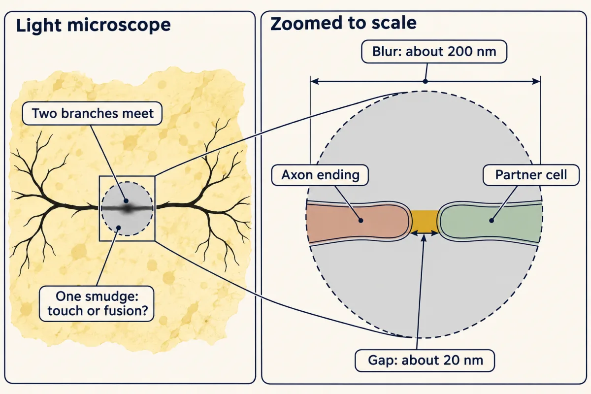
The one idea to carry through this lesson. A touch is not a fusion. Neurons are separate cells, each bounded by its own membrane, and they pass signals across specialized contacts. The difference between touch and fusion lies at the membrane where two branches meet: either an open, unrestricted cytoplasmic bridge runs through the junction, or two membranes face each other across a gap (about 20 nanometres at a chemical synapse; a few nanometres at an electrical synapse, crossed only by size-limited channels). Evidence that bears directly on the membrane at a contact, by imaging it or by testing what crosses it, can settle the question. Evidence about whole cells, embryos or signal direction can make one answer more likely, but cannot decide it.
What the rule covers.
- Which nervous systems. Vertebrates and many other animals, but not all. A few animals really do have fused neurons; see Going deeper.
- What it does not say. It does not say that neurons act alone. Coupled cells exchange ions and small molecules through channels. They do not form one unrestricted cytoplasm: each keeps its own membrane (see A pore is not a fusion).
That is why this lesson sets up the rest of the course. In vertebrates, development has to wire separate cells together, each growing its branches toward the right partners. Where neurons depend on a target’s limited supply of a factor, each separate cell can win or lose its share, the subject of the lesson on the neurotrophic hypothesis. And separate, polarized cells joined at specialized contacts are why the direction of signal flow can be traced from cell to cell.
To see one cell, stain almost none
Read the transcript of “One Stain, Two Readings”
Film transcript: One Stain, Two Readings
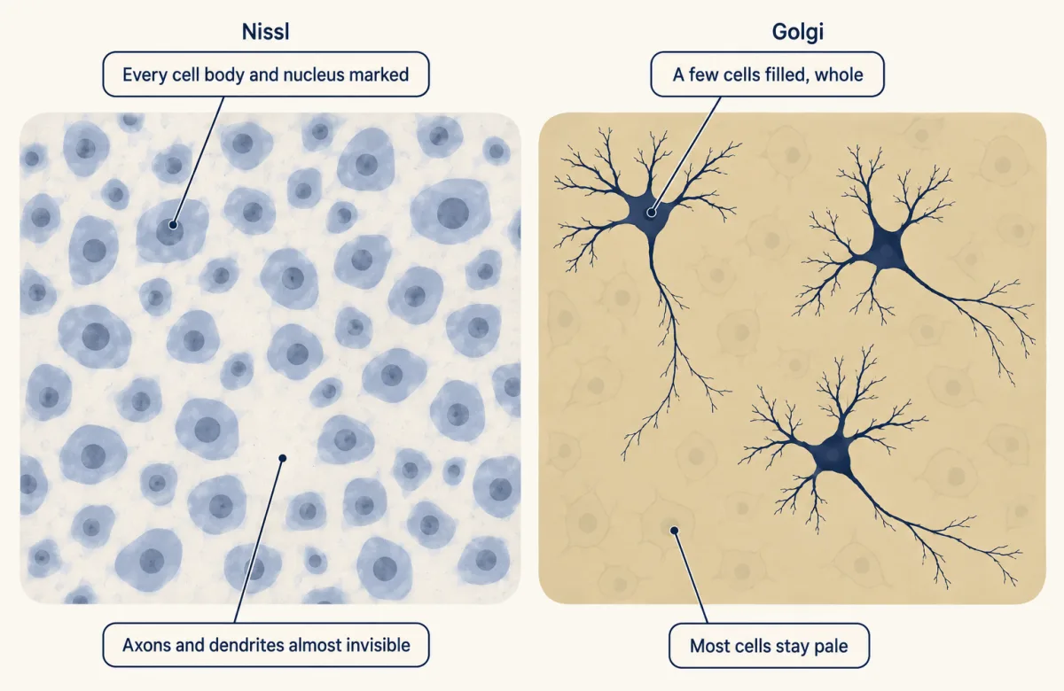
How do you see one whole cell in a crowded brain? A Nissl stain marks every cell body. But axons and dendrites, where the question of fusion lives, stay almost invisible. In 1873, Camillo Golgi found the black reaction. Most cells stay pale. A few fill with black, to the finest twig. This is sparse labelling: a few cells, filled completely, apparently at random. Picture a forest at night with a few trees lit. Each lit tree's shape stands out. Each black branch can be traced back to its own cell, where the section allows. But a contact has two sides, and both are rarely stained. Even where two black branches meet, the gap is far below what light can show.
Where two lit branches cross in the dark, you still cannot tell whether they are grafted together. In 1887, Santiago Ramón y Cajal took up the stain. Golgi read the tangle of axon branches as a continuous net. Cajal found branches ending close to other cells without joining them, again and again. Neither could win at the contact. So Cajal turned to embryos, where cells are fewer and simpler. Wilhelm His argued that each fibre grows out of a single cell, a neuroblast. In 1890, Cajal named the growth cone. He read it as the advancing end of a growing fibre. Suppose every fibre does grow out of one cell. Does that rule out Golgi's net?
No. Outgrowth shows where a fibre begins, not how it ends. A fibre could grow out, then fuse at its far end. So embryos made separate cells more likely, without settling it. Cajal added a rule. Signals enter through the dendrites and cell body, and leave through the axon. In a chain of separate cells, their order sets a direction. Continuity alone sets none. The rule has exceptions. In 1906, Golgi and Cajal shared the Nobel Prize. Golgi still defended his net. No light microscope could show the gap. Separate cells were the better explanation, but nobody had seen it.
From the film
- Golgi's black reaction fills a few cells completely, so one neuron and its branches can be traced.
- Sparse staining shows whose branch is whose, but it cannot show touch or fusion, because both partners are rarely stained and the gap is below what light can show.
- Golgi read the tangle of axon branches as a continuous net, while Cajal read the free endings he kept finding as evidence of separate cells.
- Embryo evidence and the direction of signal flow made separate cells more likely without settling the question.
Before anyone could argue about contacts, they needed to see whole cells. The stains of the nineteenth century coloured what they reached. A Nissl stain, developed by the German psychiatrist and neuropathologist Franz Nissl, marks every cell body and nucleus in the section. It is excellent for counting cells and seeing layers, but axons and dendrites, the long thin processes where the question of fusion lives, stay almost invisible.
Camillo Golgi’s black reaction worked the other way. He hardened small blocks of nervous tissue in potassium dichromate, then moved them into silver nitrate. Most cells stay pale. A few fill with a black deposit from cell body to the finest twig, so in a thick section one neuron and many of its branches can be traced against a yellowish background, although branches that leave the section are cut off.
This is sparse labelling: a few cells, filled completely, apparently at random. Golgi could not choose which cells were marked, and the fraction varies from piece to piece of tissue. Think of a forest at night with lights strung on a handful of trees. If every tree were lit you would see a glow. With only a few lit, you can see each lit tree’s shape.
Predict. If sparse staining lets you see one cell whole, could you stain even fewer cells and so see whether two touching branches are fused?
Check your prediction
No. Sparse labelling answers one question well: whose branch is this? Near the cell body, each black branch can be traced back to its own cell. It cannot answer the other question, touch or fusion, for two reasons.
- Both partners are rarely stained. A contact has two sides. As an illustration, not a measured rate, suppose cells take the stain independently and 1 cell in 20 is stained. Then both sides of a given contact are stained only 1 time in 400, and even starting from a stained cell, its partner is stained only 1 time in 20. Staining fewer cells makes this worse, not better. Where only one side is stained, the pale partner could be a separate cell the stain missed or a fused part the stain did not reach. That single cells fill without their neighbours filling is itself a hint of separateness, but while no one knows why the stain takes, it cannot decide the matter.
- Resolution does not depend on how many cells are stained. Even where two black branches do meet, the gap between them is still far below what light can show.
In the forest at night, a few lit trees show you each tree’s shape. Where two lit branches cross in the dark, though, you still cannot tell whether they are grafted together.
One slide, two readings
Santiago Ramón y Cajal came to the stain in 1887, when the Madrid histologist Luis Simarro showed him Golgi preparations. From 1888, working first on the cerebellum of birds, Cajal described axon branches that end close to other cells without joining them.
How could two careful observers look at similar preparations and disagree? Go back to the scale table: where two fine branches touch, or merely pass close at different depths in a thick section, the image cannot tell contact from fusion. Each camp filled that blind spot with what its theory predicted. Golgi read the tangle of fine axon branches as a continuous net. For Cajal, the free endings he found over and over were the evidence, and an unseen connection was no evidence at all.
Neither could win at the contact itself, so Cajal changed where he looked: to embryos.
Evidence from development: fibres grow out of single cells
An adult brain is a thicket. An embryo is a young wood: fewer cells, shorter branches, and little myelin, the fatty sheath that wraps many mature axons and can block the stain. There a whole cell fits in a section and its branches can be counted.
Cajal was not the first to argue from embryos. Wilhelm His cut embryos into complete, numbered series of sections and rebuilt their shapes in drawings and models. The neural tube, the hollow tube of tissue that becomes the brain and spinal cord, showed dividing cells next to its central cavity and young nerve cells farther out. In the late 1880s His argued that each nerve fibre grows out from a single cell, which he called a neuroblast. If a fibre is the outgrowth of one cell, it began as part of that cell and not as a strand of a net.
Predict. Suppose every nerve fibre does grow out of one cell. Does that rule out Golgi’s net?
Check your prediction
No. Outgrowth shows where a fibre begins, not how it ends. A fibre could grow out of one cell and then fuse with another at its far end. Rival developmental views also held that nerve fibres form from chains of cells, or from bridges already present between cells. So the embryo evidence made separate cells more likely without settling the question.
Where fibres come from was largely settled by watching. In 1907 Ross Harrison kept pieces of frog embryo neural tube alive in a drop of lymph and watched living nerve fibres grow out of them, the subject of the lesson on the growth cone. How a fibre ends still had to wait for an instrument that could see the membrane.
In 1890, in the spinal cord of chick embryos, Cajal described swellings at the tips of young axons, some cone-shaped, some fringed with spikes. He named the structure the growth cone and read it as the advancing end of a fibre that lengthens behind it. Every one of his specimens was fixed and dead, so the motion was an inference from a series of still shapes.
Cajal then added a rule about direction, which Arthur van Gehuchten in Belgium proposed independently. By the law of dynamic polarization, a signal enters a neuron through its dendrites and cell body and leaves through its axon. Cajal pointed to the retina and the olfactory bulb, where it is obvious which end receives the outside world and most cells line up in that order. The law is a useful generalization, not a rule without exceptions (see Going deeper). Cajal argued that a chain of separate, polarized cells explains one-way signalling better than a diffuse net. In a chain, each cell has a receiving end and a sending end, so the order of the cells sets a direction; continuity alone sets none, though a net could still have polarized parts. This matches the point from the simple reflex circuit that the direction of information flow matters.
By 1906 most anatomists favoured separate cells, though not all. That year Golgi and Cajal shared the Nobel Prize in Physiology or Medicine for their work on the structure of the nervous system. In his lecture Golgi still argued against the doctrine, and he could: no light microscope could show the gap. Separateness was the better explanation of the evidence, but nobody had yet seen it.
The direct test: look at the membrane
Read the transcript of “The Gap, Seen”
Film transcript: The Gap, Seen
Every clue so far favoured separate cells. None reached the membrane. What would it take? Suppose you could see the membrane at a contact. What would you see if Golgi were right? And if Cajal were right? If the branches are fused, the outer membrane runs on around both, and no membrane crosses the junction. If the cells are separate, two membranes face each other across a gap. Each cytoplasm stops at its own membrane.
The electron microscope could show it, in thin sections of nervous tissue. In 1955, De Robertis and Bennett examined synapses in frog and earthworm. In 1956, Sanford Palay examined the rat brain. At the chemical synapses they examined, they saw the second sketch. Two membranes stayed separate across a cleft 10 to 20 nanometres wide. Small vesicles crowded the terminal. In such images, the receiving membrane also looks thickened. Under light, that whole cleft sits inside one blur. The electron microscope replaced the blur with two membranes. The question was settled, about eighty years after the black reaction. Some neurons are joined more closely.
At an electrical synapse, channels called gap junctions cross both membranes. Ions and small molecules pass straight from one cell into the next. Does this bring back Golgi's network? What would you inject into one cell to find out?
A small tracer such as Neurobiotin passes through to coupled neighbours. A 10-kilodalton dextran, nearly 5 nanometres across, stays in the injected cell. In one shared cytoplasm, the dextran would spread too, given time. The electron microscope then confirms two membranes.
In vertebrates, Golgi's network does not exist. Coupling through pores that can close does. There are exceptions outside vertebrates. The squid giant axon forms from fused fibres. A comb jelly's nerve net shares one membrane. A touch is not a fusion, and neither is a pore. Separate cells, joined at specialized contacts, are what development has to wire.
From the film
- Electron micrographs of the chemical synapses examined in the 1950s showed two membranes staying separate across a cleft, which settled the question.
- Electrical synapses join some neurons through gap junction channels, but each cell keeps its own membrane.
- A small tracer can pass gap junctions to coupled neighbours while a large dextran stays in the injected cell, so coupling is not fusion.
- A few animals really do have fused neurons, such as the squid giant axon and the comb jelly nerve net.
None of that evidence reached the membrane at a point of contact. Settling the question took an instrument that could show it.
Predict. Suppose you could see a single membrane where an axon ending meets another cell. Sketch what you would see if Golgi were right, and what you would see if Cajal were right.
Check your prediction
If the branches are fused, the outer membrane runs on unbroken around both, no membrane crosses the junction, and the cytoplasm carries on through it. If the cells are separate, two membranes face each other across a gap, and each cell’s cytoplasm stops at its own membrane.
About half a century after the Nobel Prize, the transmission electron microscope, applied to thin sections of nervous tissue, became that instrument. In 1955 Eduardo De Robertis and H. Stanley Bennett examined synapses in frog sympathetic ganglia and the earthworm nerve cord. In 1956 Sanford Palay examined the rat brain. At the chemical synapses they examined they saw the second sketch: the membranes of two cells stay separate across a cleft that those first reports measured at 10 to 20 nanometres (about 20 nanometres is the figure usually quoted now), with small vesicles crowded in the terminal (we now know they hold the chemical messenger).
At a contact between fine branches, Golgi and Cajal had seen the same blur, and each could claim it. The electron microscope replaced the blur with two membranes, and the question was settled.
A pore is not a fusion
Some neurons are joined more closely than this. At an electrical synapse, the two membranes come within a few nanometres of each other, and channels called gap junctions cross both. Each cell supplies half of each channel, a ring of proteins called a hemichannel, and the two halves dock across the gap. In vertebrates the ring is six connexins; most invertebrates use innexins, and the innexin rings solved so far have eight. Ions and small molecules pass straight from one cell’s cytoplasm into the next. Gap junctions join some neurons, especially in development.
Predict. Does this bring back Golgi’s network? What would you inject into one cell to find out?
Check your prediction
No. Each cell keeps its own membrane, and the channels can close. A vertebrate connexin pore is roughly 1.5 nanometres across at its narrowest in the structures solved, and passes ions and small molecules, generally up to about a kilodalton; what passes depends on the connexin and the molecule (a dalton is about the mass of one hydrogen atom). Inject two tracers. A small one known to pass the junction, such as Neurobiotin, reaches coupled neighbours; Lucifer yellow passes some junctions but poorly through others. A 10-kilodalton dextran, nearly 5 nanometres across, stays in the injected cell. In a syncytium, a single cell with many nuclei sharing one cytoplasm, the dextran would spread too, given time.
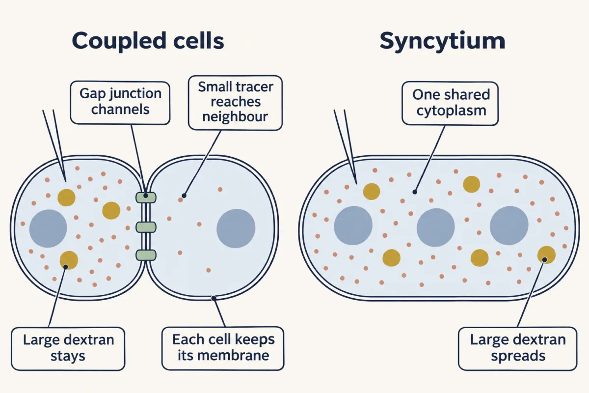
In vertebrates, Golgi’s network, in the sense he meant it, does not exist; coupling through regulated pores does. A touch is not a fusion, and neither is a pore.
Putting it together
Most of this evidence bore on the same question, at different distances from the membrane.
| Evidence | Date | Neuron theory predicts | Reticular theory predicts | What it supports | What it could not establish |
|---|---|---|---|---|---|
| Nissl stain | 1800s | cell bodies; fine processes poorly shown | the same | counts of cells and layers | the fine branches, or whether they fuse |
| Golgi’s black reaction | 1873 | whole arbors of a few cells; contacts unresolved | the same image | one cell can be traced through its branches | touch or fusion at a contact |
| Free endings in bird cerebellum (Cajal) | from 1888 | branches end near other cells | branches join other branches | separate cells, seen again and again | that no connection lies below the blur |
| Fibres grow out of single neuroblasts (His) | late 1880s | each fibre grows from one cell | a fibre could grow from one cell, then fuse at its far end | a separate origin for each fibre | how a fibre ends; it could fuse later |
| Growth cone in chick spinal cord (Cajal) | 1890 | what Cajal inferred: a growing fibre has an advancing tip | compatible: a tip could fuse on arrival | fixed tip shapes read as the advancing end of a growing fibre | the motion itself; specimens were fixed |
| Dynamic polarization (Cajal, van Gehuchten) | 1890s | with the added rule that each cell receives at its dendrites and cell body and sends from its axon, a chain of separate cells carries signals one way | continuity alone sets no direction, though a net could contain polarized parts | an interpretation of one-way circuits | membrane separation; and the rule has exceptions |
| Cut fibres and lesions (Waller; von Gudden and Forel) | 1850; 1870s and 1880s | the part of a fibre cut off from its cell body degenerates | the same for a cut nerve fibre, since no one claimed nerve fibres were fused | each cell and its fibres form a unit | fusion at grey-matter contacts; damage can also cross between separate neurons |
| Electron microscopy of chemical synapses | 1955 and 1956 | two membranes and a cleft | one cytoplasm through the junction, no membrane across it | separate cells, seen directly | only the contacts examined; a whole cell’s shape needs many serial sections |
| Dye fills across gap junctions | today | large tracer stays; a small one may reach coupled neighbours | large tracer spreads too, given time | separate compartments joined by size-limited pores | membrane structure on its own; confirm with electron microscopy |
Read down the last column. Most of the earlier methods, by light microscope or by lesion, left the same thing open, what happens at the contact, until electron microscopy reached it, first at chemical synapses in the 1950s and at electrical ones in the 1960s, with dye fills showing what the pores pass. The rest of this course builds on that answer.
Design problem
Suppose neurons were fused into a continuous network. What would you predict if you injected one neuron with a dye too large to pass through gap junctions, or if you cut one axon? Design one test that was available in 1900 and one available today, and say what each theory predicts.
Saved in this browser only.
One way to do it
The 1900 test: cut a fibre and trace the damage. Since Augustus Waller’s work in 1850 it had been known that when a nerve fibre is cut, the part beyond the cut degenerates. In the 1870s and 1880s Bernhard von Gudden and his student, the Swiss psychiatrist Auguste Forel, made lesions in young animals and followed which cells wasted away afterwards. If neurons are separate units, the wasting should stay within the cells whose fibres were cut, each cell body and its fibres behaving as one unit. If the grey matter were one fused net, there would be no reason for damage to follow the outline of single cells. Forel found that the wasting stayed confined to particular groups of cells and their processes, and took this as evidence that each nerve cell and its fibres form a unit. The test was never decisive. No reticularist claimed that neighbouring fibres in a nerve were fused, so a single cut fibre could not tell the theories apart, and damage can sometimes cross from one neuron to the next even though they are separate.
Today’s test: fill one cell and look at its edge. Use the two tracers from the gap-junction section. Under the neuron theory the 10-kilodalton dextran, too large for gap junction channels, fills the injected cell, every branch of it, and stops at its membrane; under the reticular theory it would flow on into the network. The small tracer is the control: it may reach a few coupled neighbours, and a connexin blocker should stop that. Then fix the tissue and examine the stained cell’s contacts by electron microscopy, which shows two membranes separated by a narrow cleft at each chemical synapse. A large dye that stays in one cell is strong evidence of a separate compartment, and the micrographs confirm it.
Going deeper
Stain chemistry
A Nissl dye such as cresyl violet carries a positive charge, so it sticks to negatively charged RNA and DNA. Neurons pack their cell bodies with ribosome-rich membranes, which is why the dye concentrates there. The blocks stayed in the dichromate for days before the silver bath. Where the two meet inside a cell, an insoluble salt crystallizes:
2 Ag⁺ + CrO₄²⁻ → Ag₂CrO₄ (silver chromate, which looks black in the cell)
Gerlach, Golgi and the changing reticulum
The reticular theory was older than Golgi’s stain. In the early 1870s the German anatomist Joseph von Gerlach, using other stains, proposed that the fine branches of nerve cells fuse into a continuous net. In Golgi’s preparations dendrites seemed to end freely, and he thought they mainly served nutrition. The thin branches of axons, by contrast, seemed to merge into a diffuse network through the grey matter, and that network of axon branches was his ‘reticulum’. His account changed over the years, but it would be wrong to say he thought every dendrite fused with the next.
Separate cells and the cell theory
A continuous membrane boundary is what makes a cell a separate unit (see the lesson on membrane structure), and separate compartments are what allow each one to be controlled on its own, the theme of the introductory lesson on biochemistry and cell biology.
Not everyone was convinced
From the late 1890s the doctrine was contested by Apáthy, Bethe and Held, and Franz Nissl, whose stain opens this lesson, published a book-length attack on it in 1903.
Exceptions to one-way flow
The olfactory bulb, one of Cajal’s own examples of polarized cells, was later found to have synapses from one dendrite onto another. Cajal himself later revised the law, as “axipetal polarization”, allowing the signal to bypass the cell body.
Names, priority and the three signs
Wilhelm Waldeyer is usually credited with the word ‘neuron’, in 1891. The electron-microscope observations match the ‘three signs of a synaptic specialization’: small vesicles in the presynaptic terminal, a narrow cleft, and a thickened postsynaptic membrane. Palay and George Palade had already reported vesicles in mammalian synapses at a 1954 meeting, so priority is hard to rank.
When neurons do fuse
The squid’s giant axon is formed by the fusion of axons from many cells in the giant fibre lobe, so many cell bodies share one axonal cytoplasm (J. Z. Young, 1930s). In 2023 volume electron microscopy showed that neurons in the nerve net of a comb jelly, a marine animal from one of the earliest-branching animal lineages, share one continuous membrane and form a syncytium.
Your notes
Select any text in the lesson to highlight it or add a note to it. Your highlights and notes are saved in this browser and collected in your notebook.
This browser is not letting the site save anything (a private window or strict privacy settings), so highlights and notes will be lost when you leave the page.
How we measure it
Golgi impregnation (the black reaction)
Tissue is hardened in potassium dichromate and then placed in silver nitrate, and silver chromate crystals fill a small, unpredictable fraction of cells. Because so few cells are marked, the cell body, dendrites and axon of each one can be followed through a thick section.
Nissl staining
A basic dye such as cresyl violet binds the RNA-rich material of the cell body and the DNA of the nucleus. It marks every neuron's cell body, so it is good for counting cells and seeing layers, but it leaves axons and dendrites almost unstained.
Serial sectioning and reconstruction
A fixed embryo is cut into a complete, ordered series of sections, and the shapes are rebuilt in drawings or models. It shows where cells lie and how tissues are arranged at each stage, but not how any one cell moved or changed.
Transmission electron microscopy
Electrons pass through an ultrathin section of tissue stained with heavy metals, resolving structures far smaller than light can. It showed two separate membranes and a narrow cleft at chemical synapses.
Method films · the methods library
Practice questions on this lesson (3) → MCAT-style, original, each with an explanation
Where this lesson fits
Before thisReview first
- The Wiring Problem: How a Brain Gets BuiltThis course · Lesson 1
It poses the question of how a brain gets wired, which assumes the separate cells whose evidence is weighed here.
After thisStudy next
- The Growth Cone: The Tip That Steers the AxonThis course · Lesson 3
It follows the growth cone Cajal first inferred from fixed tissue and shows how it steers an axon.
- One Axon, Many Dendrites: Neuronal PolarityThis course · Lesson 21
It explains how one axon and many dendrites arise, the polarity behind Cajal's law of dynamic polarization.
- Building the Neuromuscular Junction: Agrin, Lrp4 and MuSKThis course · Lesson 35
It shows how a specialized contact between two separate cells is built, the synapse this lesson established.
Elsewhere on the siteFilms, simulations and method films in other courses
- Counting failuresSimulation · Neuroscience
The 1950s separate-membrane synapse finding is what makes quantal chemical transmission across a gap possible.
- Blips from a Silent NerveFilm · Neuroscience
It shows the chemical relay across the cleft between two cells that the electron micrographs revealed.
- Small Space, Big ChangeFilm · Neuroscience
It shows neurons and glia as separate cells sharing a tight extracellular space, the premise of the doctrine.
Suggested by an AI model from the lessons themselves, then checked. How we use AI
Finish line
Test yourself
8 questions
Practice and MCAT-style, with explained answers.
Next · Lesson 3
The Growth Cone: The Tip That Steers the Axon
Actin at the front of a growth cone often streams backward even while the tip moves forward, and in the standard assay it turns toward a cue only 5 to 10% stronger on one side. How?
Continue →