Developmental Neuroscience · Unit 10: Glia
Where Glia Come From
Rewritten on : new text, figures, flashcards and film. Your notes and highlights stay, but the text around them may have moved.
Astrocytes and oligodendrocytes come mostly from the same pool of radial glia that made the neurons, yet the brain's own immune cells were born outside the embryo, in the yolk sac.
Pause when the reading reaches a film, and offer to play it. Saved on this device.
Watch the film now, or keep listening
Builds on:The Ventricular Zone: Radial Glia as Stem CellsThe Neural Crest: Cells That Leave HomeVentral to Dorsal: Sonic Hedgehog and BMP in the Neural TubeWhy these?
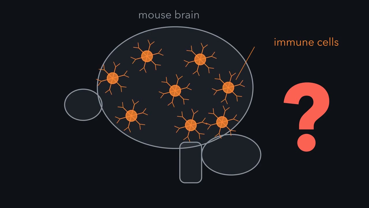 ▶ Watch One Job, Three Birthplaces · 2:20
Read the transcript of “One Job, Three Birthplaces”
Film transcript: One Job, Three Birthplaces
In a mouse, the brain's own immune cells were not made by the brain. So where were they made? They are called microglia. In mice, most of them descend from cells born in the yolk sac, a membrane outside the embryo. Glia, the non-neuronal cells among neurons, do not share one birthplace. Astrocytes, oligodendrocytes and ependymal cells come from the neural tube. Most come through the same pool of radial glia that made the neurons. Microglia, in mice, come from the yolk sac. Schwann cells and satellite glia come from the neural crest. Now take two cells with the same job. Oligodendrocytes wrap central axons in myelin. Myelinating Schwann cells wrap peripheral axons in myelin.
Mice lacking the genes Olig1 and Olig2 have no spinal cord oligodendrocytes. Should they lose the Schwann cells of their nerves too? No, not as a lineage. Schwann cells come from neural crest outside the cord, so they do not need Olig genes themselves. These mice also lack spinal motor neurons, made by the same strip of cord. The reverse holds too. Remove the receptor ErbB3, and nerves lose their Schwann cell precursors. Oligodendrocytes do not need it.
Same job, different early requirements in these two examples. But a different birthplace does not guarantee different genes. Sox10 is needed by both lineages, at different stages. So here is the idea to carry. Glia is a job, not a lineage. To predict which glia a gene removes, ask where the cell was born. Then ask what its progenitor needed from that gene, at that time. What the cell does is a weaker guide. The rule predicts the first deficit, not the final count after the animal compensates. Next question. How do you find out where a cell was born?
From the film
- Glia is a job, not a lineage: glia do not share one birthplace.
- Astrocytes and oligodendrocytes come mostly from the radial glia that made the neurons, microglia in mice come from the yolk sac, and Schwann cells and satellite glia come from the neural crest.
- To predict which glia a gene removes, ask where the cell was born and what its progenitor needed at that time.
- The rule predicts the first deficit, not the final count after the animal compensates.
Glia do not share one birthplace. Astrocytes and oligodendrocytes come mostly from the same pool of radial glia that made the neurons. Microglia, in mice, descend from cells born in the yolk sac, a membrane outside the embryo, before the brain had a blood-brain barrier. Schwann cells come from the neural crest.
“Glia” covers several kinds of non-neuronal cell that live among neurons. This lesson asks where each kind comes from, how anyone found out, and what tells a progenitor to make one. By the end you should be able to take a mutation and say which glia it is likely to remove, and what else may go with them.
One job, three birthplaces
Here is the cast. In the central nervous system (brain and spinal cord) there are astrocytes, star-shaped cells that touch blood vessels and synapses; oligodendrocytes, which wrap central axons in myelin, and their dividing oligodendrocyte precursors (OPCs); ependymal cells lining the ventricles; and microglia, the resident immune cells. In the peripheral nervous system there are Schwann cells along nerves and satellite glia around neuron cell bodies in ganglia.
Lineage methods gave this answer:
- Astrocytes, oligodendrocytes and ependymal cells come from the neuroepithelium of the neural tube, mostly through the same pool of radial glia that made the neurons. In the mouse cortex, many astrocytes are then added after birth by local division of astrocytes that have already differentiated.
- Microglia come from the yolk sac, as fate mapping in mice showed.
- Schwann cells and satellite glia come from the neural crest, the cells that leave the edges of the closing neural tube (see the neural crest lesson).
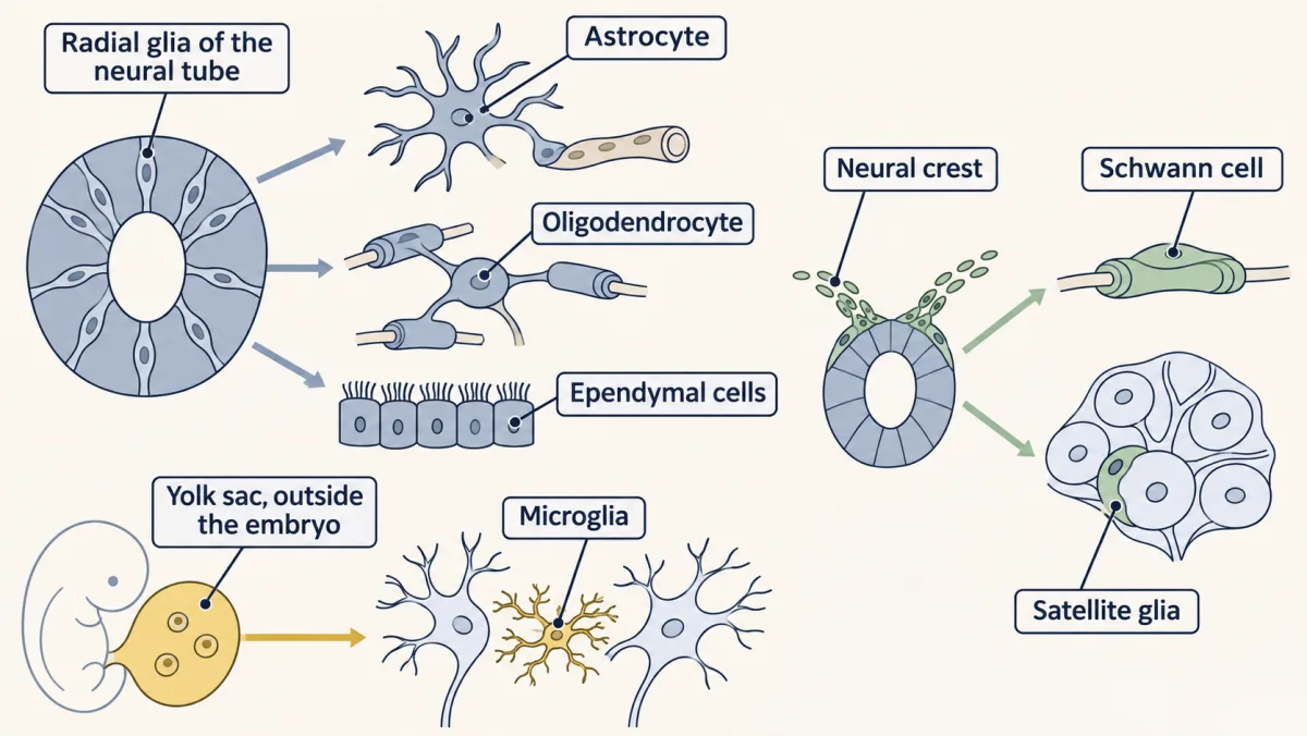
Two of these cells do the same job: oligodendrocytes and myelinating Schwann cells both wrap axons in myelin, in the central and the peripheral nervous system respectively.
Predict. Mice lacking the genes Olig1 and Olig2 have no spinal cord oligodendrocytes. Would you expect them to lose the Schwann cells of their nerves too?
Check your prediction
No, not as a lineage. Oligodendrocytes come from a strip of the spinal cord marked by Olig2, a transcription factor gene. Schwann cells come from neural crest and are not expected to need Olig genes themselves, so they are not lost as a lineage. Nerves also depend on axons, though, and these mice lack motor neurons, so Schwann cells of the ventral roots could be reduced secondarily.
The reverse holds too. Mice lacking the receptor ErbB3 lose the Schwann cell precursors of their nerves, but oligodendrocytes do not need ErbB3: oligodendrocyte lineage cells lacking it develop normally. Same job, different early requirements in these examples.
A different birthplace does not guarantee different genes, though. Sox10 is needed in both lineages, at different stages. The birthplace tells you which progenitor and which window to ask about; the gene’s known role there gives the answer.
The one idea to carry through this lesson. Glia is a job, not a lineage. To predict which glia a gene or signal removes, ask where the cell was born and what its progenitor needed at that time. What the cell does is a weaker guide: a shared job brings some shared genes, such as Sox10, but the early requirements differ.
The rule predicts the first deficit, in the window when a lineage is made; its limits follow the table at the end.
How do you learn a cell’s ancestry?
Pío del Río-Hortega’s silver stain was the first to tell microglia and oligodendroglia apart. But a stain shows a cell’s shape and position. It cannot follow a cell’s descendants, so it could not settle where any glial cell came from.
To give a cell a birthplace, you mark a progenitor and look for the mark later in everything it made. That is fate mapping, and the modern version is genetic. Cre is a recombinase, an enzyme that cuts and rejoins DNA at two short sequences called loxP sites. In a reporter mouse, two loxP sites flank a stop sequence in front of a reporter gene. Make Cre from the promoter of a gene a progenitor uses, and Cre removes the stop in that cell, switching the reporter on. The change is in the DNA, so it is permanent, and every descendant inherits it, whatever genes the descendant uses later.
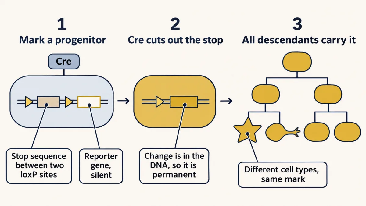
Inducible fate mapping adds a timer. Cre is fused to a fragment of the estrogen receptor, and a single dose of the drug tamoxifen lets it act for a short window, about a day in the line used for microglia. Only cells expressing the gene during that window are marked, and, as with most pulses, only a fraction of those. Every later count has to be read against that fraction.
Microglia come from outside
Read the transcript of “Marked in the Yolk Sac”
Film transcript: Marked in the Yolk Sac
How do you prove where a cell was born, when a stain shows only its shape? Mark a progenitor, then look for the mark later in everything it made. That is fate mapping. The modern version uses an enzyme called Cre. It cuts and rejoins DNA at two short sequences called loxP sites. In a reporter mouse, Cre removes a stop sequence between the sites. The reporter turns on, and every descendant inherits it. Make Cre from a gene the progenitor uses. In the line used for microglia, one dose of tamoxifen lets it act for about a day. In 2010, Florent Ginhoux, Miriam Merad and colleagues did this in mice. The Cre was driven by Runx1. A pulse at embryonic day 7.5 marked early progenitors.
Later, about a third of yolk-sac macrophages carried the mark. Call the yolk-sac labeling 30 percent. Suppose very few blood-making cells are marked, say 2 percent. What share of adult microglia should be marked? Try all from the yolk sac, half, or all from blood.
Each cell carries the mark or not. If all sources expand alike, the marked share mirrors the mix. All from the yolk sac predicts about 30 percent. Half and half, about 16. All blood, about 2. In adults, about a third of microglia carried the mark. Very few blood cells did. That is the top bar. Later pulses marked blood, but few microglia.
Read alone, a third sounds like a minority. Read against a source labeled at a third, it means most. The reading depends on the source. If the adult count is 30 percent and the true source were 40 percent labeled, about three quarters came from the yolk sac. So the right word is most, and possibly all. The data cannot give an exact number. Embryos lacking Ncx1, which the heart needs to beat, still made yolk-sac macrophages. But they had no brain microglia. So the progenitors travel in through the early circulation. Microglia also need the receptor CSF1R. Mice without it have few or none. In healthy adult mice, microglia renew themselves. Blood monocytes do not replace them.
So read a fate map against its source. In mice, most microglia were born in the yolk sac.
From the film
- Fate mapping marks a progenitor and looks for the mark in everything it made, so it can show where a cell was born.
- In mice, most microglia descend from yolk-sac cells, but the data cannot give an exact share.
- Read a fate map against the labeling of its source: a third of microglia marked, from a source marked at a third, means most.
- Microglial progenitors reach the brain through the early circulation and need CSF1R, and in healthy adult mice microglia renew themselves.
Microglia are macrophages, and for decades it was argued whether they come from blood monocytes made in the bone marrow or from an earlier source. In 1999 Fabrice Alliot, Isabelle Godin and Bernard Pessac proposed, from where and when the cells appeared in mouse embryos, that microglia arise from the yolk sac.
In 2010 Florent Ginhoux, Miriam Merad and colleagues tested this with an inducible fate map in mice. The Cre was driven by Runx1, a gene used by early blood-forming cells. A dose of tamoxifen at about embryonic day 7.5 marked cells expressing Runx1 then, which were in the yolk sac. When yolk-sac macrophages were examined later in the embryo, about a third carried the mark, not all of them.
Predict. Call the yolk-sac labeling 30 percent, and suppose the same pulse marks very few of the cells that make adult blood, say 2 percent. What fraction of adult microglia should carry the mark if they all come from the yolk sac? If half do? If they all come from blood?
Check your prediction
Each microglial cell either carries the mark or does not. If labeled and unlabeled founders expand alike, and there is no third source, the labeled share of microglia mirrors the mix of their sources, so the three hypotheses predict clearly different numbers.
| If adult microglia come from | Labeled microglia expected |
|---|---|
| the yolk sac, all of them | about 30% |
| half the yolk sac, half blood | about 16% (the average of 30% and 2%) |
| blood, all of them | about 2% |
In adults, about a third of microglia carried the mark, while very few blood cells did. That is the top row. Later pulses marked blood but few microglia.
Read alone, “a third of microglia” sounds like a minority. Read against a source labeled at a third, it means most of them. The reading is sensitive to the source fraction, though. So the right words are “most”, or “mainly”, and the data fit all of them. The comparison cannot give an exact percentage or exclude a small share from another source.
Two more results say how the cells arrive and what they need. Embryos lacking Ncx1, a sodium-calcium exchanger the heart needs to beat, made yolk-sac macrophages but had no brain microglia, so the progenitors travel in through the early circulation. Microglia also need the receptor CSF1R: mice without it have few or none.
One limit matters. In healthy adult mice, microglia renew themselves and blood monocytes do not replace them. After irradiation or some injuries, monocytes can enter the brain, which is why early bone marrow transplant experiments seemed to show replacement. Microglial roles in pruning synapses belong to the microglia and complement lesson.
Radial glia switch from neurons to glia
Read the transcript of “Same Progenitor, New Order”
Film transcript: Same Progenitor, New Order
In the mouse cortex, radial glia make neurons first, then astrocytes. Same progenitors, new output. What changes? In rodent cortex, the best-studied route to astrocytes uses cytokines. Its evidence sorts into three conditions. First, a progenitor must still be there. Proneural genes push dividing cells to become neurons. Notch turns on Hes repressors, which hold proneural genes down, so the cell stays a progenitor. Notch does not make astrocytes by itself. It keeps a reserve of progenitors for later. Second, the signal must arrive. It is a family of cytokines: CNTF, LIF and cardiotrophin-1. Their shared receptor subunit, gp130, activates JAK kinases.
JAK phosphorylates STAT3. STAT3 switches on astrocyte genes, such as GFAP. Third, the progenitor must be able to read the signal. That is competence. Give CNTF to cortical progenitors early in neurogenesis. They have the receptor, and STAT3 can be activated. What happens?
Few GFAP-positive astrocytes appear. In these early cells, the STAT3 site in the GFAP promoter is methylated. A methyl group on that site keeps STAT3 from binding there. STAT3 is active, but the GFAP gene does not answer. Later, demethylation and other changes in the cell let the same cytokine switch on GFAP.
So gliogenesis is timed partly inside the cell, not only by when the signal shows up. What unlocks it? Cells that have just committed to becoming neurons carry Notch ligands. In mouse cortex, Notch signaling in neighbouring progenitors induces a transcription factor, NFIA. NFIA helps remove the methyl groups. So Notch, which first held progenitors back, has a second job: competence. So, on this cortical cytokine route, a progenitor makes astrocytes when it is still there, given the signal, and able to read it.
From the film
- In rodent cortex, the best-studied route to astrocytes needs a progenitor still present, a cytokine signal, and the competence to read it.
- Notch keeps a reserve of progenitors and, in mouse cortex, later helps confer competence through NFIA.
- Early progenitors have the receptor, but the STAT3 site in the GFAP promoter is methylated, so gliogenesis is timed partly inside the cell.
Radial glia are the stem cells of the developing cortex (see that lesson). In the mouse cortex they make neurons first, and most astrocytes appear late in gestation and after birth. The same pool of progenitors changes what it makes. So what changes?
In rodent cortex, the best-studied route to astrocytes runs through cytokines, and the evidence for it sorts into three conditions, each tied to a molecule. They frame this route; they are not a complete account of every astrocyte.
- A progenitor must still be there. Proneural genes push a dividing cell to become a neuron (see proneural genes and lateral inhibition). Notch signaling turns on Hes repressors, which hold proneural genes down, so a cell receiving Notch stays a progenitor (see the Notch lesson). Notch does not make astrocytes by itself. It keeps a reserve of progenitors undifferentiated through the neurogenic phase, so cells are still there when the later signals arrive (whether Hes also pushes cells toward glia is less clear).
- The signal must arrive. The astrocyte signal is a family of cytokines, CNTF, LIF and cardiotrophin-1, members of the interleukin-6 family. Their receptors share the subunit gp130, which activates Janus kinase (JAK) kinases. JAK phosphorylates STAT3, which pairs up, enters the nucleus and switches on astrocyte genes such as GFAP, which encodes glial fibrillary acidic protein, the standard astrocyte marker (see the JAK-STAT lesson). In cultures of rodent cortical progenitors, CNTF drives the appearance of cells expressing GFAP.
- The progenitor must be able to read it. This is competence, a step that is easy to overlook.
Predict. Take cortical progenitors from a mouse early in neurogenesis and give them CNTF. They have the receptor, and STAT3 can be activated. What do you expect?
Check your prediction
Few GFAP-positive astrocytes. The STAT3 binding site in the GFAP promoter is methylated in these early cells: a methyl group sits on a CpG (a C followed by a G in the DNA) within the site and keeps STAT3 from binding there. STAT3 is activated, but the GFAP gene does not answer. As development goes on, the site is demethylated, other chromatin changes follow, and the cell becomes able to answer the same cytokine. Gliogenesis is timed partly inside the cell, not only by when the signal shows up.
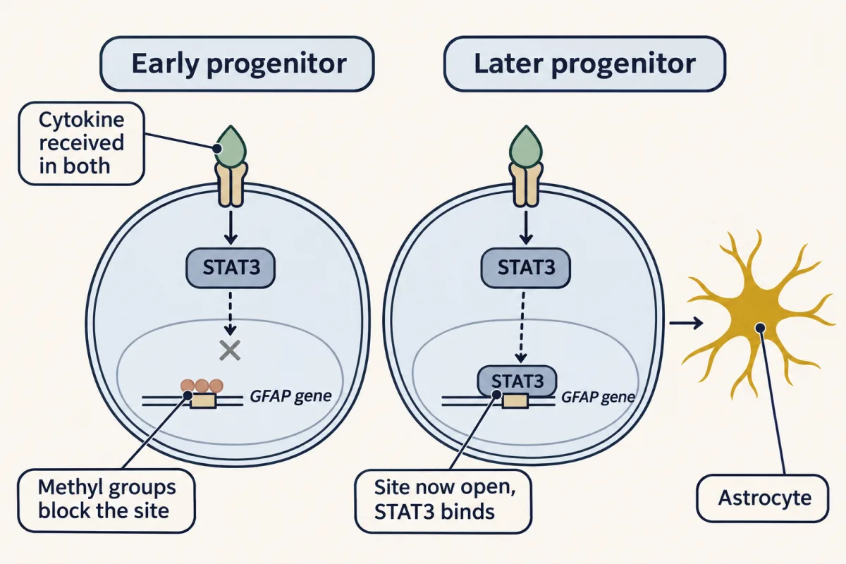
What unlocks the site? In chick and mouse spinal cord, the transcription factor NFIA comes on in progenitors just as gliogenesis begins and is needed for it to start on time. In mouse cortical precursors, cells that have just committed to becoming neurons carry Notch ligands. Notch signaling in neighbouring progenitors has been shown to induce NFIA, which helps demethylate the GFAP promoter so that cytokine-activated STAT3 can read it. So Notch, which earlier held progenitors back from becoming neurons, has a second job late in development: it helps confer competence.
Where does the cytokine come from? One proposal, from work in mouse cortex, is the neurons themselves: newborn neurons make cardiotrophin-1, and mice lacking it have been reported to make fewer cortical astrocytes. Put together, the proposed feedback is this: new neurons supply both the Notch ligands that unlock the GFAP site and the cytokine that the unlocked progenitors read. So the more neurons a region has made, the stronger its push toward glia, and glial onset is timed by the work already done. A fourth signal, BMP, works alongside the cytokines at astrocyte genes.
Oligodendrocytes start in a ventral strip
Oligodendrocyte precursors are born from specific domains, and in the spinal cord the main one is ventral. Recall from the Shh and BMP lesson that Sonic hedgehog from the floor plate divides the ventral cord into progenitor domains. One of them, pMN, expresses the bHLH factor Olig2. In mice and chicks, pMN first makes motor neurons (see the motor neuron lesson) and then switches to making oligodendrocyte precursor cells (OPCs).
Genetic fate mapping of Olig-expressing pMN cells finds both motor neurons and oligodendrocytes among their descendants. They share a birthplace, so a gene the whole pMN domain needs, such as Olig2, should be needed for both. Mice lacking both Olig1 and Olig2 have almost no spinal motor neurons and no spinal oligodendrocytes. The pMN cells are not simply lost, though. In these mutants they take other fates, first V2 interneurons and later astrocytes.
The ventral strip is not the only source. In the mouse spinal cord, a later, smaller wave of OPCs comes from dorsal domains. In the mouse forebrain, Cre lineage tracing by Kessaris and colleagues found three waves: from a ventral domain, then from the lateral and caudal ganglionic eminences (swellings in the wall of the ventral forebrain), then from the cortex itself after birth. Much of the first ventral wave is lost after birth and replaced by the later ones.
Predict. Kill one forebrain wave of OPCs as it forms. The rule says that wave’s cells are lost first. What do you expect to find in the adult forebrain?
Check your prediction
Normal numbers of OPCs. When Kessaris and colleagues killed any one wave, the other waves expanded and filled the gap. The rule predicted the first deficit correctly, but not the final count, because the animal compensated. Whether the waves are interchangeable in what they do as adults is still open.
Once born, OPCs migrate widely and divide in response to PDGF-A, acting on the receptor tyrosine kinase PDGFRα (see receptor tyrosine kinases). How they then wrap axons belongs to the myelination lesson.
One cell, two futures
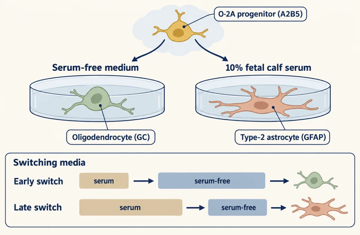
In 1983 Martin Raff, Robert Miller and Mark Noble cultured cells from the optic nerve of seven-day-old rats. Cells carrying the surface marker A2B5 became oligodendrocytes in serum-free medium, carrying galactocerebroside (GC). In 10% fetal calf serum they became GFAP-positive cells with an unusual shape, called type-2 astrocytes to set them apart from the more common type-1 astrocytes of brain cultures. They named the progenitor O-2A. A clear counterpart of the type-2 astrocyte in the living animal has been hard to find, so treat it as a fate seen in a dish.
Instruct or select? A design problem
A fate that follows the medium can mean two things. Serum might instruct each cell, or it might only select which already-decided cells survive. How would you tell them apart? The logic is the same as the three controls in the design problem of the neurotrophic hypothesis lesson: count across the window, mark division, and check identity.
One way to do it
Follow single cells or clones, not proportions of a mixed culture: plate cells at a density where each can be followed, and count every cell at the start and the end in both media.
If serum selects, cells headed for the wrong fate die or stop dividing in each medium, so the cells left in each medium are a different subset. If serum instructs, the same starting cells survive in both media, and the fate of each clone follows the medium.
The stronger test is to move clones between media at different times. A clone moved from serum to serum-free medium on day two that ends as oligodendrocytes, with no loss of cells and no extra divisions to make up for losses, was redirected, not culled. Repeating the move on later days tells you when the decision becomes fixed. A reciprocal move, from serum-free to serum, tells you whether the window closes at the same time in both directions.
In 1984 Raff, Brenda Williams and Miller ran the timed version. They switched media at different times and counted the recovered cells, to argue that the shift was not explained by death. The choice could still be reversed after one to two days in serum, after which it became fixed (the MCAT question below works through illustrative numbers).
Schwann cells come from the crest, in steps
In the peripheral nervous system, satellite glia and the enteric glia of the gut wall are crest derivatives. Schwann cells pass through stages that Kristján Jessen, Rhona Mirsky and colleagues defined in embryonic rat nerves: crest cell, then Schwann cell precursor, then immature Schwann cell, then myelinating or non-myelinating Schwann cell.
Each step has a different relationship to the axon. Schwann cell precursors die in culture unless neurons are present, and in a 1995 paper the group reported that neuregulin, a growth factor on axons read by ErbB2 and ErbB3, can keep them alive and drive their maturation into Schwann cells. Mouse genetics agreed: mice without ErbB3 lack Schwann cell precursors along their nerves.
This is the logic of the neurotrophic hypothesis turned toward glia, though these experiments did not test whether the supply is limiting and competed for, as nerve growth factor (NGF) is for sympathetic neurons.
When is ErbB2 needed? A conditional knockout deletes a gene only in cells that make Cre. In one such mouse, Cre came on early in the spinal roots, about embryonic day 11, but in the sciatic nerve only around birth, and ErbB2 was deleted with it.
Predict. If ErbB2 is needed early in the precursor pool and much less later, what should the Schwann cell counts look like a few days after birth, in the roots and in the sciatic nerve?
Check your prediction
At postnatal day 3.5, ventral-root Schwann cells fell from 51 to 5 per section, a loss of about 90%. Sciatic nerve counts barely changed, 588 in controls against 564, about 4% lower and not a significant change, though their myelin was thin.
That fits a need for ErbB2 early in the precursor pool, but the comparison is between two places as well as two times. Root Schwann cells come partly from boundary cap cells, which sit where the roots meet the cord, and the Cre line is active in them early. So the result does not fully separate when from where.
How glia later feed axons is in the metabolic partners lesson.
Putting it together
Each row below removes one progenitor, receptor or route, and asks what goes first.
| Removed | Glia lost first | Also lost | Spared | Caution |
|---|---|---|---|---|
| Olig1 and Olig2 (spinal cord, from the start) | spinal oligodendrocytes | spinal motor neurons | microglia; Schwann cell lineage (no direct Olig requirement) | pMN cells take other fates rather than simply die; ventral-root Schwann cells depend on motor axons, which are also lost |
| CSF1R (embryo and adult) | microglia (few or none) | many other tissue macrophages | neural-tube glia still form, though their numbers can shift secondarily | needing the receptor does not say where microglia come from |
| ErbB3 (throughout) | Schwann cell precursors | some other crest derivatives | oligodendrocytes (they do not need ErbB3) | does not show that axonal supply is limiting |
| ErbB2, Cre early in roots (about embryonic day 11) | about 90% of ventral-root Schwann cells | — | — | timing and place are not separated |
| ErbB2, Cre late in sciatic nerve (around birth) | no significant loss (564 vs 588); myelin thin | — | Schwann cell number | — |
| One forebrain OPC wave, killed as it forms (embryonic or around birth) | that wave’s OPCs | — | adult OPC number (other waves fill in) | does not show that each wave has a distinct job |
| Ncx1 (no heartbeat, before circulation starts) | brain microglia | circulation everywhere | yolk-sac macrophages | does not test a role for Ncx1 in the brain |
Every row asks the same two questions: where was the cell born, and what did its progenitor need then? Where the answer falls short, common causes are compensation, a change of fate, secondary effects of losing neighbouring cells or axons, a gene needed by both lineages, and a design that cannot separate timing from place. These rows do not test whether job or birthplace predicts better.
What the rule covers.
- Which cells. The glia and birthplaces listed at the start, in the mouse, with the chick for the spinal cord. The yolk-sac origin of microglia is a mouse result. In zebrafish, adult microglia come mainly from later, definitive blood-forming sites rather than from the earliest wave, so the birthplace has to be shown in each species.
- Which manipulation. Loss of a progenitor, or of a signal it reads, during the window when that lineage is being made. Later losses behave differently. Removing ErbB2 near birth barely changes Schwann cell number. Microglia lost in adults are rebuilt mostly by surviving microglia; blood-derived cells engraft only after irradiation, injury or particular depletion conditions, and whether they become true microglia is open.
- What it predicts. The first deficit, together with everything else the same progenitor makes, for a gene that progenitor needs at that stage. It does not predict the final count after the animal compensates, or whether a progenitor dies or switches fate.
Going deeper
How glia were first told apart
Cajal’s stains showed astrocytes well but left many small nuclei with no visible branches, which he grouped as a “third element”. Pío del Río-Hortega, using a silver carbonate stain he kept refining, split that group in two: microglia, described in 1919, and oligodendroglia, the “glia with few processes”, named in 1921. He saw many oligodendroglia lying between bundles of nerve fibres and, in a paper of 1922, proposed that they were the central counterpart of Schwann cells.
Instruction or selection: the 1984 medium-switch and later work
The serum signal itself was not identified in 1984; later work showed that bone morphogenetic proteins in serum can push these cells toward the type-2 astrocyte fate, as can CNTF together with extracellular matrix. Separately, in 1988, Raff’s group and Noble’s group showed that platelet-derived growth factor (PDGF) made by astrocytes keeps O-2A progenitors dividing in culture.
BMP at astrocyte genes
In fetal neural progenitors, BMP’s effector Smad1 and STAT3 are bridged by the coactivator p300, and together they induce astrocytes.
Does Hes push progenitors toward glia?
Hes1 maintains neural stem cells: forced Hes1 expression in mice keeps stem cells going and prolongs the making of later-born neurons. Whether Hes itself pushes cortical progenitors toward glia is less clear. In mouse telencephalon, forced Hes1 and Hes5 kept cells as stem cells, and those cells later made both neurons and astrocytes.
How sure is the microglia fate map?
Later work resolved the yolk-sac source as c-kit-positive erythromyeloid precursors, which depend on the transcription factors PU.1 and Irf8. That refinement carries a caveat for the arithmetic above. The one-third labeling was measured on yolk-sac macrophages, not on these precursors. Take the adult count as 30 percent and blood as 2 percent. If the precursors had been labeled at 40 percent while the macrophages were at 30, the yolk-sac share would be (30 − 2) ÷ (40 − 2), about 74 percent, or three-quarters: still most, but further from all. This is an illustration of sensitivity, not a measured contribution.
A job that hides a birthplace
In zebrafish, motor exit point glia are glia born in the central nervous system that leave it and ensheathe peripheral motor roots. They do a peripheral job with a central birthplace.
What else Schwann cell precursors make
Schwann cell precursors are not committed only to Schwann cells. They also give rise to melanocytes, endoneurial fibroblasts and parasympathetic neurons, so a mutation that removes the precursors removes more than glia.
Your notes
Select any text in the lesson to highlight it or add a note to it. Your highlights and notes are saved in this browser and collected in your notebook.
This browser is not letting the site save anything (a private window or strict privacy settings), so highlights and notes will be lost when you leave the page.
How we measure it
Clonal culture with medium switching
Single progenitors, or cultures of known composition, are grown in two defined media and moved from one to the other at chosen times. Counting all surviving cells as well as their markers separates a signal that instructs a fate from one that only selects survivors.
Inducible genetic fate mapping
A Cre recombinase fused to an estrogen-receptor fragment is made from a lineage gene's promoter. A single dose of tamoxifen lets Cre act for a short window, about a day in the Runx1 line used for microglia, so it switches on a permanent reporter only in cells expressing the gene then, and in all their descendants. The pulse marks only some of those cells, so a labeled fraction is read against the fraction marked in the source.
Bone marrow chimeras and parabiosis
Marked bone marrow is transplanted into a host, or two mice are joined so that they share blood. If a tissue's resident cells are replaced from blood, they take on the donor's mark. Irradiation before transplantation can itself let blood cells enter, so parabiosis is the cleaner test.
Conditional knockout with Cre lines
A gene is flanked by loxP sites so that it is deleted only in cells that make Cre, the recombinase that cuts and rejoins DNA at loxP sites, removing the stretch between them. Choosing a Cre line active in one place and time shows when and where a receptor such as ErbB2 is needed.
Practice questions on this lesson (3) → MCAT-style, original, each with an explanation
Where this lesson fits
Before thisReview first
- The Ventricular Zone: Radial Glia as Stem CellsThis course · Lesson 13
Radial glia as cortical stem cells are the pool that later makes astrocytes and oligodendrocytes here.
- The Neural Crest: Cells That Leave HomeThis course · Lesson 20
You need the crest's origin and migration to follow Schwann cells and satellite glia.
- Ventral to Dorsal: Sonic Hedgehog and BMP in the Neural TubeThis course · Lesson 10
Shh divides the ventral cord into domains, including pMN, where oligodendrocyte precursors begin.
After thisStudy next
- Myelination: Wrapping the Axon So the Signal JumpsThis course · Lesson 44
It takes the oligodendrocytes and Schwann cells born here and shows how they wrap axons.
- Feeding the Axon: Glia as Metabolic PartnersThis course · Lesson 45
It shows what astrocytes and oligodendrocytes do for axons once they exist.
Elsewhere on the siteFilms, simulations and method films in other courses
- BioassaysMethod film
The O-2A culture work uses a living cell response to separate instruction from selection by a factor.
- Targeted cell ablationMethod film
Predicting which glia are lost when a progenitor or survival signal is removed rests on killing chosen cells.
- One Protein From Receptor to GeneFilm · Cell Signaling
The cytokine route to astrocytes is read through JAK-STAT, which this film shows from receptor to gene.
- The Receptor Is the MessageFilm · Cell Signaling
Notch holds back progenitors and later confers competence for glia, which this film explains.
Suggested by an AI model from the lessons themselves, then checked. How we use AI
Finish line
Test yourself
8 questions
Practice and MCAT-style, with explained answers.
Next · Lesson 44
Myelination: Wrapping the Axon So the Signal Jumps
Wrap an axon in tight layers of glial membrane, leave short bare gaps, and its signal jumps from gap to gap instead of creeping.
Continue →← Lesson 42 · Sensitive Periods in Behavior: Song, Language and Sight