Developmental Neuroscience · Unit 6: Axons find their way
One Axon, Many Dendrites: Neuronal Polarity
Rewritten on : new text, flashcards and film. Your notes and highlights stay, but the text around them may have moved.
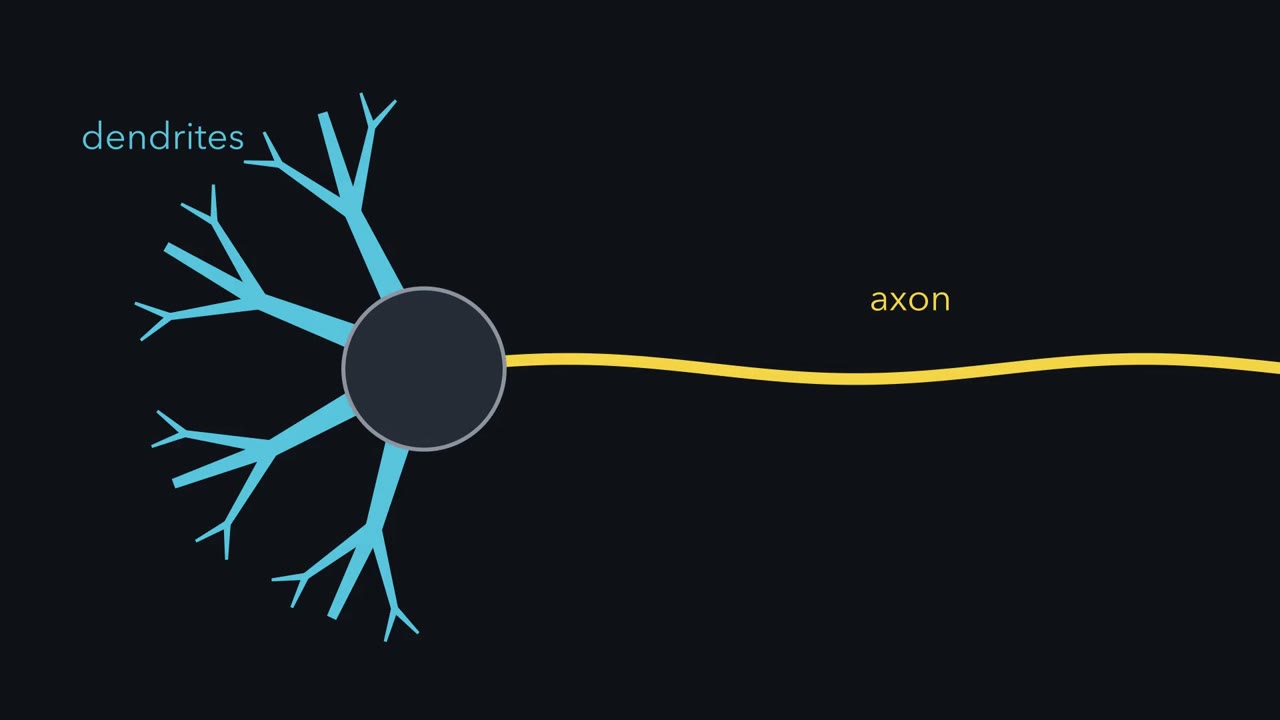
A neuron takes in signals on dendrites and sends them out along one axon. Cut that axon close to the cell body in a dish and one of its dendrites can become a new axon. How does a neuron choose its axon, and keep it?
Pause when the reading reaches a film, and offer to play it. Saved on this device.
Watch the film now, or keep listening
Builds on:The Growth Cone: The Tip That Steers the AxonSeeing Neurons: Golgi, Cajal and the Neuron DoctrineWhy these?
 ▶ Watch Cut the Axon · 2:47
Read the transcript of “Cut the Axon”
Film transcript: Cut the Axon
A neuron listens with its dendrites and speaks through its axon. How does it keep the two apart? Dendrites are usually several, short and branched. They receive input from other cells. The axon is usually single, even in width and often very long. It carries the action potential to other cells. Polarity is the word for a cell with two distinct domains, different in structure and molecules. The difference is molecular. Dendrites carry the protein MAP2, receptors for neurotransmitters, and often spines. The axon carries other proteins, such as tau, and its own channels and vesicle machinery. A neuron must sort thousands of proteins to the right side, and keep them there for decades.
Now a puzzle from a dish. Take a mature neuron, and cut its axon close to the cell body. Cut another neuron's axon far from the cell body. What grows out in each case? Cut close, and one of the dendrites can grow out as a new axon. Cut far, and the old axon simply regrows. So axon is not a permanent label. It is a state the cell keeps, and losing most of the axon can reset it. That raises two questions. How is the axon won? And once won, how is it held? Here is the map: a young neuron in a dish, as one neurite pulls ahead. In the middle sits the cell body. Several short minor neurites grow out from it, all alike. Each neurite ends in a growth cone, the motile tip of a growing neurite.
One has pulled ahead, and is becoming the axon. The others will become dendrites. The axon's first few tens of micrometres, the proximal axon, is where the initial segment will form. The idea to carry through: an axon is won in a race, and then held. In a useful model, positive feedback in each neurite, strong enough to compound a lead, plus competition among them, decides the race. Growth in simple proportion to length picks no winner. Then structures in the axon help hold the win: its microtubule tracks, and a gate near its start. Loosen the hold, and the axon can lose features. Cut almost all of it away, and the race reopens. Won, then held. But this race was seen in a dish.
Next, we watch it begin.
From the film
- An axon is not a permanent label: in a dish, cutting it close to the cell body can let a dendrite grow out as a new axon.
- An axon is won in a race among young neurites, and then held.
- Cut almost all of the axon away and the race can reopen.
In a dish, cut a neuron’s axon close to the cell body, and one of its dendrites can grow out as a new axon. Cut it farther away, and the old axon regrows.
A neuron takes in signals on its dendrites and sends them out along its axon, so the two kinds of process must stay different. The cut-axon result tells you that “axon” is not a permanent label stamped on a process when it first sprouts. It is a state the cell keeps, and losing most of the axon can reset it. This lesson asks how an axon is formed, and how it then stays an axon.
One cell, two kinds of process
Most neurons in a vertebrate brain have two kinds of process. Dendrites are usually several, short, branched and tapering; they receive input from other cells. The axon is usually single, of even width and often very long; it carries the neuron’s output, the action potential, to other cells. Polarity is the word for a cell having two distinct ends or domains that differ in structure and molecules.
The difference is molecular as well as visible. Dendrites contain the microtubule-associated protein MAP2, receptors for neurotransmitters, and, in many neurons, spines where synapses form. The axon contains other proteins, such as the microtubule-associated protein tau, and its own channels and vesicle machinery. A neuron must sort thousands of proteins to the right side and keep them there for decades. That is what makes the cell biology special.
The map
Most arguments in this lesson happen on one drawing, which the first film draws: a young neuron in a dish at the moment one of its neurites pulls ahead.
- The cell body sits in the middle.
- Minor neurites, several short processes that all look alike, grow out from it. Each ends in a growth cone, the motile tip of a growing neurite (see the lesson on the growth cone).
- The axon is the one neurite that has pulled ahead. Its first few tens of micrometres, the proximal axon, is where the axon initial segment will later form.
- The dendrites are what the other minor neurites will become.
The cut in the opening was made in fully polarized, mature neurons, and it returns near the end. The first experiment below is a different cut, made in young neurons just as the axon appears.
The map raises two puzzles.
- How is the axon won? The neurites start alike. What makes one of them the axon?
- How is it held? Once one is chosen, what keeps the axon an axon, and the dendrites dendrites?
The one idea to carry through this lesson. An axon is won in a race and then held. Young neurites compete, and a useful model says the race is decided by positive feedback in each neurite plus competition among them, with feedback strong enough to compound a lead; growth in simple proportion to length, even from a shared supply, picks no winner. Once one neurite is ahead, structures in the axon help hold the win: its microtubule tracks and its initial segment, a gate near its start. Loosen the hold and the axon can lose features; cut almost all of it away and the race can open again.
What the rule covers. Neurons dissociated from the embryonic hippocampus and grown in a dish, where the race was seen and where the drug and cut tests below were done. Three limits. In the brain the race may be tilted or skipped. The hold differs along the axon: the initial segment holds only its proximal part. And some neurons break the pattern: anaxonic neurons, such as some retinal amacrine cells, have no clear axon, and many sensory neurons have a single process that splits into two axon branches.
Five stages and a race
Read the transcript of “Five Stages and a Race”
Film transcript: Five Stages and a Race
Are young neuron's neurites truly equal? Or is one of them fixed as the axon from the start? Gary Banker's laboratory grew neurons from the embryonic rat hippocampus. Most are pyramidal cells of one broad type. Grown at low density, each neuron can be watched on its own. In 1988, Carlos Dotti, C. A. Sullivan and Banker described five stages. Stage one. Within hours of plating, the round cell spreads thin, flat veils of membrane. Stage two. The veils condense into several short neurites, all alike. Stage three. After a day or two, one neurite grows much faster than the rest. It becomes the axon. Stage four. Over the next few days, the other neurites grow, taper and branch as dendrites.
Stage five. In the second week and later, spines form, and synapses multiply and mature. Back to stage two. The neurites look alike. That could mean two things. Perhaps one was fixed for good as the axon. Or perhaps the choice is open, and they are competing. A young neuron's cut tests whether the choice can be undone. Take a neuron just at stage three. Cut its leader back to the length of its siblings, erasing its lead. If the axon was fixed for good, which process should grow out as the axon? And if the neurites compete?
If the choice were fixed, the stump should regrow as the axon, whatever its length. Often, it did not. In 1987, Carlos Dotti and Gary Banker found that another neurite, a future dendrite, could grow out as the axon. Then Kimberly Goslin and Banker cut axons to different lengths. When one remaining process was more than about 10 micrometres longer than all the others, it became the axon. When the remaining processes were nearly equal, the new axon could not be predicted. It could be any of them, the cut one included. So the choice can be undone. It behaves like a race, and length is part of the score. Goslin and Banker proposed that uncut neurons choose the same way.
The 10 micrometre figure belongs to these experiments in cultured neurons. It is not a rule for every neuron. The logic does not depend on the number. Get far enough ahead, and you win. Stage two is a race. Next question: what kind of race has only one winner?
From the film
- Cultured rat hippocampal neurons polarize in five stages, and the axon appears at stage three when one neurite grows much faster than the rest.
- Cutting the leader can hand the axon to another neurite, so the choice can be undone and behaves like a race in which length is part of the score.
- The 10 micrometre lead belongs to these cultured-neuron experiments and is not a rule for every neuron.
The clearest view of how a neuron polarizes comes from a dish. Neurons taken from the embryonic rat hippocampus survive and grow processes in culture, and because most hippocampal neurons are pyramidal cells of one broad type, a dish holds a fairly uniform population. In 1988 Carlos Dotti, C. A. Sullivan and Gary Banker followed single neurons grown at low density. They described five stages.
- Lamellipodia. Within hours of plating, the round cell spreads thin, flat veils of membrane.
- Minor neurites. The veils condense into several short neurites, all alike.
- The axon. After a day or two, one neurite starts to grow much faster than the rest. It becomes the axon.
- Dendrites. Over the next few days the other neurites grow, taper and branch as dendrites.
- Maturation. In the second week and later, dendritic spines form and synapses, first seen within days, multiply and mature. (Synapses get their own lesson later: the lesson on synapses in the brain.)
Is it a race? Cut the leader
At stage 2 the neurites look alike. That could mean two different things. Perhaps one of them was fixed for good as the future axon. Or perhaps the choice is open, and they are competing. A young neuron’s cut tests whether the choice can be undone.
Predict. A young neuron has just reached stage 3: one neurite has pulled ahead and is becoming the axon. Cut it so that its stump is no longer than its siblings, erasing its lead. If the axon was fixed for good, which process should grow out as the axon? What if the neurites are competing?
Check your prediction
If the choice were fixed for good, the stump should regrow as the axon whatever its length. Often it did not. In 1987 Carlos Dotti and Gary Banker found that after such a cut, a different neurite, one that would otherwise have become a dendrite, could grow out as the axon. Kimberly Goslin and Banker then cut axons to different lengths. When one remaining process, the stump or a sibling, was more than about 10 micrometres longer than all the others, it became the axon. When the remaining processes were nearly equal in length, the new axon could not be predicted: it could come from any of them, the cut one included.
So the choice at stage 2 can be undone: whatever early bias a neurite may carry, a cut can hand the axon to another. It behaves like a race, and length is part of the score. Goslin and Banker proposed that uncut neurons choose the same way. The 10 micrometre figure belongs to these experiments in cultured neurons, not to every neuron (Going deeper), but the logic does not depend on the exact number: get far enough ahead and you win.
What a race with one winner needs
Read the transcript of “What Makes a Winner”
Film transcript: What Makes a Winner
A longer neurite grows faster. Is that enough to make one winner? Here is a model, not a measurement. Start with five neurites. Four are 15 micrometres long, and one is 20, 5 micrometres ahead. Each neurite grows in proportion to its own length. A longer one grows faster. Run the rule for two days. Does one neurite win?
No. All five grow. The leader stays the same fraction ahead, a third longer than each sibling. The gap in micrometres widens, but nothing holds the others back. You end with five long neurites, and no single winner. Growing faster the more it grows is true. But growth in simple proportion to length picks no winner. The leader's gain must come at the others' cost. Two ingredients do this. First, positive feedback within a neurite. Growth recruits more of whatever drives growth. Second, competition between neurites. One proposal: they draw on a limited supply from the cell body. So one neurite's gain is the others' loss. But even a shared supply is not quite enough.
If each neurite takes a share in proportion to its length, the fractions again stay fixed. A long neurite's growth, relative to its own length, has to be larger than a short neurite's. There are two ways. The feedback could be steeper than proportional. Or every neurite could pay the same fixed running cost, such as retraction. That cost takes a bigger bite out of a short neurite's growth. With either one, a small lead grows into a large one. The others grow about fourfold, then slow to crawl. With both, the others shrink back to stubs. Now there is one winner. Now place the molecules in the loop. PI3-kinase makes the lipid PIP3. It builds up at the tip of the future axon.
PIP3 is a mark at one spot on the membrane. It draws proteins with PH domains to that spot. The Par3/Par6/aPKC complex, which also marks the top of an epithelial cell, gathers there. At the tip, the switches Cdc42 and Rac reorganize actin. Loosened actin is proposed to let microtubules push forward into the growth cone. Part of the loop is thought to be physical. A faster neurite draws more material and more transport. Then it grows faster still. The loop closes. There is also a brake. RhoA tends to oppose neurite growth. What holds the losers back is less settled. A limited supply and an inhibitory signal are both proposed. So a winner needs competition, plus something that compounds a lead.
Next, can we rig the race?
From the film
- Growth in simple proportion to length picks no winner, because the leader stays the same fraction ahead.
- One winner needs positive feedback within each neurite plus competition between neurites.
- In the proposed loop, PIP3 builds up at the tip of the future axon, and the race model is a thought experiment, not a measurement.
A race needs a rule for how the runners speed up. Try the simplest rule first.
Predict. Start with five neurites: four 15 micrometres long and one 20 micrometres long, 5 micrometres ahead. Suppose each grows in proportion to its own length, so a longer neurite grows faster. Run the rule for two days. Has one neurite won?
Check your prediction
No. All five grow, and the leader stays exactly the same fraction ahead, a third longer than each sibling, for as long as you run the rule. The gap in micrometres widens, but nothing holds the others back. You end with five long neurites and no single winner.
So the leader’s gain has to come at the others’ cost. Two ingredients are needed, though even together they are not quite enough.
- Positive feedback within a neurite. Growth recruits more of whatever drives growth.
- Competition between neurites. One proposal is that the neurites draw on a limited supply from the cell body, such as membrane, tubulin or signalling proteins. Another is a spreading inhibitory signal that holds the others back. Either way, one neurite’s gain is the others’ loss.
If every neurite simply takes a share of a shared supply in proportion to its length, the fractions again stay fixed. Something must make a long neurite’s growth, relative to its own length, larger than a short neurite’s. Steeper-than-proportional feedback does this; so does a fixed running cost that every neurite pays, such as retraction, which takes a bigger bite out of a short neurite’s growth. With either one, a small lead grows into a large one; with both, the other neurites shrink back to stubs.
| Rule (four neurites at 15 µm, one at 20 µm, 48 hours; illustrative model) | Leader : next sibling | The other four |
|---|---|---|
| Each grows in proportion to its own length | 1.33, unchanged | keep growing |
| A shared supply, split in proportion to length | 1.33, unchanged | keep growing |
| The same, plus a fixed cost for every neurite | about 5 | grow about fourfold, then crawl |
| A shared supply, split in proportion to length squared | about 15 | grow about fourfold, then crawl |
| Length squared plus a fixed cost | about 400 (about 820 µm against the 2 µm floor) | shrink to the floor by about 14 hours |
The model is a thought experiment, not a measurement. Its supply (20 micrometres of growth an hour, shared among five neurites), its cost (2 micrometres an hour for each neurite) and its floor (no neurite shrinks below a 2 micrometre stub) are illustrative values.
The players, sorted by job
The molecules reported in cultured neurons now have places to go.
- The positive loop at the tip of the future axon.
- PI3-kinase makes the lipid phosphatidylinositol 3,4,5-trisphosphate (PIP3), a mark at one spot on the membrane that draws proteins with PH domains to that spot (see the lesson on PI3-kinase and Akt and the lesson on lipid anchors). PIP3 builds up at the tip of the future axon.
- The Par3/Par6/aPKC complex, the same polarity complex that marks the top of an epithelial cell, gathers there.
- At the tip, the Rho-family GTPases Cdc42 and Rac, molecular switches that are on when bound to GTP (see the lesson on GTPase switches), reorganize actin.
- Loosened actin is proposed to let microtubules push forward into the growth cone, and microtubules in the future axon are more stable than in its siblings: two polymers with opposite jobs at the tip, one giving way, the other advancing. Part of the loop is thought to be physical: a neurite that grows faster draws more building material and more transport, and then grows faster still, which brings the loop back to the start.
- A brake. RhoA tends to oppose neurite growth (see the lesson on integrins and Rho GTPases).
- Holding the others back. Less settled: the shared supply or the spreading inhibitory signal proposed above.
Rig the race
Read the transcript of “Rig the Race”
Film transcript: Rig the Race
If the race reads the cytoskeleton at a tip, can you rig it? Start with what the winner's tip looks like. In 1999, Frank Bradke and Carlos Dotti looked at growth cones in cultured hippocampal neurons. The future axon's growth cone had a looser, more dynamic actin network than its siblings. But is loose actin a cause of winning, or a consequence? Test it. Cytochalasin D is a drug that interferes with actin assembly. At stage two, before any neurite pulls ahead, apply a low dose to one growth cone only. What happens to that neurite? And what if you bathe the whole cell in the drug?
The treated neurite was likely to become the axon. On screen is an idealized tally. By chance, one neurite in five would win. The drugged one wins far more often. And bathed in the drug, a neuron could grow several axons.
In 2008, Harald Witte, Dorothee Neukirchen and Frank Bradke did the same with microtubules. Taxol is a drug that stabilizes microtubules. A flash of light released a taxol analogue in one neurite. That neurite became more likely to be the axon. Low-dose taxol given to the whole neuron led several neurites to grow as axons.
Now block the positive loop everywhere, with PI3-kinase blocked throughout. This is reported to leave no neurite becoming an axon. It does not free the others. It stops all of them. Two results fit the race at once. A local boost rigs it. Blocking the loop everywhere leaves no winner. The several-axon result needs a second look. If every neurite's supply is tripled in this model, one still wins. So a plain speed-up of this kind does not match the several-axon result. One possible reading is a threshold. The model does not test it. On that reading, the drug pushes each neurite past whatever level of loose actin or stable microtubules marks an axon.
Each one crosses that line without having to win the race. Either way, the race reads the cytoskeleton at the tip. Next, the microtubules become tracks.
From the film
- The growth cone of the future axon has a looser, more dynamic actin network than its siblings.
- Boosting loose actin or stable microtubules in one neurite made it more likely to become the axon, and blocking PI3-kinase throughout is reported to leave no axon.
- Whole-cell drug doses led several neurites to grow as axons, which a plain speed-up of the race model does not predict.
If the race reads the state of the cytoskeleton at a tip, the tip of the winner should look different, and you should be able to rig the race. In 1999 Frank Bradke and Carlos Dotti reported the first half: the growth cone of the future axon has a looser, more dynamic actin network than the growth cones of its siblings. Then they tested whether that was cause or consequence.
Predict. At stage 2, before any neurite has pulled ahead, apply a low dose of cytochalasin D, a drug that interferes with actin assembly, to the growth cone of one neurite only. What happens to that neurite? What should happen if you bathe the whole cell in the drug instead?
Check your prediction
The treated neurite was likely to become the axon. Bathed in the drug, a neuron could grow several axons. In 2008 Harald Witte, Dorothee Neukirchen and Frank Bradke did the same with microtubules. Releasing a light-activated taxol analogue in one neurite made that neurite more likely to become the axon, and low-dose taxol given to the whole neuron led several neurites to grow as axons. Blocking PI3-kinase throughout is reported to do the opposite: no neurite becomes an axon.
Two of these results fit the race directly. A local boost rigs it, and blocking the positive loop everywhere leaves no winner.
The several-axon result needs a second look. Raise the supply in the full race model, with steep feedback and a fixed cost, or multiply every rate by the same factor, and one neurite still wins. The whole-cell drug results are therefore not what a simple across-the-board speed-up of this model predicts. One possible reading is that the drugs push each neurite past whatever level of actin looseness or microtubule stability marks an axon, without its having to win the race; the model does not test that.
Either way, the race reads the cytoskeleton at the tip.
Tracks that point two ways
Read the transcript of “Tracks That Point Two Ways”
Film transcript: Tracks That Point Two Ways
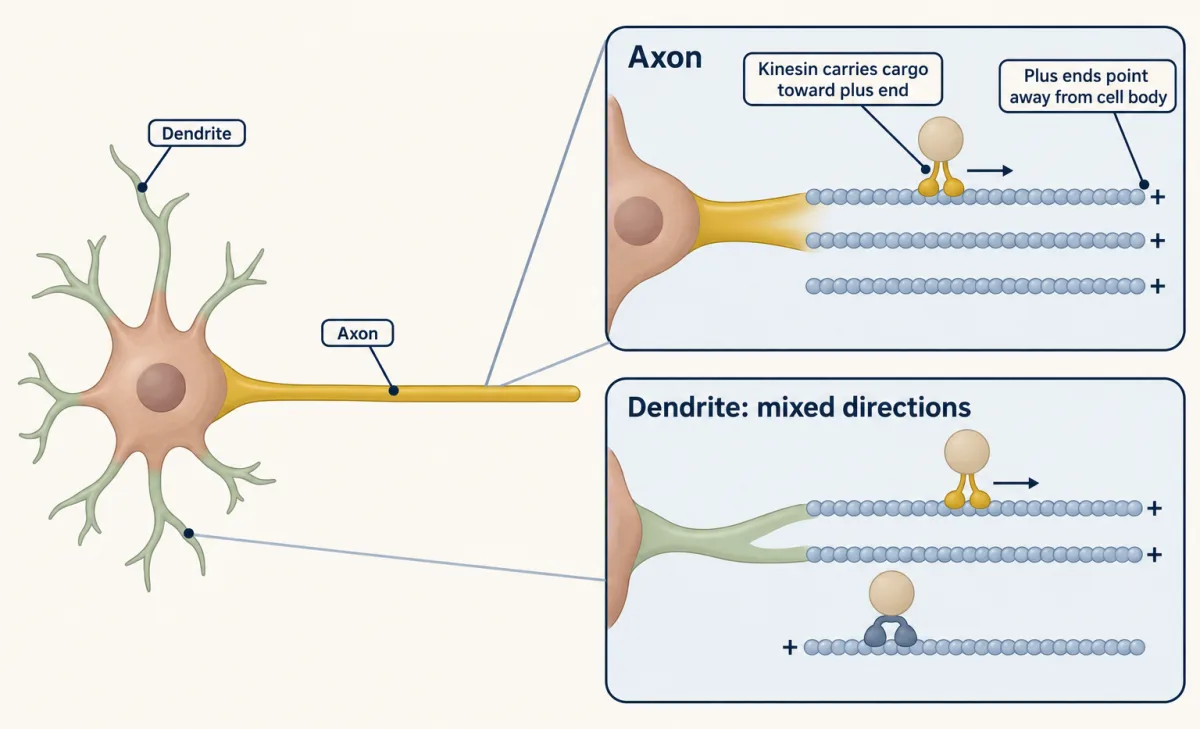
Once an axon is chosen, how does cargo know which branch to go into? Microtubules link choosing the axon to holding it. A microtubule has a fast-growing plus end and a slower minus end. Motor proteins read that direction. Most kinesins walk toward plus ends. Dynein walks toward minus ends. Picture a plus-end-out track. Its plus end points away from the cell body. On that track, kinesin walks outward. Dynein walks home, toward the cell body. On a minus-end-out track, it is the reverse. So which way do the tracks point in a real neuron? In 1988, Peter Baas and colleagues used hook decoration on cultured rat hippocampal neurons. Extra tubulin adds curved sheets to each microtubule's wall.
In cross section, the hooks curve one way or the other. The curve shows which way that microtubule points. In the axon, nearly all microtubules point their plus ends away from the cell body. In dendrites the orientation is mixed. A large fraction point each way. So which motor can carry cargo outward into a dendrite, but hardly ever into the axon?
Dynein. It can carry cargo outward only on minus-end-out tracks. Vertebrate dendrites have many of those tracks. The axon has almost none. Kinesin can walk outward in both, since both have plus-end-out tracks. But a kinesin that prefers stable, plus-end-out microtubules can be steered into the axon. So the way the tracks point limits which motors can carry cargo outward into a process. That gives the neuron a sorting rule. The uniform plus-end-out array may form as the axon forms. If so, the tracks may help choose the axon, as well as keep it. Flies differ. Their dendrites are mostly minus-end-out. So the details depend on the animal. But in both, axon and dendrites offer different tracks.
Different tracks, different motors, different cargo. The tracks may be part of the hold. Next, the gate at the start of the axon.
From the film
- In cultured rat hippocampal neurons, axon microtubules nearly all point plus end out, while dendrite microtubules point both ways.
- The way the tracks point limits which motors can carry cargo outward: dynein can do so into dendrites far more readily than into the axon.
- In fly neurons the dendrites are mostly minus end out, so the details depend on the animal.
Microtubules may link choosing the axon to holding it. A microtubule has a fast-growing plus end and a slower minus end. Motor proteins read that direction. Most kinesins walk toward plus ends; dynein walks toward minus ends. A plus-end-out track is one whose plus end points away from the cell body.
| On a track that is | Kinesin walks | Dynein walks |
|---|---|---|
| plus-end-out | outward | toward the cell body |
| minus-end-out | toward the cell body | outward |
In 1988 Peter Baas and colleagues used hook decoration to score microtubule orientation in cultured rat hippocampal neurons. In the axon, nearly all microtubules point their plus ends away from the cell body. In dendrites the orientation is mixed: a large fraction point each way.
Predict. Given those two patterns, which motor can carry cargo outward into a dendrite far more readily than into the axon?
Check your prediction
Dynein. Cargo carried by dynein can travel outward only on minus-end-out tracks, which vertebrate dendrites have in large numbers and the axon almost lacks. Kinesin can walk outward in both, since both have plus-end-out tracks, but a kinesin that prefers stable plus-end-out microtubules can be steered into the axon.
So the orientation of microtubules in a process limits which motors can carry cargo outward into it. That gives the neuron a sorting rule. The uniform plus-end-out array may form as the axon forms, so this rule may help choose the axon as well as keep it (Going deeper). In fly neurons, dendrites are instead mostly minus-end-out, so the details depend on the animal. In both, axon and dendrites offer different tracks.
The initial segment: a gate that holds the win
Read the transcript of “The Gate, and the Race Reopened”
Film transcript: The Gate, and the Race Reopened
The axon has been chosen. What keeps dendrite proteins from wandering into it? The first few tens of micrometres of the axon form the axon initial segment. Under its membrane sits a dense coat, built on a scaffold called ankyrin G. Ankyrin G binds beta-IV spectrin, which links it to rings of actin under the membrane. Through its ANK repeats, it also binds sodium channels, KCNQ2/3 potassium channels and neurofascin-186. The initial segment has two jobs. The first is electrical. A high density of sodium channels, among other features, gives it a low threshold. So in many neurons, this is where the action potential starts. In 1999, Bettina Winckler, Paul Forscher and Ira Mellman tested whether the membrane itself is divided there.
They attached small beads to membrane proteins, and dragged them with a laser trap. the cell body and dendrites the beads moved freely. At the initial segment they were held back. The hold depended on actin. How the barrier works is still argued. Now test the scaffold. Lower ankyrin G after the initial segment has formed. The axon is already chosen. What happens to the initial segment? And does MAP2, the dendrite protein, stay out of the axon?
In 2008, Kristian Hedstrom, Yasuhiro Ogawa and Matthew Rasband did this. The initial segment fell apart. MAP2 spread into the axon. Near the cell body, the axon began to look like a dendrite, with dendritic spines. Farther out, it kept axonal proteins. So identity has to be actively held. Near the cell body, the initial segment holds it. Farther out, something else does, not yet identified. In 2008, Frank Bradke's laboratory, with Susana Gomis-Rüth and Corette Wierenga, made the mature neuron's cut. When the stump was shorter than 35 micrometres, a dendrite became the axon. Longer stumps regrew. The authors linked this to stable microtubules in the distal axon.
They did not test the initial segment. So a young neuron's cut reopens the race easily. A mature neuron's cut reopens it only when almost the whole axon is removed. One caution: this was all in a dish. In the brain, the race may be tilted or skipped. In the developing cortex, the trailing process of a migrating neuron often becomes its axon. One pathway is clearly required there: the kinase LKB1, and the SAD kinases it activates. Cortical neurons lacking LKB1, or both SAD kinases, fail to form axons. An axon is won in a race, then held. Loosen the hold, and the axon can lose features. Cut almost all of it away, and the race reopens.
From the film
- The axon initial segment is a coat built on ankyrin G, and in many neurons it is where the action potential starts.
- Lowering ankyrin G in cultured neurons after the axon was chosen broke up the initial segment and let MAP2 spread into the axon, so identity has to be actively held.
- A mature neuron's cut reopens the race only when almost the whole axon is removed, in the cultures studied.
- In the developing cortex, neurons lacking LKB1 or both SAD kinases fail to form axons.
The first few tens of micrometres of the axon form a specialized zone, the axon initial segment (AIS). Under its membrane sits a dense coat built on the scaffold ankyrin G. Ankyrin G binds beta-IV spectrin, which links it to rings of actin under the membrane. Through its ANK repeats it binds voltage-gated sodium channels, KCNQ2/3 potassium channels and the adhesion molecule neurofascin-186, and it also binds the adhesion molecule NrCAM. In many neurons, such as cortical pyramidal cells, Kv1 potassium channels gather there too.
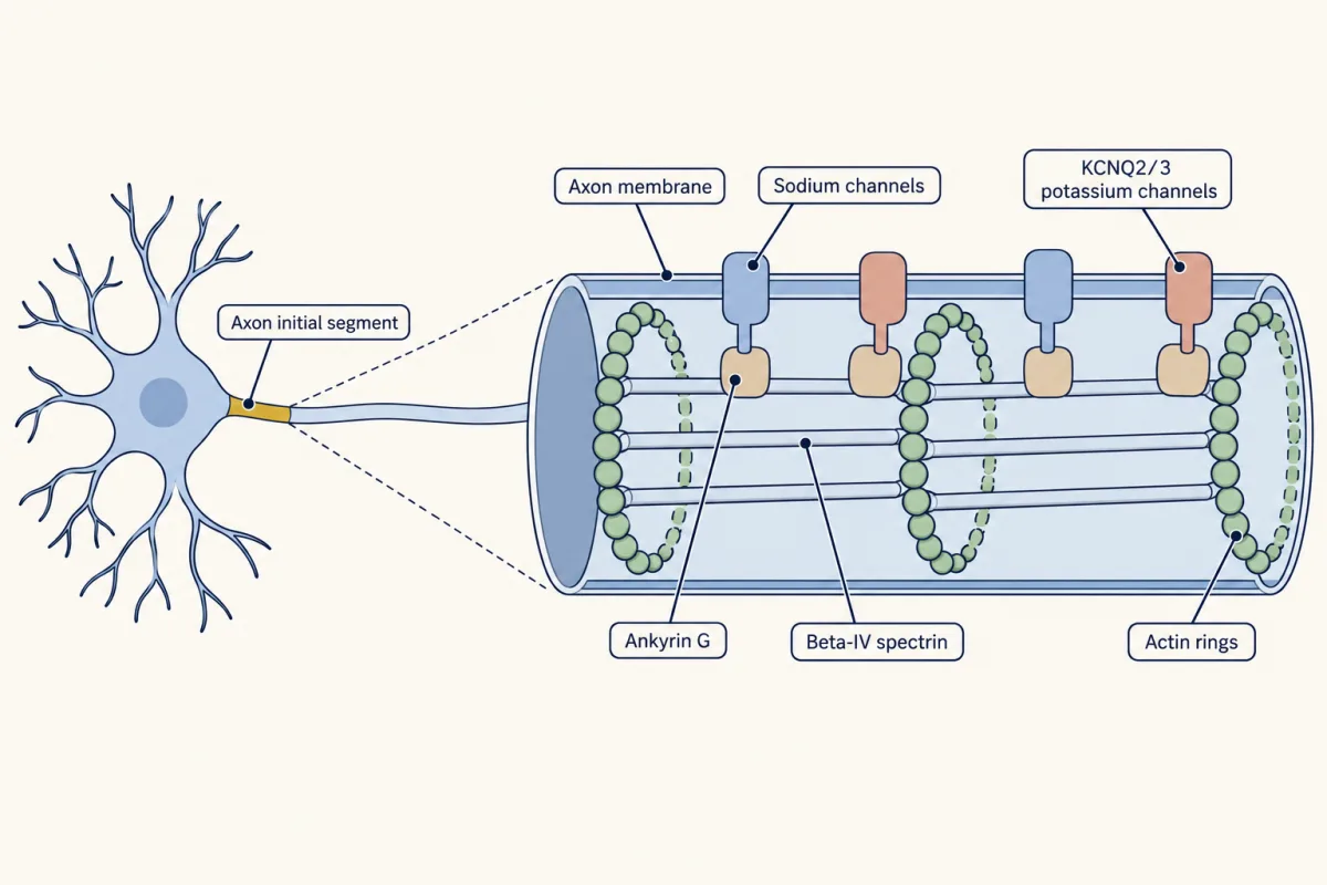
The AIS has two jobs. The first is electrical. A high density of sodium channels, among other features, gives it a low threshold, so in many neurons it is where inputs summed from the dendrites and cell body start the action potential. The second is to hold polarity.
How does the AIS hold polarity? In 1999 Bettina Winckler, Paul Forscher and Ira Mellman dragged membrane proteins with a laser trap and found that they moved freely in the cell body and dendrites but were held back at the initial segment, and that the hold depended on actin. How the barrier works is still argued (Going deeper).
Predict. Lower ankyrin G in cultured hippocampal neurons after the AIS has formed, so the axon has already been chosen. What happens to the AIS, and does MAP2, normally kept out of the axon, stay out?
Check your prediction
In 2008 Kristian Hedstrom, Yasuhiro Ogawa and Matthew Rasband did this. The AIS fell apart, and MAP2 spread into the axon. Near the cell body the axon began to look like a dendrite, with dendritic spines, though farther out it kept axonal proteins.
Ankyrin G is required to maintain neuronal polarity. Because the axon was already chosen, the experiment tests maintenance, not choice: identity has to be actively held. The AIS holds the proximal axon; the distal axon keeps its features without it, so something else must also help hold polarity, though these experiments do not say what. MAP2 is not a membrane protein but binds microtubules inside the cell, so the hold reaches beyond the membrane that the laser trap tested; how the segment keeps MAP2 out is not settled.
Design problem: the mature neuron’s cut
This is the cut from the opening. You grow mouse hippocampal neurons until they are fully polarized. In one neuron you cut the axon so close to the cell body that only a short stump remains (the experiment used less than 35 micrometres). In another you cut it much farther out, leaving a long stump. Predict what grows out in each case, and say what the pair of results tells you about where axon identity is stored.
One way to do it
Start with what each cut removes. The distant cut leaves the cell body, the dendrites and a long axon stump, with its stable microtubules, untouched. You expect the original axon to regrow from its stump, because nothing about its identity has been lost.
The close cut leaves only a short stump, with little of the axon’s stable microtubules. Now no process has a clear axonal advantage, which is a little like stage 2, though the dendrites are mature. You expect a dendrite to take over and grow out as the axon. That is what Susana Gomis-Rüth, Corette Wierenga and Frank Bradke reported in 2008.
So identity is held, not stamped: remove nearly all of the axon and the label can be reassigned; leave enough of it and the axon simply repairs itself. A follow-up test: lower ankyrin G first, then make the distant cut, and ask whether a dendrite now competes with the regrowing stump.
The authors linked the difference between short and long stumps to stable microtubules in the distal axon and did not test the initial segment. Myelin and nodes of Ranvier, which repeat much of the AIS machinery along the axon, come in the lesson on myelination.
Does the brain run the same race?
One caution. The five stages describe dissociated neurons in culture, which have lost their neighbours and their history. In the brain many neurons arrive with polarity already set by their migration, so the race may be tilted or skipped. In the developing cortex, for example, the trailing process of a migrating neuron often becomes its axon. The molecules overlap, but the starting point differs, and in tissue, cues from the environment also help orient the same machinery. One pathway is clearly required in the living cortex: the kinase LKB1 and the SAD-A and SAD-B kinases it activates. Cortical neurons lacking LKB1, or both SAD kinases, fail to form axons.
Putting it together
Go back to the two cuts. A young neuron’s cut reopens the race easily, and relative length is part of the score: a process more than about 10 micrometres ahead tends to win, and when the remaining processes are nearly equal the winner cannot be predicted. A mature neuron’s cut reopens it only when almost the whole axon is removed, leaving a stump shorter than about 35 micrometres in the cultures studied. The mature neuron has had time to build more that could hold the axon, such as a mature initial segment and further-stabilized microtubules. Which change makes the difference is not settled, since the initial segment was not tested. Won in a race, then held.
| Manipulation | When | What grows as the axon | What it shows |
|---|---|---|---|
| Cut so one remaining process is more than about 10 µm longer than the rest | young (stage 3) | that process, old axon or not | length is part of the score |
| Cut so the remaining processes are nearly equal | young | unpredictable: the stump or another neurite | the choice can be undone |
| Cytochalasin D on one growth cone | stage 2 | more often that neurite than its siblings | loose actin at a tip can tip the race |
| Cytochalasin D or low-dose taxol, whole cell | stage 2 | several neurites | not what a plain speed-up of the race model predicts |
| Light-released taxol at one neurite | stage 2 | more often that neurite or one near it | stable microtubules can tip the race |
| PI3-kinase blocked throughout | stage 2 | none reported | the winner’s feedback cannot start |
| Ankyrin G lowered after the initial segment forms | mature | the old axon stays, but its proximal part gains dendritic features | the win must be held |
| Cut so the stump is under 35 µm | mature | usually a dendrite | removing nearly all the axon reopens the race |
| Cut so the stump is longer | mature | usually the old axon | the stump keeps axon identity (the authors pointed to its stable microtubules) |
| LKB1, or SAD-A plus SAD-B, deleted in cortex | in the brain | no axon in most neurons | in tissue one kinase pathway is required |
Going deeper
Length thresholds in the cut-axon experiments
Dotti and Banker’s 1987 cuts were made in young neurons within a day of the axon’s appearance. The 35 micrometre stump threshold in mature neurons, like the 10 micrometre lead in young ones, belongs to those cultured-neuron experiments, not to every neuron.
The race model shows why a measured threshold need not carry over. Add random extension and retraction to the version with squared-length allocation and a fixed cost, and the chance that the neurite given a head start wins rises steadily with the head start, from one in five with no head start, the level set by symmetry among five equal neurites. A threshold-like curve appears, but where it sits depends on how noisy growth is, how steep the feedback is and how a winner is defined.
When do the tracks sort?
Hook decoration in 1989 found mixed tracks appearing only as dendrites matured, after the axon was chosen. Live imaging in 2016 found mixed tracks already in the young, equal neurites, with the uniform plus-end-out array forming as the axon forms.
How the barrier might work
Proposed models, which may all contribute: a fence of anchored membrane proteins that slows the diffusion of lipids and proteins; an actin-based filter that blocks vesicles carrying dendritic cargo; and sorting by motors that read microtubule orientation and modifications.
Is the AIS ancient?
Comparative sequence work has been read as suggesting that the ankyrin-G binding motif on sodium channels arose in early chordates, and that the motif on KCNQ2/3 channels arose later, around the time myelin appeared, though that dating rests on which animals have been sequenced. The ankyrin scaffold may be older still: fly neurons have a diffusion barrier in the proximal axon that needs a giant ankyrin.
Your notes
Select any text in the lesson to highlight it or add a note to it. Your highlights and notes are saved in this browser and collected in your notebook.
This browser is not letting the site save anything (a private window or strict privacy settings), so highlights and notes will be lost when you leave the page.
How we measure it
Dissociated hippocampal neuron culture
Hippocampi from late-embryonic rats are broken into single cells and plated at low density, so each neuron can be watched as it grows. Because the timeline of polarization is reproducible, a drug, a knockdown or a cut can be timed to a known stage.
Hook decoration of microtubules
Permeabilized, unfixed neurons are bathed in extra tubulin, which adds curved sheets to the walls of existing microtubules. In a cross-section seen by electron microscopy, the hooks curve one way or the other depending on which way the microtubule points, so each microtubule's orientation can be scored.
RNA interference knockdown
A short hairpin RNA that matches one messenger RNA is expressed in a neuron, and the cell destroys that messenger, lowering the protein. Starting the knockdown after a structure has formed separates what a protein does to build the structure from what it does to keep it.
Axotomy in culture
A single axon is cut with a fine glass needle or a focused laser at a measured distance from the cell body. Which process grows out afterwards, and how fast, shows where axon identity is stored and whether it can be reassigned.
Practice questions on this lesson (3) → MCAT-style, original, each with an explanation
Where this lesson fits
Before thisReview first
- The Growth Cone: The Tip That Steers the AxonThis course · Lesson 3
The competing neurites each end in a growth cone, and its actin and microtubule dynamics are what the race reads.
- Seeing Neurons: Golgi, Cajal and the Neuron DoctrineThis course · Lesson 2
You need the picture of the neuron as one cell with dendrites and a single axon from there.
After thisStudy next
- Pioneers, Guideposts and the Logic of Guidance CuesThis course · Lesson 22
Once one axon is chosen and held, this lesson asks how its growth cone is steered to a target.
- Myelination: Wrapping the Axon So the Signal JumpsThis course · Lesson 44
Myelin and nodes of Ranvier repeat the initial segment's ankyrin G machinery along the axon.
Elsewhere on the siteFilms, simulations and method films in other courses
- Grip, Swing, Let GoFilm · Cell Biology
It shows how kinesin and dynein walk microtubule tracks of fixed direction, which limits cargo entry into axon versus dendrite.
- Tight Ends, Loose EndsFilm · Cell Biology
It shows how microtubule ends grow and shrink, the stabilization difference that favours one neurite.
Suggested by an AI model from the lessons themselves, then checked. How we use AI
Finish line
Test yourself
8 questions
Practice and MCAT-style, with explained answers.
Next · Lesson 22
Pioneers, Guideposts and the Logic of Guidance Cues
Kill a guidepost cell on a grasshopper embryo's leg, and the first axons to grow past it lose their way.
Continue →