Developmental Neuroscience · Unit 10: Glia
Myelination: Wrapping the Axon So the Signal Jumps
Rewritten on : new text, figures, flashcards and film. Your notes and highlights stay, but the text around them may have moved.
Wrap an axon in tight layers of glial membrane, leave short bare gaps, and its signal jumps from gap to gap instead of creeping.
Pause when the reading reaches a film, and offer to play it. Saved on this device.
Watch the film now, or keep listening
Builds on:Where Glia Come FromNeurotrophins, Trk Receptors and p75The Growth Cone: The Tip That Steers the AxonWhy these?
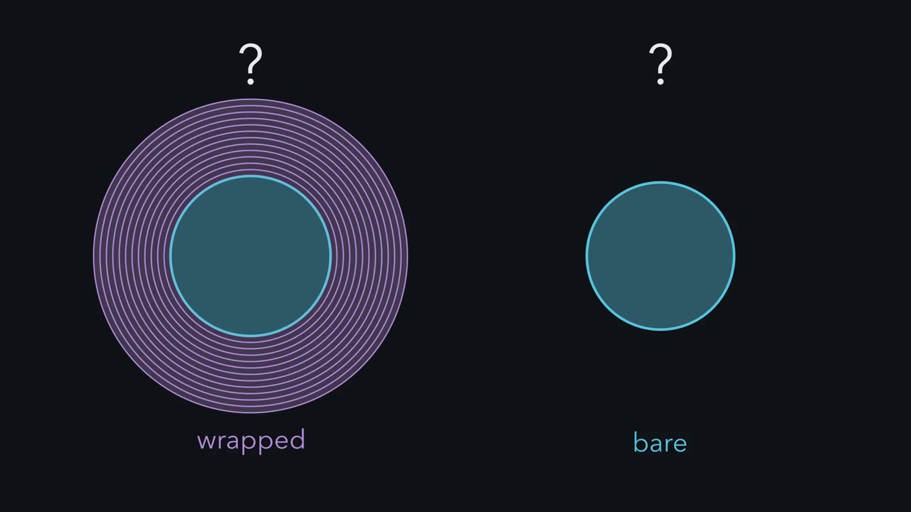 ▶ Watch Why Wrap, and How Much · 2:39
Read the transcript of “Why Wrap, and How Much”
Film transcript: Why Wrap, and How Much
Why would a glial cell wrap an axon in dozens of layers of membrane? And why are some axons never wrapped? Start with one patch of membrane. Current spreads to it from the active stretch behind. Before it can fire, that current must charge it. In a bare axon, current also leaks out at every point. So the signal is rebuilt on every patch. Less current is left to charge the patch ahead. Myelin is glial membrane, wound many times around the axon and squeezed tight.
Each turn adds two membranes in series. So the wall's capacitance falls. And its resistance rises. Far more of the current now reaches the next gap. Those gaps are the nodes of Ranvier. The axon packs its sodium channels there. The signal is rebuilt at the nodes, so it seems to jump. This is saltatory conduction.
In a bare axon, speed rises with the square root of diameter. In a myelinated fiber, it rises in proportion to diameter. So which axon gains more from a wrap, a thin one or a thick one? The thick one. In a simple model, the gain grows with the axon's size. The axon diameter is the axon alone. The fiber diameter adds the myelin. Their ratio is the g ratio. A thinner sheath on the same axon gives a higher g ratio. Healthy peripheral fibers sit near 0.6 to 0.7. That is close to the fastest value, but the optimum is broad. Now the puzzle. In peripheral nerve, axons up to about 1 micrometer stay bare. Some cable models of small central fibers put the break-even for myelin much lower, so speed alone may not explain the cutoff.
Space and energy cost may matter too. Something in the tissue may decide. Here is the idea to carry. The speed gain is largest on large axons, and nerves do match the wrap to the axon. In nerve, the axon decides. In the brain and spinal cord, the oligodendrocyte brings much of its own program.
From the film
- Myelin lets the signal jump between nodes of Ranvier, and the speed gain is largest on large axons.
- In peripheral nerve the axon decides how the Schwann cell wraps it; in the brain and spinal cord the oligodendrocyte brings much of its own program.
- Nerves do match the wrap to the axon, so the next question is how.
Wrap an axon in tight layers of glial membrane, leave short bare gaps between the wraps, and its action potential stops creeping. It jumps from gap to gap. The largest myelinated fibers in mammals conduct at about 100 meters per second or a little more, while thin bare fibers manage about a meter per second or less.
Yet many thin axons in your peripheral nerves are never wrapped, while their large neighbours carry dozens of turns. Two questions run through this lesson. Why does wrapping pay, and where does it pay most? And how does a developing nerve decide which axons get the wrap, and how much?
Why wrap an axon?
Myelin is glial plasma membrane, made by oligodendrocytes in the brain and spinal cord and by Schwann cells in peripheral nerve. It is wound many times around an axon and squeezed until almost no cytoplasm is left between the turns. To see why that speeds the signal, take it in three steps.
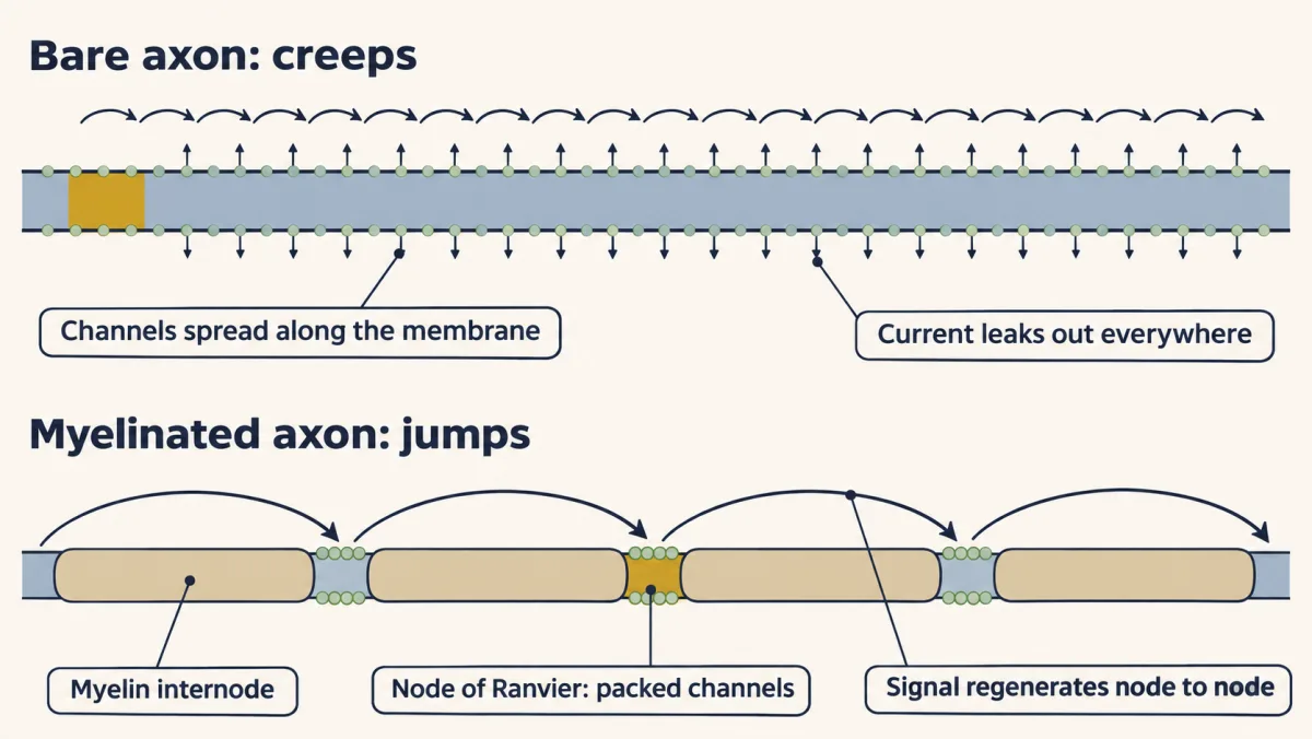
- A patch must be charged before it fires. Current from the active stretch spreads along the axon’s inside to the patch ahead. That patch of membrane is a capacitor, which the current must charge to threshold before it can fire. In a bare axon, current also leaks out at every point, so the action potential has to be rebuilt on every patch. It creeps.
- Charging and leaking both slow the signal ahead. Leaked current is lost; current that charges the membrane on the way is held there for the moment. Either way, less is left for the next stretch, so it reaches threshold later.
- Wrapping cuts both. Each turn of the sheath adds two membranes in series, one after another across the current’s path. A 5 µm axon at g = 0.65 (defined below), with an illustrative 17 nm per turn, would carry about 80 turns, so about 160 membranes; in an ideal sheath of identical compact membranes, the capacitance of the wall falls, and its resistance rises, by roughly that factor. Far more of the current now reaches the next bare gap and charges it to threshold quickly.
The axon exploits this by packing its voltage-gated sodium channels into short gaps between sheaths, the nodes of Ranvier. Each wrapped stretch is an internode. The action potential is regenerated at the nodes, where sodium channels are densely packed, and seems to leap from one to the next: saltatory conduction, from the Latin for “to jump”.
Two diameters, and the g ratio
Myelin work uses two diameters: the axon diameter (the axon alone) and the fiber diameter (axon plus myelin).
g ratio = axon diameter ÷ fiber diameter. A 6 µm axon inside a 10 µm fiber has g = 6 ÷ 10 = 0.6. A thinner sheath on the same axon gives a higher g ratio.
For large myelinated mammalian fibers (several µm and up), velocity ≈ 6 m/s × fiber diameter in µm (Hursh, 1939, an estimate from mammalian peripheral nerve).
In unmyelinated axons, conduction velocity rises roughly with the square root of diameter. In myelinated fibers it rises roughly in proportion to diameter. Part of the reason: as the axon widens, the sheath thickens in step, so the capacitance per unit length of the internode stays about the same while axial resistance falls. Internodes also lengthen roughly in proportion to fiber diameter. The time to cross one internode therefore stays about constant while the distance covered per jump grows, so speed rises in proportion to diameter.
- Unmyelinated axon
- rises with √diameter
- Doubling the diameter multiplies velocity by about
- 1.4
Rule to keep: velocity rises roughly with the square root of diameter in a bare axon and roughly in proportion to diameter once it is myelinated, so the gap widens as diameter grows. The bare-axon curve is a sketch in arbitrary units, not a measurement, and is not read against the m/s axis.
Predict. In a simple model, a bare 1 µm axon conducts at 1 m/s, so bare speed is √d m/s (d in µm). Compare a 2 µm axon with a 10 µm axon. Which gains more, as a multiple of its bare speed, from being wrapped?
Check your prediction
The larger axon gains more. Wrap each axon at g = 0.65 and apply Hursh’s 6 m/s per µm of fiber diameter.
| Axon diameter | Fiber diameter at g = 0.65 | Bare (m/s) | Myelinated (m/s) | Gain |
|---|---|---|---|---|
| 2 µm | about 3 µm | 1.4 | 18.5 | about 13× |
| 5 µm | about 8 µm | 2.2 | 46 | about 21× |
| 10 µm | about 15 µm | 3.2 | 92 | about 29× |
The gain itself grows with the square root of diameter, because one speed rises with d and the other with √d. Hursh’s line is a rule for large fibers, so the 2 µm row is the least reliable; the trend, not the exact numbers, is the point.
Healthy peripheral fibers usually have g ratios near 0.6 to 0.7, close to the value theory predicts gives the fastest conduction for a fiber of a given size. That optimum is broad, though, and fibers in the brain and spinal cord sit somewhat above it (see Going deeper).
Now the puzzle. In peripheral nerve, axons up to about 1 µm across stay bare. Some cable models of small central fibers (Waxman and Bennett, 1972) put the speed break-even for myelin near 0.2 µm, and the estimate depends on the assumptions, so speed alone may not explain the peripheral cutoff (see Going deeper). Space and energy cost may matter too, and something in the tissue may decide.
The one idea to carry through this lesson. At a fixed g ratio, myelin makes speed grow in proportion to diameter rather than with its square root, so the speed gain from wrapping is largest on large axons.
Nerves do match wrap to axon: large axons carry thick myelin and the thinnest carry none. How does development produce that match? In peripheral nerve, the axon decides: the amount of neuregulin-1 type III on its surface is the main axonal signal for whether a Schwann cell wraps it and how thickly, working together with the basal lamina and Gpr126. In the brain and spinal cord, the oligodendrocyte brings much of its own program, and axon cues such as caliber and activity bias it.
Two wrappers
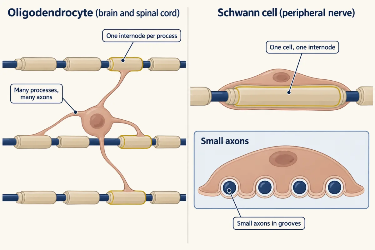
Two kinds of glia do the wrapping. In the brain and spinal cord, an oligodendrocyte sends out many processes, and each one makes an internode, usually on a different axon. In peripheral nerves, a myelinating Schwann cell makes a single internode on a single axon. Schwann cells that meet small axons do not myelinate; they hold several of them in grooves of their membrane, an arrangement called a Remak bundle. (Where both cell types come from is the subject of that lesson.)
In nerve, the axon decides
Read the transcript of “The Axon Sets the Dose”
Film transcript: The Axon Sets the Dose
A Schwann cell meets a large axon and wraps it in dozens of turns. Beside a thin axon, it wraps nothing. Who decides? In the 1970s, two laboratories swapped Schwann cells between nerves. Schwann cells from a nerve with bare axons now meet axons that are normally myelinated. What do they do?
They myelinated their new axons. The reverse swap gave the reverse result. In peripheral nerve, the axon decides. So the axon must carry a signal. The main one is neuregulin-1 type III, a protein on the axon's surface. It stays tethered near the axon membrane, so a Schwann cell reads it mainly by touch. Two receptors on the Schwann cell read it, ErbB3 and ErbB2. Paired, they signal. A large axon has more membrane per unit length. If the density is similar, it carries more type III. Could the amount be the message? Give mice half the normal type III. What happens to the sheath, and to the g ratio?
The sheaths were thinner for the axon's size, so the g ratio went up. Now let neurons make extra type III. Predict the sheath. Thicker myelin, and a lower g ratio. The dose changed in both directions, and the sheath followed both times.
Sympathetic axons normally stay bare. Make their neurons produce extra type III, and add Schwann cells. What then? The Schwann cells myelinated them. Giving extra signal to axons that would stay bare changed what the glia did. The outcome followed the delivery. That makes type III instructive. It is the same permissive-versus-instructive test as in the neurotrophic hypothesis lesson. Below a threshold of type III, an axon stays bare. Above it, more type III helps thicken the sheath. If big axons carry more of it, one readout could match the wrap to the axon. That is a proposal, not yet shown. The signal is decisive, but not enough alone. The Schwann cell also needs its basal lamina, and its own receptor, Gpr126.
Both are required, and not mere on-switches. The basal lamina also tunes the response to neuregulin. In nerve, the axon sets the dose, and the dose is the main signal for the wrap.
From the film
- In peripheral nerve the axon decides: the amount of neuregulin-1 type III on its surface is the main signal for whether a Schwann cell wraps it and how thickly.
- Halving the signal thinned the myelin and extra signal thickened it, and extra signal made normally bare sympathetic axons get wrapped.
- The Schwann cell also needs its basal lamina and its receptor Gpr126, so the signal is decisive but not enough alone.
Schwann cells give large axons dozens of turns and tuck thin ones, unwrapped, into Remak bundles. Who decides: the glial cell or the axon?
Predict. Take Schwann cells from a nerve whose axons are all bare, and let them settle on axons that are normally myelinated. If the Schwann cell decides, what do you expect? If the axon decides?
Check your prediction
In grafting experiments in the 1970s, by Albert Aguayo’s laboratory and by Weinberg and Spencer, Schwann cells from unmyelinated nerves myelinated the axons they met in their new nerve, and the reverse swap gave the reverse result. The Schwann cells did what their new axons called for. In peripheral nerve, the axon decides.
So the axon must carry a signal the Schwann cell can read. The main one turned out to be neuregulin-1 type III, a protein on the axon’s surface. Neuregulin-1 comes in several forms. Type III keeps its active, EGF-like part tethered near the axon membrane, so a Schwann cell reads it mainly by touching the axon. Two receptor tyrosine kinases of the epidermal growth factor (EGF) receptor family read it (the lesson on receptor tyrosine kinases). ErbB3 binds neuregulin but has a weak kinase. ErbB2 binds no ligand but has a strong kinase. Paired, they signal.
Now the key question. A large axon has more membrane per unit length than a small one. If type III sits at similar density on both, the large axon carries more of it. Could the amount be what the Schwann cell reads?
Predict. What should happen to the g ratio in mice with half the normal type III? In mice whose neurons make extra? And what should happen to sympathetic axons, which normally stay bare, if you make their neurons produce extra type III and grow them with Schwann cells?
Check your prediction
| Type III on the axon | Experiment | What the Schwann cell did |
|---|---|---|
| none | sensory neurons lacking type III, in culture (Taveggia et al., 2005) | did not myelinate them |
| about half | mice with reduced neuregulin-1 (Michailov et al., 2004) or one working copy of the type III gene (Taveggia et al., 2005) | myelinated, but thinner for the axon’s size: a higher g ratio |
| normal | control mice | g ratio near 0.6 to 0.7 |
| extra | mice whose neurons overproduce type III (Michailov et al., 2004) | thicker myelin: a lower g ratio |
| extra, on axons that are normally bare | sympathetic neurons made to produce more, cultured with Schwann cells (Taveggia et al., 2005) | myelinated them |
The 2004 work was by Galin Michailov, Klaus-Armin Nave and colleagues; the 2005 work by Carla Taveggia and colleagues in James Salzer’s laboratory.
Below a threshold amount of type III, an axon stays bare. Above it, more type III helps make a thicker sheath. The dose was changed in both directions, and the sheath followed both times.
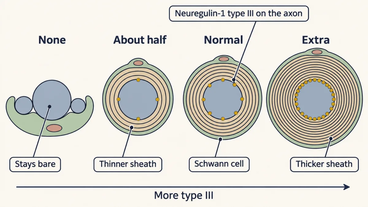
Could the same readout match the sheath to the axon’s size? To hold g at 0.65, the sheath must thicken in step with the axon. Its thickness is (fiber − axon) ÷ 2: about 0.54 µm on a 2 µm axon and 1.35 µm on a 5 µm axon. At an illustrative spacing of about 17 nm per turn, that is about 32 and 79 turns. If type III density is similar on every axon, a Schwann cell that reads the amount gets that size signal for free. This proposal is consistent with the dose data (see Michailov and colleagues, 2004, and later reviews). No one has shown that Schwann cells measure size this way, or that the readout is strictly proportional.
In the terms of the lesson on the neurotrophic hypothesis, type III is instructive here: giving extra to axons that would stay bare made Schwann cells myelinate them, so supplying it changes the outcome rather than just allowing it.
Inside the Schwann cell, ErbB2/ErbB3 signaling runs through phosphoinositide 3-kinase (PI3K)–Akt and other routes to turn on a transcription factor Schwann cells need in order to myelinate, Krox20 (also called Egr2). Which routes matter most is still argued.
The axon’s signal is decisive but not enough on its own. Two other inputs are also required, and they are not mere on-switches. First, the Schwann cell must lay down its basal lamina, the sheet of extracellular matrix around it. In the Bunges’ cultures, Schwann cells needed contact with an extracellular matrix, and in later purified cocultures ascorbic acid, which cells need to build collagen, was required for both the basal lamina and myelin. Through laminin-211, the basal lamina also restrains myelination of small axons and tunes the response to neuregulin. Second, the Schwann cell must signal through a receptor of its own, Gpr126, which raises cAMP inside it; in zebrafish lacking Gpr126, Schwann cells stop just short of myelinating (they reach a single axon but do not wrap it), and Gpr126 also takes part in sorting axons.
Keep the scope. This is the rule for Schwann cells in peripheral nerve, and it covers two outputs: whether an axon is wrapped and how thick the sheath is. Sheath length, node assembly and compaction need other machinery too, though neuregulin signaling can affect some of them.
In the brain, the oligodendrocyte brings its own program
Read the transcript of “The Brain Brings Its Own Program”
Film transcript: The Brain Brings Its Own Program
Does the peripheral rule hold in the brain and spinal cord? Does the axon decide there too? In culture, oligodendrocytes will wrap inert plastic fibers of a suitable diameter. No axon, and still a wrap. In mice, Nave's group removed neuregulin-1 and ErbB signaling in the brain and spinal cord. Myelin still formed.
Yet extra neuregulin from neurons could still thicken central myelin. The signal can tilt the wrap. It is not needed to start it. So in the brain, the glial cell brings much of the program itself. Axon cues such as activity, and the caliber of the fiber, bias it. Zebrafish larvae have transparent spinal cords, so the first sheaths can be filmed as they form. Most early wraps are withdrawn. Jacob Hines, Bruce Appel and colleagues blocked vesicle release in one axon among normal neighbours. Will oligodendrocytes treat it differently?
Yes. The release-blocked axon was wrapped less often, and its sheaths were shorter. Release from the axon biases the choice. In mouse cortex, stimulating neurons with light added new oligodendrocytes and thickened myelin in that circuit. These are larval fish and mouse cortex. Whether such myelin helps adult humans learn is still open. So the idea has two halves. In nerve, the axon decides. In the brain, the glia bring much of the program, and the axon tunes it.
From the film
- In the brain and spinal cord the oligodendrocyte brings much of its own program, and in culture it will wrap inert plastic fibers of a suitable diameter.
- Myelin still formed in mice without neuregulin-1 and ErbB signaling in the brain and spinal cord, though extra neuregulin could thicken it.
- Axon cues such as caliber and activity bias the wrap, as when a release-blocked axon in zebrafish larvae was wrapped less often.
Myelination in the central nervous system is late and slow. In humans it begins before birth in the spinal cord and brainstem and continues through childhood and adolescence, and some association areas of the cerebral cortex are still gaining myelin in early adulthood. That leaves a long window in which activity could shape it.
In the brain and spinal cord the balance differs. In culture, oligodendrocytes will wrap inert plastic fibers of a suitable diameter. In the mouse brain and spinal cord, Nave’s group found that oligodendrocytes still myelinate when neuregulin-1 and ErbB signaling are removed (Brinkmann and colleagues, 2008). Yet extra neuregulin-1 made by neurons can still thicken central myelin. So neuregulin-1/ErbB signaling is not needed to start wrapping, and the oligodendrocyte brings much of the program itself. Axon cues, such as activity and the caliber of the fiber, do not decide alone; they tilt where it wraps and how much.
Once an oligodendrocyte can wrap, which axons does it keep? Zebrafish larvae have transparent spinal cords, so the first sheaths can be filmed as they form. Jacob Hines, Bruce Appel and colleagues found that most early wraps were withdrawn, many within about an hour and a half.
Predict. Hines and colleagues blocked vesicle release from one labeled axon among normal neighbours by making it express tetanus toxin. If oligodendrocytes ignore the axon’s release, what should they do with the release-blocked axon? If they prefer axons that release?
Check your prediction
The release-blocked axons were less often myelinated (about 32 percent of the time, against 55 percent for control axons) and carried shorter sheaths. Release from the axon, which depends on its activity, is one of the cues that biases the choice. Because the release-blocked axon sat among normal neighbours in the same animal, the comparison controls for the shared surroundings, though not for every property of the axon.
In a companion paper (both appeared in Nature Neuroscience in 2015), Sigrid Mensch, David Lyons and colleagues found that blocking vesicle release throughout the nervous system cut the number of sheaths each oligodendrocyte made by about a third, and that raising activity with a convulsant drug increased it unless release was blocked. Activity biased how many sheaths formed and where they stayed.
In mice, Erin Gibson, Michelle Monje and colleagues reported in 2014 that light-driven stimulation of neurons in premotor cortex increased the division of oligodendrocyte precursor cells and the making of new oligodendrocytes, and thickened myelin within the stimulated circuit. Together these results describe a developmental program that stays open to adjustment by activity, in larval fish and mouse cortex. How much activity-dependent myelin contributes to learning in adult humans is still open.
What holds the layers together
The membrane itself suits the job. Like any membrane it is a lipid bilayer with proteins in it (see that lesson), but it is unusually rich in lipid, roughly 70 to 80 percent of its dry weight, with a large share of cholesterol and of galactolipids such as galactocerebroside (see that lesson).
In the electron microscope compact myelin shows a repeating pattern. A dark major dense line marks where the cytoplasmic faces of successive turns meet; a fainter intraperiod line marks where their outer faces meet.
Myelin basic protein (MBP) is small, strongly positive and sits on the cytoplasmic side of the membrane. It binds the negatively charged lipids of two facing cytoplasmic leaflets and pulls them together, squeezing out the cytoplasm. The seam it leaves is the major dense line. Other proteins hold the outer faces: proteolipid protein (PLP) in the central nervous system, and protein zero (P0), an immunoglobulin-like adhesion protein, in peripheral nerve. The two are not related in structure.
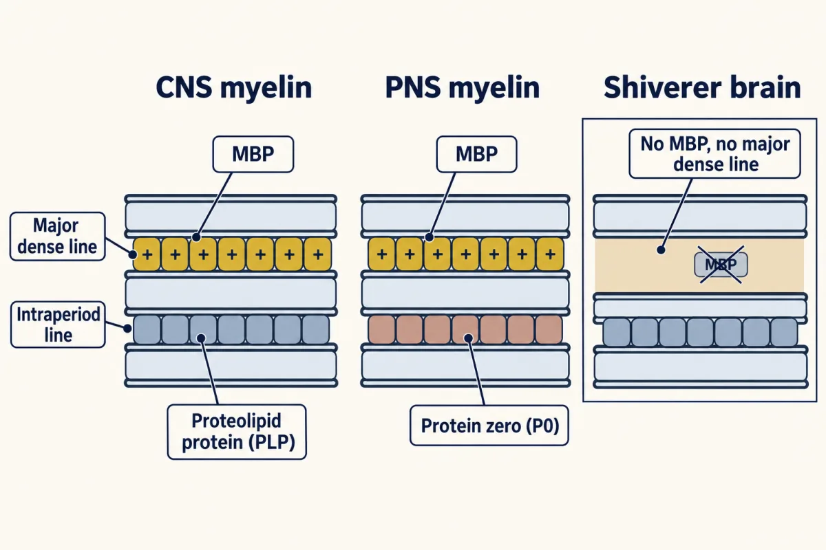
A mutant mouse called shiverer shows what this one protein is worth. Its neurons and axons are present, but from the second or third week after birth it trembles. Its brain myelin lacks the major dense line and nearly all its MBP (first reported by a French group in 1979), and the mutant MBP gene has lost about 20 kilobases of DNA, including most of its exons. Peripheral nerve in shiverer is far less affected, because P0 can also hold the cytoplasmic faces together there.
Predict. The mutant points to necessity: without MBP, no major dense line. But a large deletion could also have damaged DNA next to the gene. What experiment would show that losing MBP alone explains shiverer, and what result would you need?
Check your prediction
Put the gene back and nothing else. A normal MBP transgene bred into shiverer mice was enough to restore compact central myelin with major dense lines and stop the tremor, even at about a quarter of normal MBP (Readhead and colleagues, 1987). So loss of MBP is enough to explain those defects; other features were not tested. This is the necessity-then-sufficiency logic of the lesson on the neurotrophic hypothesis, applied to a structural protein.
A second job: feeding the axon
Insulation is not myelin’s only job. In the brain and spinal cord, oligodendrocytes also help keep the axons they wrap alive, and broad reviews now count this support as a second major function.
The first evidence came from mouse mutants. Mice lacking the myelin membrane protein PLP, or the oligodendrocyte enzyme CNP, show long-term degeneration of their axons even though their myelin sheaths look normal or nearly so. The sheath was there; something else the glial cell provides was missing. In people, Pelizaeus-Merzbacher disease, caused by mutations in the PLP gene, also involves axon loss.
What does the glial cell supply? Part of the answer is fuel. A sheath covers most of the axon’s surface, which limits the axon’s access to nutrients from the fluid outside. Oligodendrocytes pass energy metabolites to the axon through a monocarboxylate transporter, monocarboxylate transporter 1 (MCT1) (Lee and colleagues, 2012). One proposal is that the oligodendrocyte breaks glucose down to lactate and hands the lactate to the axon, whose mitochondria use it to make ATP. The lactate would travel through narrow channels of cytoplasm left in the non-compact parts of the sheath, leave the oligodendrocyte through MCT1, and enter the axon through a related transporter. The axon’s dependence on this support grows with age. How much of the support is lactate, and what else passes from glia to axon, is still being worked out.
Myelin lost, and repaired
Multiple sclerosis shows what losing central myelin costs. In its usual account, the immune system attacks myelin and oligodendrocytes in the brain, optic nerves and spinal cord, leaving scattered lesions, and axons are damaged too. It is a leading cause of non-traumatic neurological disability in young adults. Most patients begin with relapsing-remitting disease, many later enter a secondary progressive course, and a minority progress from the start (primary progressive).
A lesion takes away both of myelin’s jobs. Without the sheath, saltatory conduction is lost. Without healthy oligodendrocytes, the axon loses its support. In long-standing lesions, axons keep degenerating, and several causes are thought to act together: lost sheath and oligodendrocyte support; inflammation that persists inside the brain behind the blood-brain barrier, which treatments that quiet relapses may control only incompletely; and mitochondrial and energy failure. Together they help explain why disability can keep growing after relapses have been quieted.
The nervous system tries to remyelinate lesions, and remyelination restores saltatory conduction and helps keep axons healthy. In animal models, oligodendrocyte precursor cells move in, mature and make new sheaths, which are typically thinner with shorter internodes. Repair reuses much of the developmental program, but in an inflamed adult lesion. In people the source is less certain: carbon dating of MS tissue found that oligodendrocytes in remyelinated-looking lesions were old cells, so surviving oligodendrocytes may make some of the new myelin. Repair often falls short, especially in long-standing lesions and in older patients.
Putting it together
| Experiment | System | Observed result | What it supports | What it does not establish |
|---|---|---|---|---|
| Swap Schwann cells between nerves | nerve grafts, 1970s | Schwann cells myelinate, or not, as their new axons call for | in peripheral nerve, the axon decides | which signal |
| Remove type III | sensory neurons in culture | no myelin | type III is needed to start | how thick |
| Halve type III | mice | thinner sheaths, higher g ratio | dose sets thickness | that the readout is strictly proportional |
| Add type III | mice; sympathetic neurons in culture | thicker sheaths; normally bare axons myelinated | type III is instructive | that it acts alone (basal lamina and Gpr126 are also needed) |
| Remove neuregulin-1/ErbB signaling | mouse brain and spinal cord | myelin still forms | the CNS rule differs | what the oligodendrocyte does read |
| Block one axon’s vesicle release (tetanus toxin) | zebrafish larva | wrapped less often, shorter sheaths | release from the axon biases the choice | that activity is needed in mammals |
| Stimulate premotor neurons | mouse cortex | more new oligodendrocytes, thicker myelin in the circuit | activity can add myelin | a role in human learning |
| Add MBP back to shiverer | mouse | compact myelin, tremor stopped | MBP loss is enough to explain the missing major dense line and the tremor | that every other feature of the mutant was restored; the rescue used about a quarter of normal MBP |
| Lose PLP or CNP | mouse | axons degenerate under near-normal myelin | oligodendrocytes support axons beyond insulation | which supports (energy metabolites or others) matter, or whether the sheath’s subtle defects contribute |
The guiding idea holds with its scope, and the table adds one more point: because, in the brain and spinal cord, the cell that makes the sheath also helps keep the axon alive, losing myelin costs more than speed.
Design problem
In long-standing multiple sclerosis lesions, axons keep degenerating. Take two of the possible causes. The immune attack could damage axons directly, or axons could die because they have lost their oligodendrocytes. In a patient both happen at once. Design a mouse experiment that separates the two, and say what result would show that losing oligodendrocytes is enough to harm axons. (Hint: mouse strains that lack mature T and B cells exist.)
Saved in this browser only.
One way to do it
Take the adaptive immune system out of the question. Use mice that have no mature T or B cells, such as mice lacking the gene Rag1. Kill their oligodendrocytes with a genetic method that acts only in those cells, and compare them with Rag1-deficient mice whose oligodendrocytes are left alone.
If axons degenerate only in the mice that lost their oligodendrocytes, loss of the glia is enough to harm axons without an adaptive immune attack. Two limits remain. Microglia and other innate immune cells are still present, and dying oligodendrocytes activate them. And killing oligodendrocytes removes the myelin and its support together, so this test cannot say which loss matters. If the axons survive, oligodendrocyte loss was not enough under these conditions; check how completely the cells were killed and how long the mice were followed before concluding more.
A sharper test leaves the oligodendrocytes alive, removes one thing they supply, such as the transporter MCT1, checks that the sheath stays intact, and asks whether axons still degenerate.
Pohl, Suter, Franklin and colleagues reported in 2011 that genetically induced death of adult oligodendrocytes was followed by axon damage even in Rag1-deficient mice. Deleting MCT1 from cells of the oligodendrocyte lineage was later reported to cause late-onset hypomyelination and axon degeneration (Philips and colleagues, 2021). Because the sheaths also thinned, that study could not fully separate the two effects.
Going deeper
Growing myelin in a dish
Edith Peterson and Margaret Murray reported in 1955 that sensory ganglia from chick embryos made myelin in a dish. In 1967 Mary Bunge, Richard Bunge, Peterson and Murray described long-term cultures of dorsal root ganglia from fetal rats. Over several weeks, Schwann cells in these cultures wrapped sensory axons in compact myelin with nodes, one cell to one axon, much as in a real nerve. Because the medium, the matrix and the cells could each be changed one at a time, the dish became an experiment, and the matrix and ascorbic acid requirements described above came from such cultures.
Where myelin stops paying in speed
Extended down to very small fibers, Hursh’s straight line would have myelin paying down to a few hundredths of a micrometer, which is physically meaningless; the linear rule holds only for large fibers. The Waxman and Bennett crossover near 0.2 µm fits the brain and spinal cord, where axons as small as about 0.2 µm can be myelinated, though a caliber that permits wrapping does not decide which axons are wrapped. It does not fit the bare peripheral axons up to about 1 µm. The peripheral cutoff looks like a biological threshold rather than the speed crossover, and a threshold amount of type III is one candidate, alongside basal-lamina signals that restrain myelination of small axons.
How sharp is the g ratio optimum?
Rushton’s 1951 analysis, for a fiber of fixed total diameter, puts the fastest conduction at a g ratio of about 0.61. The peak is flat. Relative to the maximum, speed is about 97 percent at g = 0.5, 99.5 percent at 0.65, 97.5 percent at 0.7, 94 percent at 0.75 and 88 percent at 0.8. In this model, central fibers, which typically run 0.75 to 0.8, would give up only about 6 to 12 percent of the maximum speed. The optimum also depends on what is held fixed: in this simple model, for a fixed axon diameter, more myelin always adds speed, so the limit is set by other costs, such as space.
Signaling inside the Schwann cell
Downstream of ErbB2/ErbB3, the signal fans out along routes you have met:
- Phosphoinositide 3-kinase (PI3K) and Akt, which in other cells promote growth and survival, support myelin growth (see that lesson).
- The Ras-ERK cascade is activated as well (see that lesson).
- PLC-gamma raises calcium, which activates the phosphatase calcineurin and frees the transcription factor nuclear factor of activated T cells (NFAT). Work from Gerald Crabtree’s laboratory proposed that NFAT joins Sox10 in the nucleus in turning on Krox20, but that study removed calcineurin from all neural crest cells. When a later study removed it from Schwann cells alone, myelination and Krox20 were close to normal.
Oligodendrocytes remember where they came from
On inert fibers, with no axon to instruct them, oligodendrocytes taken from different regions of the central nervous system made sheaths of different lengths, much as in the tissue they came from (Bechler and colleagues, 2015). Sheath length, too, is partly the glial cell’s own program.
Shiverer, step by step
The deletion was found in the mid-1980s by laboratories including those of Leroy Hood and Richard Lazzarini. Carol Readhead, Hood, Richard Sidman and colleagues then injected a normal copy of the mouse myelin basic protein (MBP) gene, with its own regulatory DNA so that it would be turned on in oligodendrocytes, into fertilized mouse eggs, and bred the resulting transgenic mice with shiverer mice until they had animals homozygous for the shiverer deletion that also carried the transgene. Compared with shiverer littermates without the transgene and with normal mice, the transgenic shiverer mice no longer shivered or died young. Had a transgene made enough MBP, in the right forms and in oligodendrocytes, without improving the myelin or the tremor, something else in the deletion would probably have mattered. The 1987 study was one of the early uses of a transgene to cure a mouse mutant.
Multiple sclerosis: history, numbers and risk
Jean-Martin Charcot’s description of multiple sclerosis is usually dated to 1868. The disease is diagnosed most often between about 20 and 40 and affects women two to three times as often as men. A fourth category, progressive-relapsing, was dropped in the 2013 revision of the clinical course descriptions. Risk combines genes, with variants of the immune gene HLA-DRB1 the strongest, and environment, notably earlier infection with Epstein-Barr virus.
Your notes
Select any text in the lesson to highlight it or add a note to it. Your highlights and notes are saved in this browser and collected in your notebook.
This browser is not letting the site save anything (a private window or strict privacy settings), so highlights and notes will be lost when you leave the page.
How we measure it
Myelinating dorsal root ganglion cultures
Sensory ganglia, or purified sensory neurons with Schwann cells, are grown for weeks until the Schwann cells myelinate the axons. Because the medium, the matrix and the cells can be changed one at a time, the culture shows what each contributes to myelin formation.
Electron microscopy of myelin
Thin sections of fixed nerve are imaged at high magnification so that single membrane layers are visible. The method shows whether myelin is compact, whether the major dense and intraperiod lines are present, and how thick the sheath is relative to its axon (the g ratio).
Transgenic rescue
A normal copy of a gene is injected into fertilized eggs, and the transgene is bred into a mutant strain. If the added gene restores the normal structure and behavior, loss of that gene is enough to explain the mutant's defect.
Mosaic live imaging in zebrafish
Single neurons in a transparent larva are made to express a fluorescent protein, with or without a blocker such as tetanus toxin, among unaltered neighbours. Time-lapse imaging then compares how glia treat the altered and the normal axons in the same animal.
Practice questions on this lesson (3) → MCAT-style, original, each with an explanation
Where this lesson fits
Before thisReview first
- Where Glia Come FromThis course · Lesson 43
It explains where oligodendrocytes and Schwann cells come from, and this lesson assumes you know the two wrappers.
- Neurotrophins, Trk Receptors and p75This course · Lesson 30
It introduces the target-derived signals neurons use, which helps in reading the neuregulin signal an axon presents to Schwann cells.
- The Growth Cone: The Tip That Steers the AxonThis course · Lesson 3
It covers the axon whose surface signals the glial cell reads when deciding whether to wrap.
After thisStudy next
- Feeding the Axon: Glia as Metabolic PartnersThis course · Lesson 45
It takes the oligodendrocyte's second job, passing lactate to axons through MCT1, further.
- Injury and Regeneration: Why the Brain Does Not RegrowThis course · Lesson 46
It uses myelin loss and repair, and axon dependence on glia, to ask why the injured brain does not regrow.
Elsewhere on the siteFilms, simulations and method films in other courses
- Run the 1952 equationsSimulation · Neuroscience
Running the model of the spike shows how the signal travels along an axon, which the node-to-node jump speeds up.
- The Spike and Its PuzzlesFilm · Neuroscience
It shows how sodium channels produce and conduct the impulse that myelin makes jump between nodes.
- A Threshold Is Not a CountFilm · Sensory Systems
It shows that a normal threshold test can hide lost synapses, a lesson about hidden damage like axon loss under normal-looking myelin.
Suggested by an AI model from the lessons themselves, then checked. How we use AI
Finish line
Test yourself
8 questions
Practice and MCAT-style, with explained answers.
Next · Lesson 45
Feeding the Axon: Glia as Metabolic Partners
The brain is about 2 percent of your weight and uses about 20 percent of your resting energy. Who delivers the fuel to the far end of an axon?
Continue →