Neuroscience · Skim version
Neurons and Glia at Work
Read the full lesson → Skim the whole course →
The guiding idea
Let potassium pile up outside neurons during seizure-like firing and E_K can move by more than 30 mV. Who cleans it up?
The space outside brain cells is small and holds little potassium, so activity can change it strongly, the shift in E_K comes mostly from outside, and glial cells such as astrocytes keep clearing it.
From the film: Small Space, Big Change
- The space outside brain cells is only about a fifth of the tissue, so in a simple model a little released potassium changes the concentration outside much more, in proportion, than inside.
- In cat cortex, strong repetitive stimulation raised outside potassium to a ceiling of about 10 mM, which in the lesson's example moves E_K about 32 mV.
- A modest rise in outside potassium can make a neuron fire more easily, which can feed a seizure, so its resting potential depends on cells that keep clearing that space.
From the film: Who Puts the Potassium Back
- Glial cells in the mudpuppy optic nerve depolarized as neighbouring axons fired, because they were sensing the potassium the axons released.
- In a simplified model of spatial buffering, potassium enters astrocytes through Kir4.1 where it is high and leaves where it is low, as one loop of current through astrocytes joined by gap junctions.
- Spatial buffering spreads potassium thinner and leaves it outside, while sodium-potassium pumps in astrocytes and neurons put it back inside cells, and how much each contributes is still debated.
- In mouse hippocampal slices, astrocytes lacking Kir4.1 took up potassium poorly, though the deletion began in development and also reached oligodendrocytes.
From the film: Settled and Argued
- That astrocytes clear potassium and glutamate is settled: GLT-1 is mostly astrocytic, and deleting it from astrocytes in mice caused seizures and early death.
- Whether astrocytes also release glutamate that acts quickly on synapses, called gliotransmission, is still argued.
- A null result from deleting the IP3 receptor type 2 cannot exclude effects of the calcium that remains, and the positive evidence so far is for one subgroup of adult mouse hippocampal astrocytes.
-
The puzzle: a small space outside
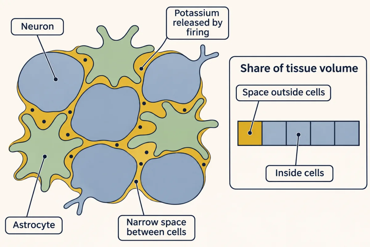
The space between brain cells is a thin web, about a fifth of the tissue, so potassium released into it by firing can change its concentration there strongly. AI-generated illustration -
A map of the neuron
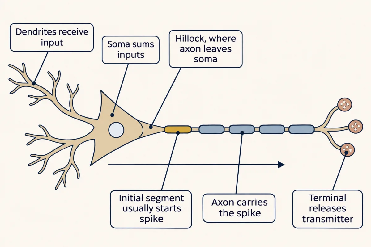
Each compartment of the canonical neuron mainly does one job, from dendrites receiving input to terminals releasing transmitter, and the spike usually starts at the initial segment just beyond the hillock. AI-generated illustration -
How far EK moves
-
Astrocytes clear the potassium
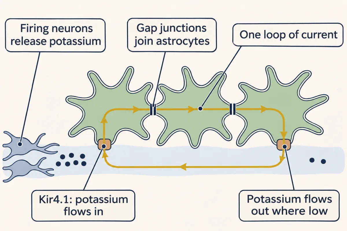
In spatial buffering, potassium enters astrocytes through Kir4.1 where it is high and leaves where it is low, as one loop of current through astrocytes joined by gap junctions. AI-generated illustration -
Glutamate: the same rule, more sharply
-
The axon’s other partners: myelin, fuel and microglia
-
What is still argued: gliotransmission
-
Going deeper
Where this lesson fits
Before thisReview first
- The Map: How the Nervous System Is OrganizedThis course · Lesson 1
It introduces the neurons and glia and the barrier around brain tissue that this lesson then follows in action.
After thisStudy next
- The Resting Membrane and Passive SignalsThis course · Lesson 3
It uses E_K and potassium leak to explain the resting voltage, and how passive signals spread along dendrites.
- The Action Potential and ConductionThis course · Lesson 4
It explains how spikes begin and how potassium leaves the axon, the source of the potassium that glia clear here.
- Synaptic Transmission and Quantal ReleaseThis course · Lesson 5
It follows glutamate release at the terminal, the transmitter that astrocyte GLT-1 clears afterwards.
Elsewhere on the siteFilms, simulations and method films in other courses
- Two Pushes, One HillFilm · Cell Biology
It shows how concentration difference and voltage together set which way potassium flows.
- Cut the AxonFilm · Developmental Neuroscience
It shows how a neuron sets up one axon and many dendrites, the compartments mapped here.
- Why Wrap, and How MuchFilm · Developmental Neuroscience
It shows how glial wrapping makes the signal jump between nodes of Ranvier.
- When the Body Pushes BackFilm · Biology
It shows how opposing responses hold internal conditions in range, as astrocytes do for outside potassium.
Suggested by an AI model from the lessons themselves, then checked. How we use AI