Developmental Neuroscience · Unit 9: Experience refines the wiring
Activity Before Experience: Spontaneous Retinal Waves
Rewritten on : new text, figures, flashcards and film. Your notes and highlights stay, but the text around them may have moved.
Weeks before vision begins, the retina already fires in waves. What are they for?
Pause when the reading reaches a film, and offer to play it. Saved on this device.
Watch the film now, or keep listening
Builds on:Topographic Maps: Sperry's Eye and Eph GradientsThe Neurotrophic Hypothesis TestedCompetition: How Neurons Win a TargetWhy these?
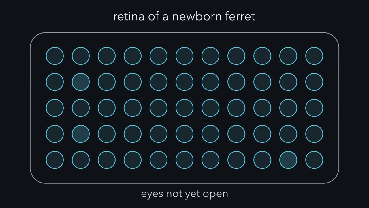 ▶ Watch Timing Is the Address · 3:02
Read the transcript of “Timing Is the Address”
Film transcript: Timing Is the Address
A newborn ferret's eyes have not opened yet. Even so, its retina is firing. Its ganglion cells fire bursts that sweep across the tissue as waves. Nothing is being seen. Why send this to the brain? The brain is still wiring itself. Molecules have drawn only a rough first map. In the thalamus, the two eyes' axons at first overlap. Later they separate. Two cells side by side in one eye sit at almost the same point on the gradients. So does a cell at the matching point in the other eye, though molecular cues also differ somewhat between the eyes. It still has to end up in a different territory, and the cues alone do not finish the job. Timing can sort them.
Watch a wave cross the retina at about 100 micrometres a second. Two cells this close fire almost together. Their bursts overlap almost completely. A cell a millimetre farther along the wave's path fires ten seconds later. Their bursts do not overlap. Now predict. What about a cell at the same point in the other eye?
Its waves are not timed to this eye's, so it matches only by chance, a few percent of the time. But waves start in many places and run in varying directions. So position is carried not by one cell's timing, but by how closely two cells' firing matches, over many waves. How could a target cell use that match? In 1949, the psychologist Donald Hebb proposed a rule. In paraphrase: when cell A repeatedly takes part in firing cell B, A becomes more effective at firing B. Take one thalamic cell with inputs from both eyes, each firing in its own waves. Predict: what happens to the right eye's inputs?
If they are strong enough to fire the cell at their own times, they strengthen too. Hebb's rule only strengthens, so the cell stays binocular. Segregation needs a second half: competition. Picture the inputs sharing a fixed budget. Each round, both eyes grow, then the same amount is taken back from each. The eye that starts slightly ahead gains more than it gives back. So it pulls further ahead. In this toy model, the other eye's inputs are pushed out of that cell. Rescale both eyes in proportion instead, and the split never changes. The kind of sharing matters. Takeaway: timing is the address. A cell keeps inputs that fire in step with its strong inputs, and they crowd out the rest.
From the film
- Timing is the address: a target cell keeps the inputs that fire in step with its other strong inputs, and they crowd out the rest.
- Near neighbours in one eye fire almost together in a wave, while the other eye's waves match only by chance.
- Hebb's rule alone only strengthens inputs, so separating the eyes also needs competition between inputs.
In the retina of a newborn ferret, whose eyes have not yet opened, ganglion cells fire bursts lasting a few seconds that sweep across the tissue at about 100 micrometres per second, separated by a minute or two of silence.
Nothing is being seen. Ganglion cells are the retina’s output neurons; their axons make up the optic nerve. Here they fire on their own, in groups, and the group moves. Why would a retina that cannot yet sense an image bother to send this pattern to the brain? The answer is that the brain is still wiring itself, and this pattern carries information the wiring can use.
The problem: a rough first draft
Spontaneous activity is activity that circuits generate on their own, without being driven by sensory input. Developing neurons in many parts of the nervous system produce it, often as bursts separated by long silences, and the developing retina produces it as waves. The waves arrive at a time when the brain’s first draft of its connections is rough.
That draft is laid out by molecules. In the lesson on topographic maps you saw gradients of Eph receptors and ephrins steer retinal axons to roughly the right place, so that neighbouring points in the retina project to neighbouring points in the target. Early in development many axons branch over a broad area, and in mammals such as cats, ferrets and mice the axons from the two eyes at first overlap in the lateral geniculate nucleus (LGN), the relay in the thalamus that passes visual signals to the cortex. In the adult they end in separate, eye-specific territories, which in cats, ferrets and primates are distinct layers. Getting from overlap to separate territories, and from broad branches to focal ones, is called refinement.
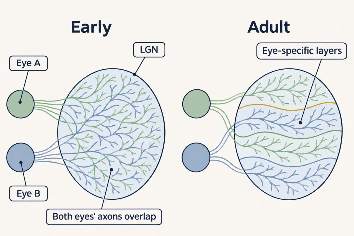
Why activity? Two neighbours look the same to a gradient
Take two ganglion cells 20 micrometres apart in one retina. On the Eph and ephrin gradients they sit at almost the same value, so the gradients give their axons almost the same instruction. Take a third cell at the matching point in the other eye. Its retinotopic label is nearly the same. Yet the first two must end side by side and the third in a different territory. Molecular cues set rough order and help place each eye’s axons, but they do not by themselves produce the sharp territories of the adult; something else has to finish the job.
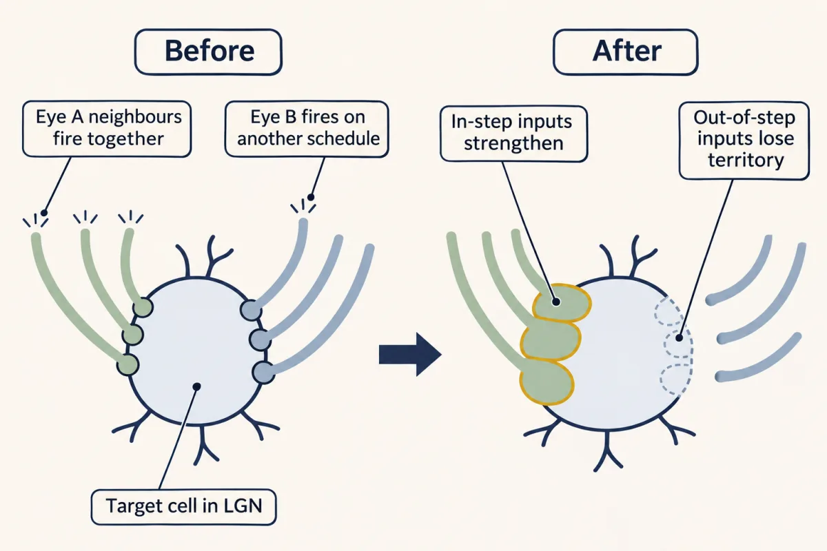
Timing can. The two neighbours fire together; the cell in the other eye fires on its own schedule. A target that groups its inputs by when they fire can keep the first two together and set the third apart.
The one idea to carry through this lesson. Timing is the address. A target cell keeps the inputs that fire in step with its other strong inputs, and because the inputs compete, the ones it keeps crowd out the rest. Waves make “in step” mean “a near neighbour in the same eye”.
Hebb’s rule, and the half he did not write
In 1949 the psychologist Donald Hebb proposed a rule for how connections might change. In paraphrase: when an axon of cell A is near enough to excite cell B and repeatedly takes part in firing it, some growth or metabolic change makes A more effective at firing B. The summary “Cells that fire together, wire together” is associated with Carla Shatz; it is a paraphrase of Hebb, not his words.
Predict. A neuron in the lateral geniculate nucleus (LGN) receives inputs from a few neighbouring cells of the left eye and a few of the right. Each eye’s neighbours fire together in that eye’s waves, and the two eyes’ waves are independent. Apply Hebb’s rule exactly as stated. What happens to the right-eye inputs?
Check your prediction
Nothing bad. The left-eye neighbours fire together and drive the cell, so they strengthen. But the right-eye neighbours also fire together, in their own waves, and if they are strong enough to help fire the cell at those other times, they strengthen too. Hebb’s rule as written only strengthens, so every active input grows and the cell stays binocular.
Segregation needs a second half, which Hebb did not write. If you predicted that the out-of-step right-eye inputs weaken, you supplied it yourself. Later theorists added that inputs which fire out of step with the cell’s main drive weaken (Stent 1973), and correlation-based models of ocular dominance and eye segregation need inputs to compete, so that one input’s gain is another’s loss (Miller, Keller and Stryker 1989, for cortical ocular dominance columns). Not every way of sharing does the job, as the toy model below shows.
Here is the second half worked through as a toy model, not a measurement. One cell’s inputs share 10 units of strength, and the left eye starts slightly ahead, 5.5 units to 4.5. Each eye’s inputs fire together, so in each round each eye first gains 20 percent of its own strength, giving 6.6 and 5.4, a total of 12. The rows differ only in how the total is brought back to 10. No strength can fall below zero.
| How the total is brought back to 10 | Left after one round | Right after one round | After many rounds |
|---|---|---|---|
| It is not (Hebb alone) | 6.6 | 5.4 | both keep growing; the cell stays binocular |
| Shrink each eye by the same fraction (divide by 1.2) | 5.5 | 4.5 | the split never changes; the cell stays binocular |
| Take the same amount, 1 unit, from each eye | 5.6 | 4.4 | the gap widens each round; in round 13 the right eye reaches zero, and the left eye is then held at 10 |
Only the last row segregates. Shrinking by the same fraction keeps the ratio. Taking back equal amounts costs the smaller eye a bigger share of what it has: 1 unit of 5.4 against 1 unit of 6.6. So the gap grows from 1.0 to 1.2 units in one round; the eye that is ahead gains more than it gives back. Whichever eye starts ahead wins; exactly equal eyes stay equal, but any small difference grows, so which eye wins a given cell depends on small early differences. Real territories shift in a graded way rather than all or none. Territory is a property of a population of cells, and cues other than activity may also limit how far any takeover goes.
This is the same shape of argument as the neurotrophic hypothesis: there, neurons share a limited supply of survival factor; here, inputs share a limited amount of synaptic territory. In both cases a shortage turns “keep what works” into “keep what works better than its rivals”.
Repeat this for every LGN neuron whose inputs come from cells close enough to fire together, and most end up driven mainly by one eye, from a small patch of that eye. The waves have carried two pieces of information: which eye, and roughly where in that eye. The rule does not by itself make neighbouring LGN cells pick the same eye, or decide where each eye’s cells end up within the nucleus; molecular cues and interactions between neighbouring target cells help decide that.
A detector for “together”
The target cell needs a way to detect that two inputs were active together. A good candidate is the N-methyl-D-aspartate (NMDA) receptor, a glutamate-gated channel that passes little current unless glutamate (with a co-agonist such as glycine or D-serine) is bound and the membrane is depolarized enough to relieve magnesium block of its pore. So it passes most current when glutamate arrives while the cell is already depolarized, for example by other inputs firing at the same moment; that is how it reports coincident input (see the lesson on ion channels as receptors). It admits calcium, which can set off lasting changes in the synapse (see the lesson on calcium signaling). The three-eyed frog, at the end of the lesson, tests whether it does this job.
What the waves look like
In 1991 Markus Meister, Rachel Wong, Denis Baylor and Carla Shatz laid isolated retinas from newborn ferrets and fetal cats on an array of electrodes and recorded up to 100 ganglion cells at once. The cells fired in nearly synchronous bursts, and each burst was a wave several hundred micrometres wide crossing the retina. Cells near each other fired close together in time, and cells far apart did not. Later, in 2012, James Ackman, Timothy Burbridge and Michael Crair used calcium imaging in live neonatal mice and saw waves of retinal activity propagate through the visual system before the eyes opened, so the pattern is not an artefact of a dish.
Does a wave really make “in step” mean “a near neighbour”? Work it out from the opening numbers.
Predict. A wave front moves at about 100 micrometres per second, and each cell bursts for a few seconds as it passes; take 2 seconds. Cell X and cell Y are 20 micrometres apart, cell Z is 1,000 micrometres from X along the wave’s path, and cell W is in the other eye. How far apart in time do Y, Z and W fire from X?
Check your prediction
Time apart is distance divided by speed. The overlap is the part of the 2-second burst left after that lag: for X and Y, (2 − 0.2) ÷ 2 = 90 percent.
| Pair, if the front runs from one cell toward the other | Distance | Time apart | Share of X’s 2-second burst that overlaps |
|---|---|---|---|
| X and Y | 20 micrometres | 0.2 seconds | 90 percent |
| X and a cell 100 micrometres away | 100 micrometres | 1 second | 50 percent |
| X and a cell 200 micrometres away | 200 micrometres | 2 seconds | none |
| X and Z | 1,000 micrometres | 10 seconds | none |
| X and W, other eye | — | unrelated | only by chance, a few percent of the time |
Z fires long after X’s burst has ended, so being in the same eye is not enough. W’s waves are not timed to this eye’s at all; each W burst lasts 2 seconds out of the minute or two between waves, so it overlaps X’s by chance only about 2 to 3 percent of the time.
Along a wave’s path, overlap runs out at wave speed times burst length, here 200 micrometres. (One of the MCAT questions uses a stricter test, onsets within 1 second, which along the path is about 100 micrometres.) That cutoff holds only for two cells lined up along the direction of travel. Real waves start in many places, run in varying directions, with some biases, and spread only so far, so in a real retina the correlation between two cells in one eye fades gradually with distance rather than stopping at a line.
For the same reason, a single cell’s firing time does not reliably tell you where it sits. Two neighbours fire together whichever way a wave runs, though the moment they fire changes from wave to wave. Only how closely two cells’ firing matches, averaged over many waves, reflects how far apart they are. That match is what the target cell can read.
What the rule covers, now that you have seen it work.
- Which job. Refinement inside a map that molecules have already drawn roughly: separating the two eyes and sharpening where each eye’s axons end. It does not draw the first map.
- Which inputs. Inputs to one target cell that come from cells close together in the retina, within the range where their firing still overlaps.
- What it needs. Competition of a kind that lets the leader pull away; rescaling every input in proportion does not.
- What it does not claim. Which feature of the waves the target reads, and which detector does the reading in the mammalian lateral geniculate nucleus (LGN). Those questions are open.
What makes a wave, briefly
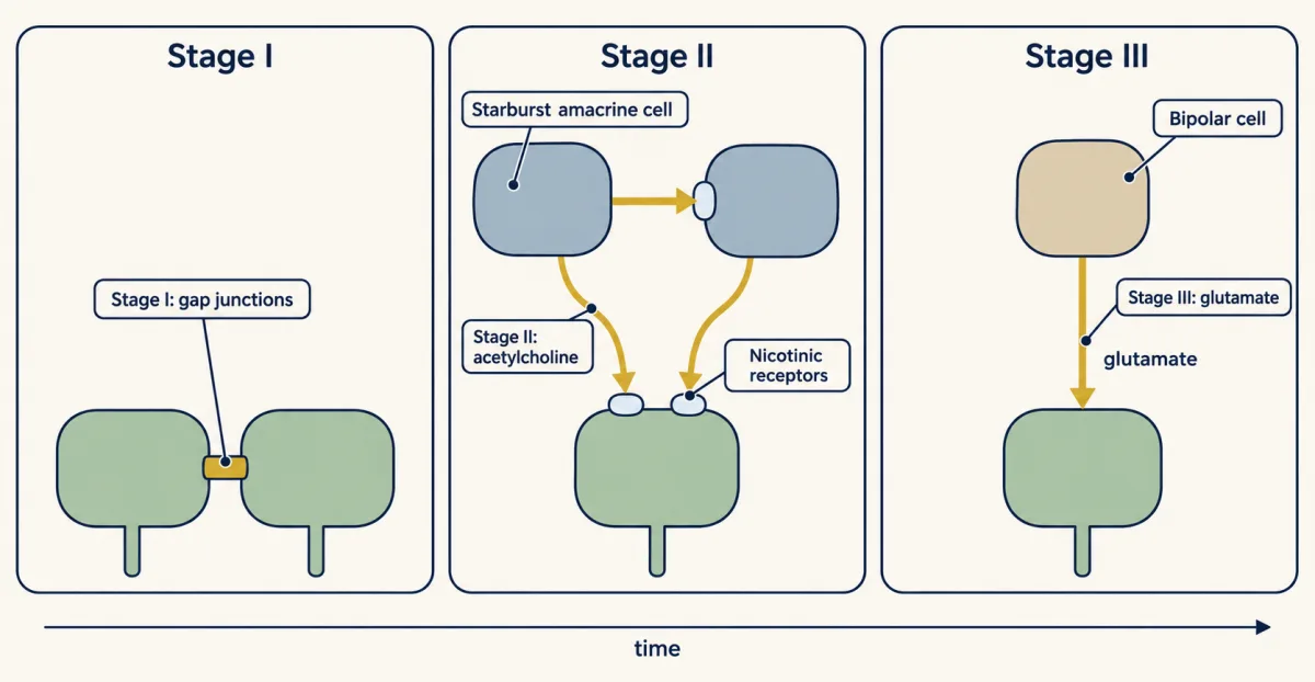
The waves are not driven by one mechanism throughout development. The dominant driver changes in three stages, best worked out in mice, rabbits and ferrets; cat and primate retinas also make waves. The mouse ages below are approximate. What matters for this lesson is the right-hand column: which tool removes or alters the waves at which age.
| Stage | Age in the mouse | Main driver | Tools that remove or alter it |
|---|---|---|---|
| I | before birth, from roughly embryonic day 16 | gap junctions, channels that connect the cytoplasm of neighbouring cells directly | gap junction blockers |
| II | from around birth to about postnatal day 10 | acetylcholine from starburst amacrine cells, interneurons of the inner retina, onto nicotinic receptors on one another and on ganglion cells | nicotinic drugs such as epibatidine; the beta2 knockout |
| III | from about day 10 until the eyes open, around day 12 to 14 | glutamate from bipolar cells, which will later carry the photoreceptor signal | glutamate receptor blockers |
In 1996 Marla Feller and colleagues showed in the developing mammalian retina that waves require synaptic activation of nicotinic receptors, and that the only cholinergic cells present were the starburst amacrine cells. Many of the receptors on ganglion cells contain the beta2 subunit. Stage II is the one most of the key experiments below target.
The stages overlap, and their boundaries blur. What every stage shares is the output: correlated bursts among neighbouring ganglion cells, with the two eyes’ waves largely independent of each other.
Block it: silence everything
Read the transcript of “Block It, Tilt It”
Film transcript: Block It, Tilt It
What happens when you take activity away? And what happens when you make the two eyes unequal? In 1988, Carla Shatz and Michael Stryker asked the first question in fetal cats. Pumps infused tetrodotoxin into the brain for two weeks. It blocks sodium channels, so it stops action potentials. Predict. With spikes blocked, do the two eyes' projections separate?
They did not. The two projections stayed mixed. Did the drug reach one eye's axons, or both? Both, and likely the thalamic cells as well. So the result shows that spikes are needed. It cannot show that the eyes compete by their activity. Removing a signal shows that it is needed. It cannot show whether the signal's pattern tells the wiring what to do. To show that a signal instructs, change where it is. Here, that means making the eyes unequal. In young ferrets, the drug epibatidine was injected into one eye, disrupting its waves. The other eye kept normal waves. Predict. Which eye wins territory in the thalamus?
The active eye. In 1998, Anna Penn, Carla Shatz and colleagues reported it. The active eye's projection expanded into the other's territory, and the treated eye's shrank. David Stellwagen and Shatz then made waves more frequent, with drugs that raise cyclic AMP. When one eye was made more active, its layer was larger. Predict. Now make waves more frequent in both eyes. What happens to the layers?
Neither changes. The layers were normal in size, which fits the idea that the eyes divide a shared target. Territory follows the difference between the eyes, not the total. One caveat. These drugs change wave frequency and pattern, not only the number of spikes. Takeaway: blocking activity shows it is needed. Tilting it shows that relative activity between the eyes shifts the territory.
From the film
- Blocking spikes with tetrodotoxin in fetal cats kept the two eyes' projections mixed, so spikes are needed.
- In young ferrets, the more active eye's projection expanded and the other's shrank, so relative activity shifts territory.
- Making waves more frequent in both eyes left the layers normal, so territory follows the difference between the eyes, not the total.
In 1988 Shatz and Michael Stryker implanted osmotic pumps in fetal cats and infused tetrodotoxin (TTX), which blocks voltage-gated sodium channels and so stops action potentials (spikes), into the brain for two weeks during the period when segregation normally happens. Tracers injected into the eyes showed that the two projections had not separated.
A student once asked: “Does TTX cut off only one eye or both?” Both, and more: infused into the brain, the drug would be expected to reach axons from both eyes and the lateral geniculate nucleus (LGN) cells themselves. So the experiment shows that spikes are needed, but cannot tell you whether the eyes compete by their activity, or whether the spikes that matter are retinal.
A permissive signal only has to be present for the wiring to form; an instructive signal’s pattern tells the wiring what to do. As in the neurotrophic hypothesis, removing a signal can show that it is needed, but not which of the two it is. To show that a signal instructs, you change where or when it is delivered and see whether the outcome follows. For activity, one such test makes the two eyes unequal.
Tilt it: make the eyes unequal
You can lower or raise wave activity in one eye only, or in both, in young ferrets. Making one eye differ tests competition most directly; changing both tests whether what matters is the difference.
Predict. Inject epibatidine, a nicotinic agonist thought to disrupt cholinergic waves by persistently activating and desensitizing the receptors (the treated retina keeps firing, without normal waves), into the left eye and leave the right one alone. What happens to each eye’s territory in the lateral geniculate nucleus (LGN)?
Check your prediction
If inputs compete by activity, the active right eye should win. That is what Anna Penn, Patricio Riquelme, Marla Feller and Carla Shatz reported in ferrets in 1998: the projection from the active retina expanded into territory that normally belongs to the other eye, and the treated eye’s projection shrank. The shift was graded, not the toy model’s all-or-none takeover.
David Stellwagen and Shatz turned the tilt the other way. They made waves more frequent with drugs that raise cAMP. When one eye was made more active, its layer was larger even though the other eye was normal.
Predict. Now make waves more frequent in both eyes. Do both layers grow, both shrink, or neither change?
Check your prediction
Neither. When both eyes were made more active, the layers were normally sized. That fits a model in which the eyes divide a shared target, so raising both equally gives neither more of it: territory follows the difference between the eyes, not the total.
These are the instructive results: relative activity between the eyes shifts which eye gets more territory. One caveat: these drugs change wave frequency and pattern as well as the number of spikes, so they do not say which feature of the activity counts.
Disrupt the correlation, keep plenty of spikes
Read the transcript of “Correlation, Not Just Spikes”
Film transcript: Correlation, Not Just Spikes
Can a retina full of spikes still fail to sharpen the brain's map? This test reads a different map. Not which eye, but where in the eye. Label a few neighbouring ganglion cells. In the superior colliculus, in the midbrain, their axons end in a zone, small if the map is sharp. Todd McLaughlin, Christine Torborg, Marla Feller and Dennis O'Leary studied mice lacking the beta2 nicotinic subunit. In their recordings, these mice had no waves in the first week. Yet their ganglion cells fired often, without correlation. Predict. By day 8, are the mutants' zones small, or broad?
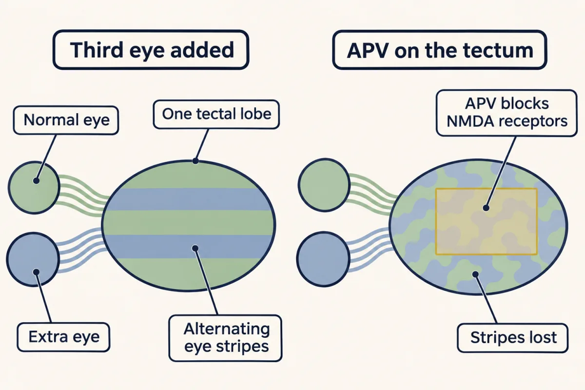
Broad. Normal mice had small zones; the mutants' stayed broad. Later waves and vision did not fix them, which points to an early window. In these recordings they had plenty of spikes, but not the normal correlation between neighbours. Later labs found altered, not absent, waves in these mice. So the safe claim is narrower. Normally patterned activity, not just spiking, is needed early for the map to sharpen. Last, a detector for together. A frog's tectum normally gets axons from one eye. Martha Constantine-Paton and Margaret Law gave frog embryos an extra eye, so two eyes shared one tectum. The two projections formed alternating stripes, where none normally exist.
Hollis Cline, Elizabeth Debski and Constantine-Paton then laid APV on the tectum. It blocks NMDA receptors. These receptors pass little current unless glutamate is bound and the membrane is already depolarized. So they can report coincident input. Predict. What happens to the stripes while APV is on the tectum?
The stripes were lost, while the retinal axons still responded. After APV was removed, the stripes returned. In the frog, keeping the stripes needed these receptors. Which detector sorts the two eyes in the mammalian thalamus is less clear. Takeaway: timing is the address. In the models, inputs compete, and the evidence so far says that patterned firing, not just spike count, matters for which inputs stay.
From the film
- In beta2 knockout mice, plenty of uncorrelated spiking in the first week left the termination zones broad.
- Normally patterned activity, not just spiking, is needed early for the map to sharpen.
- In the frog tectum, a third eye makes stripes where none normally exist, and keeping them needs NMDA receptors.
So far the question was how much activity each eye has. The next question is whether its pattern matters too. This experiment reads a different map: not which eye, but where in the eye. Label a small group of neighbouring ganglion cells, and in the superior colliculus, the midbrain visual target, their axons end in a termination zone. The zone is small if the map is sharp and broad if it is not.
Todd McLaughlin, Christine Torborg, Marla Feller and Dennis O’Leary used mice lacking the beta2 nicotinic subunit. In their recordings these mice had no waves in the first postnatal week, yet their ganglion cells fired at high rates without correlation.
Predict. By postnatal day 8, will the termination zones in the mutants be small and focal, like normal, or broad? And if correlated glutamatergic waves and then vision arrive later, will the zones catch up?
Check your prediction
By day 8, axons of neighbouring ganglion cells in normal mice had converged on small termination zones. In the mutants the zones stayed broad, and they still failed to remodel after glutamatergic waves and then vision arrived.
The mutants had plenty of spikes, so what they lacked was the normal pattern of correlation between neighbours. That points to an early window: in this study, correlated activity arriving later did not sharpen zones that had already stayed broad.
Later laboratories recorded correlated but altered waves in beta2 knockout retinas under some conditions, so the mutants lack normal waves rather than all waves. The claim that survives is narrower: normally patterned activity, not just spiking, is needed early for the map to sharpen, though the knockout alone does not show which feature of the pattern counts. Other caveats, and a closer test of pattern, are in Going deeper.
Neighbours that fire together keep their axons together; where firing is not correlated, the termination zone stays broad. Times and sizes are in relative units and patch size is arbitrary, and the picture is a sketch of the logic, not a measurement.
Add an eye: the three-eyed frog
A frog’s optic tectum, its main visual target, normally receives retinal axons from one eye only, so it has no reason to make eye-specific stripes. In 1978 Martha Constantine-Paton and Margaret Law implanted an extra eye primordium into frog embryos, and the normal and extra eyes sent axons into a single tectal lobe. The two projections formed alternating bands, so interaction between the inputs created stripes where none normally exist.
In 1987 Hollis Cline, Elizabeth Debski and Constantine-Paton laid slow-release polymer containing APV, a N-methyl-D-aspartate (NMDA) receptor antagonist, on the tecta of three-eyed tadpoles. The active form of APV desegregated the stripes, while the retinal axons still responded; the inactive form left them normal; NMDA itself sharpened their borders; and after APV was removed the stripes returned within two weeks. In the frog tectum, keeping the stripes needed NMDA receptor activity, which fits the coincidence detector the Hebbian account calls for. Whether the mammalian lateral geniculate nucleus (LGN) uses the same detector to sort the two eyes is less clear (Going deeper).
Putting it together
| Experiment | Animal and age | Observed result | What it supports | What it does not establish |
|---|---|---|---|---|
| Multielectrode recording (1991) | newborn ferret and fetal cat retina, isolated | bursts sweep across as waves several hundred micrometres wide | neighbours fire together before vision | that the brain uses it |
| Calcium imaging (2012) | live neonatal mice, before eye opening | waves propagate through the visual system | the waves are not an artefact of a dish | what they do there |
| TTX infused into the brain (1988) | fetal cat | the two eyes’ projections stay mixed | spikes are needed: necessity | competition, or that the spikes that matter are retinal |
| Epibatidine in one eye (1998) | young ferret | active eye’s projection expands, treated eye’s shrinks | the eyes compete by activity: instructive | which feature of the activity counts |
| More frequent waves in one eye, or both (2002) | young ferret | one eye: its layer larger; both: layers normal | territory follows the difference, not the total | which feature of the activity counts |
| Beta2 knockout (2003) | mouse, first postnatal week | no normal waves, plenty of uncorrelated spikes; broad termination zones that later waves and vision do not fix | normally patterned activity, not just spiking, is needed in an early window | which feature of the pattern counts; that waves were wholly absent; that the retina alone is responsible |
| Third eye (1978) | frog embryo | two eyes in one tectum form stripes | stripes arise from interaction between inputs | the mechanism |
| APV on the tectum (1987) | three-eyed tadpole | stripes lost, return after APV is removed | NMDA receptors are needed to keep the stripes in the frog | the same for the mammalian LGN |
Read down the “supports” column and the evidence builds in steps. Activity is necessary. Relative activity between the eyes shifts territory. Normally patterned early activity, not just the spike count, matters. A coincidence-sensing receptor maintains segregation where it has been tested. Timing as the address, with competition making the address stick, is the account these results fit best, not one any of them isolates; the sections below record where it is still contested.
Going deeper
The stages in more detail
Stage I waves, before birth, depend on gap junctions and also on nicotinic receptors; embryonic waves are less well studied than later ones. Details such as wave size and the relative timing of ON and OFF cells (ganglion cells that respond to light coming on and light going off) change from stage to stage.
Why Hebb needed a second half
The form of the competitive constraint matters (Miller and MacKay 1994), as the worked table shows. A simulation written for this lesson checks the same logic with wave-like correlations instead of a fixed 20 percent. It is an illustrative one-dimensional model with linear, covariance-based updates: cells lie along the wave’s path, waves run in two directions, and correlations drop to zero beyond about 200 micrometres. Each simulated target cell gets four inputs per eye from cells spanning 100 micrometres, with strengths bounded between 0 and 1. With Hebb alone, or with proportional rescaling, none of 300 cells became eye-dominated (more than 90 percent of input strength from one eye); with equal amounts taken back, all 300 did. The same model matches the key experiments in sign: giving one eye stronger correlated drive makes it win every cell, raising both equally leaves the population balanced, and removing the correlation within each eye while keeping the same variance leaves almost every cell mixed. When one cell’s inputs instead come from cells spread over 300 micrometres, wider than the 200-micrometre range over which the modelled firing overlaps, the rule drops the distant inputs, so each eye keeps a compact patch, but no cell becomes eye-dominated. These are results of one simple model, and they depend on the geometry, the input span and the rule.
The beta2 knockout, more carefully
McLaughlin and colleagues’ title states their reading of the result: “Retinotopic map refinement requires spontaneous retinal waves during a brief critical period of development”. A knockout removes the subunit everywhere, not only in the retina. Later work that restored beta2 in retinal ganglion cells alone has kept the retina at the centre of the explanation. Transgenic mice with an altered wave pattern but unchanged overall activity showed that the pattern itself instructs map development (Xu and colleagues, 2011), the closer test of “pattern, with the spikes kept”.
Are the waves instructive? A long debate
Depleting starburst amacrine cells with an immunotoxin in ferrets was reported not to prevent eye-specific segregation in the lateral geniculate nucleus (LGN) (Huberman and colleagues, Science, 2003). Others argued that enough correlated activity remained in those retinas to do the job; two reviews published side by side set out the case for and against an instructive role (Feller, and Chalupa, Neural Development, 2009). The debate over which features of the waves are needed, and whether they instruct or only permit, continued for years.
NMDA receptors in the mammalian LGN
The mammalian picture is less tidy than the frog’s. In ferrets, a N-methyl-D-aspartate (NMDA) receptor blocker did not prevent the two eyes from segregating in the LGN (Smetters, Hahm and Sur 1994), although NMDA receptor blockers did disrupt the later split of each eye’s layers into ON and OFF sublayers (Hahm, Langdon and Sur 1991). Which detector reads the waves when the eyes segregate, and whether retinal waves act on LGN synapses through NMDA-receptor-dependent plasticity on the timescale of a wave lasting seconds, remain open questions.
Your notes
Select any text in the lesson to highlight it or add a note to it. Your highlights and notes are saved in this browser and collected in your notebook.
This browser is not letting the site save anything (a private window or strict privacy settings), so highlights and notes will be lost when you leave the page.
How we measure it
Multielectrode array recording
A piece of retina is laid on a grid of electrodes, each picking up the action potentials of nearby ganglion cells. Recording many cells at once shows how correlated activity is spread out and how it travels across the tissue, which a single pair of electrodes cannot map.
Chronic drug delivery in the developing animal
A drug is supplied for days to weeks from an osmotic pump, repeated injections into one eye, or a slow-release polymer laid on the brain. Where the drug acts, and whether it reaches one eye or both, decides which question the experiment can answer.
Anterograde tracing of each eye's projection
A tracer injected into one eye is carried down the ganglion cell axons and labels their endings in the brain. Labelling the two eyes differently shows whether their territories in the target overlap or have separated.
Knockout mice
A conventional knockout mouse lacks one gene in every cell. Removing the beta2 nicotinic subunit changes the retina's spontaneous activity, so the map can be compared with normal, though the gene is also lost outside the retina.
Practice questions on this lesson (3) → MCAT-style, original, each with an explanation
Where this lesson fits
Before thisReview first
- Topographic Maps: Sperry's Eye and Eph GradientsThis course · Lesson 26
Eph and ephrin gradients draw the rough first map that waves then refine, and this lesson builds directly on that.
- The Neurotrophic Hypothesis TestedThis course · Lesson 29
Inputs competing for a limited supply of survival factor is the same argument as competition for synaptic territory here.
- Competition: How Neurons Win a TargetThis course · Lesson 32
You need the idea that a small early lead becomes a win under competition, which the Hebb-plus-competition model reuses.
After thisStudy next
- Ocular Dominance and the Critical PeriodThis course · Lesson 39
It tests how relative activity between the two eyes shifts cortical territory, taking the competition idea into the critical period.
- Opening and Closing a Critical PeriodThis course · Lesson 40
It asks what opens and closes the window in which patterned activity can refine the wiring.
Elsewhere on the siteFilms, simulations and method films in other courses
- Depletion and the paired-pulse ratioSimulation · Neuroscience
Hebb's rule, strengthening a synapse when input and target fire together, is the core rule used here.
- A Synapse That Could LearnFilm · Neuroscience
It shows how Hebb's rule was tested through long-term potentiation, the synaptic change that correlation drives.
Suggested by an AI model from the lessons themselves, then checked. How we use AI
Finish line
Test yourself
8 questions
Practice and MCAT-style, with explained answers.
Next · Lesson 39
Ocular Dominance and the Critical Period
Why does closing one eye of a kitten for a week rewire its cortex, when closing both eyes shifts the balance much less?
Continue →← Lesson 37 · Synapse Elimination: One Muscle Fiber, One Axon