Developmental Neuroscience · Unit 9: Experience refines the wiring
Opening and Closing a Critical Period
Rewritten on : new text, flashcards and film. Your notes and highlights stay, but the text around them may have moved.
A mouse that lacks one enzyme for making GABA keeps its critical period waiting for months. One drug starts it.
Pause when the reading reaches a film, and offer to play it. Saved on this device.
Watch the film now, or keep listening
Builds on:Ocular Dominance and the Critical PeriodTangential Migration: Interneurons and the Long Way RoundWhy these?
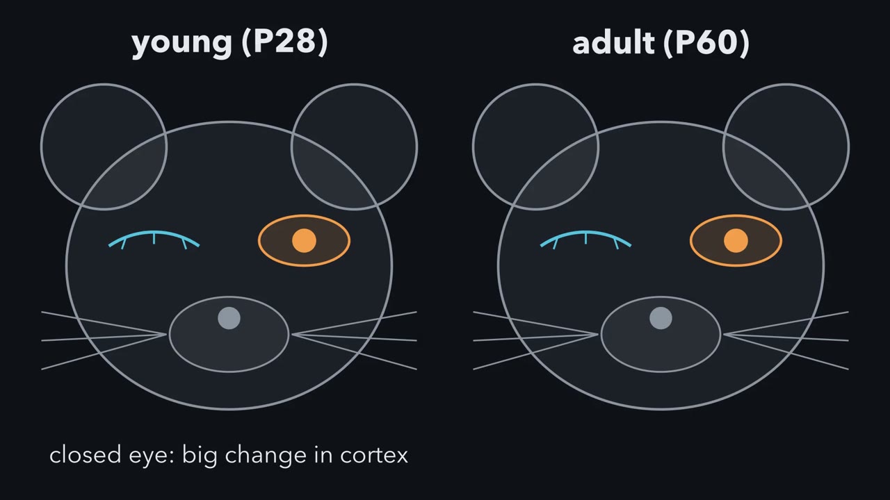 ▶ Watch A Window That Follows the Cells · 2:11
Read the transcript of “A Window That Follows the Cells”
Film transcript: A Window That Follows the Cells
Why can a few days with one eye closed reshape a young mouse's cortex, but change an adult's much less? In the binocular zone of mouse visual cortex, most neurons answer both eyes, and some answer only one. Most cells answer the eye on the opposite side of the head more strongly. The ocular dominance index puts a number on that preference. This example cell scores 0.50. Now close the opposite eye for about four days, at postnatal day 28. Responses shift toward the open eye. This example cell's score falls below zero. In young mice, most of that shift comes from the closed eye's responses weakening.
Pause and predict. Do the same thing to a two-month-old mouse. What happens to the score? Much less. In mice the shift is largest near postnatal day 28. It is much smaller before about day 19, or after about day 32. So the window lies in a few weeks of youth. Is that age itself, or something age brings? Here is the idea to carry through. The window follows the inhibitory cells, not the calendar.
A working model: maturing inhibition, especially from parvalbumin basket cells, opens the window once it passes a threshold. As inhibition keeps maturing, and brakes build up around these cells, it closes. In young cortex, a brief boost of inhibition was enough to start a window. The boost did not have to last.
The rule covers this young shift in mouse and rat visual cortex. Adults keep a weaker, different form. It does not cover people, or song and language. Takeaway: the window is set by the state of a circuit, not simply by age.
From the film
- In mouse visual cortex, a few days with one eye closed reshape the cortex strongly in youth and much less in adults.
- The window follows the inhibitory cells, not the calendar: in the working model, maturing inhibition, especially from parvalbumin basket cells, opens it once it passes a threshold.
- As inhibition keeps maturing and brakes build up around these cells, the window closes.
A mouse that lacks one of its two enzymes for making GABA can go through the whole of youth without the critical period that should have reshaped its visual cortex. Give it a few days of a benzodiazepine, even as an adult, and the window opens.
Start with the picture behind the word “window”. Close one eye of a four-week-old mouse for a few days, and its visual cortex shifts: it now answers that eye much less. Do the same to a two-month-old mouse, and the cortex changes much less. You met this result, in kittens as well as mice, in the lesson on ocular dominance. A stretch of time like this is a window of opportunity and of vulnerability, and it is one reason amblyopia, a weak eye from childhood, is best treated early. This lesson asks how such a window is regulated, and the knockout mouse in the bold line is where the answer starts.
The readout, step by step
Most of the evidence comes from one system: mouse primary visual cortex, with monocular deprivation as the readout. The mouse earns its place because you can change its genes.
- Where to record. In the binocular zone of visual cortex, most neurons answer both eyes, though some answer only one. In the mouse, most cells answer the contralateral eye, on the opposite side of the head, more strongly than the ipsilateral eye, on the same side.
- Put a number on it. The ocular dominance index is (C − I)/(C + I), where C and I are a cell’s responses to the contralateral and the ipsilateral eye. An illustrative cell that fires 30 spikes per second to the contralateral eye and 10 to the ipsilateral eye has an index of 20/40 = 0.50. For responses of zero or more, the index runs from +1 (contralateral eye only) to −1 (ipsilateral eye only).
- Deprive. Suture the contralateral eyelid shut for about four days at postnatal day 28 (P28), then record again. Responses shift toward the open eye, so the index falls, and a cell like this one can fall below zero: it now prefers the eye that stayed open. In young mice, most of the shift comes from the closed eye’s responses weakening.
- Vary the age. The shift is largest around P28, and much smaller before about P19 or after about P32 (Gordon and Stryker, 1996).
The first MCAT question at the end works one cell through. For this lesson, then, the window is the range of ages in which four days with one eye closed move the index toward the open eye. Is that range set by age itself, or by something that age brings with it?
The one idea to carry through this lesson. The window follows the inhibitory cells, not the calendar. In the working model, when maturing inhibition, especially in circuits involving parvalbumin basket cells (fast-firing inhibitory neurons), passes a threshold, it helps start the window. As inhibition goes on maturing, and other brakes build up around these cells, the window closes. In a normal mouse the two agree: inhibition reaches the threshold at a typical age.
What the rule covers.
- Which plasticity. The juvenile ocular dominance shift in mouse and rat visual cortex, in which a few days of monocular deprivation weaken the closed eye’s responses. Adults keep a weaker, different form. Song, language and people are outside it.
- What “threshold” means. In young cortex, crossing it starts a window; the evidence below is that a brief drug boost was enough. Whether normal inhibition must stay high to keep a window open has not been tested. Once the window has closed, lowering inhibition helped reopen it in adult rodents.
- What it does not say. Inhibition is not the only clock; other parts of the circuit mature too. Nor does it say which eye wins.
Is it the calendar? The GAD65 test
Read the transcript of “Switch It On with a Drug”
Film transcript: Switch It On with a Drug
If the window waits for inhibition, what happens in a mouse that cannot release enough GABA? Inhibitory neurons make GABA with two enzymes. One, called GAD65, sits at the synaptic terminal. It tops up the supply when the neuron fires hard. A knockout lacks a working copy of that gene. Pause and predict. Inhibition quiets neurons. Should a knockout's cortex shift more than normal when one eye closes?
It does not shift at all. Its neurons still respond to vision. What failed was the change. Now add diazepam. It binds the GABA receptor at a site between an alpha and a gamma subunit. It makes the receptor respond more strongly to GABA. But it cannot open the channel alone. So it amplifies the GABA the knockout still releases. Given during deprivation, it restored the shift. The drug did not shift the cortex by itself. The shift went toward the eye that stayed open. Inhibition is permissive. The difference between the eyes is instructive. Pause and predict. Raise a knockout to day 60, long past the normal window. Give diazepam and close one eye.
It shifts. The capacity waited into adulthood. Age had not closed a window that had never opened. Give diazepam to a normal mouse at about day 15, before its window. It shifts early. Give a second group two days of diazepam after eye opening, then close one eye at day 28. Much less shift is left. The window opened early and was gone by day 28. In knockouts, two days of diazepam near the start of deprivation switched plasticity fully on. So in young cortex, a brief boost of inhibition can start a window without having to stay present. This should feel backward. Why would less inhibition make a cortex harder to change? The inhibition implicated here is thought to come largely from basket cells.
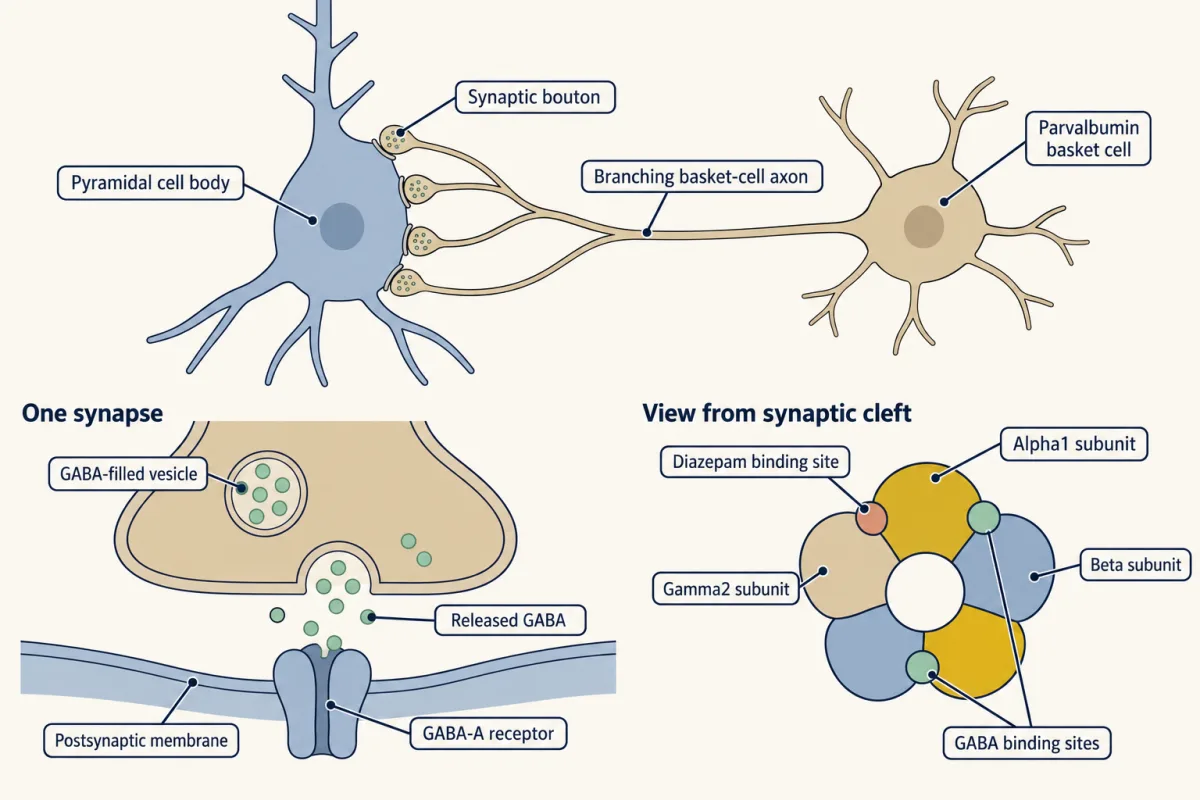
Their synapses cluster on the cell bodies of pyramidal neurons. One leading idea: maturing basket-cell inhibition sharpens when pyramidal cells fire. That could let the circuit tell which eye reliably drives each cell. This is a hypothesis, not a settled mechanism. The experiments establish the order: inhibition first, then plasticity. Takeaway: inhibition opens the window like a trigger. The open eye still decides which way the cortex shifts.
From the film
- GAD65 knockout mice, which release too little GABA, showed no ocular dominance shift at the normal age, and diazepam restored it.
- Inhibition is permissive and the difference between the eyes is instructive: the shift went toward the eye that stayed open.
- In young cortex, a brief boost of inhibition can start a window without having to stay present.
If the window simply followed age, every mouse would have it from about P19 to P32, whatever else differed. The first test came from a mouse with weaker inhibition.
A knockout mouse lacks a working copy of one gene. Inhibitory neurons make GABA, the main inhibitory transmitter of the cortex, from glutamate with two enzymes. GAD67 makes most of the GABA present at rest. GAD65 sits at the synaptic terminal and adds to the supply when the neuron fires hard. A GAD65 knockout still has GABA, but it releases too little during strong activity.
Predict. Inhibition quiets neurons. Close one eye of a GAD65 knockout at P28. Should its cortex shift more than a normal mouse’s, about the same, or less?
Check your prediction
Less: not at all. In 1998 Takao Hensch, Michela Fagiolini, Michael Stryker and colleagues reported that GAD65 knockouts showed no ocular dominance shift after deprivation at the normal age. Their neurons still responded to vision, with normal basic response properties. What failed was the change.
If you guessed “more”, that is a natural guess: a less inhibited cortex sounds easier to change. A section below takes up why it is wrong.
Then the rescue. Diazepam is a benzodiazepine, a positive modulator of the GABA-A receptor. It binds at a site between an alpha and a gamma subunit and makes the receptor’s chloride channel respond more strongly to GABA, without opening it alone (see the lesson on ion channels as receptors, and the lesson on drugs that act on one part of a receptor). So it amplifies whatever GABA the knockout still releases. Infused into the visual cortex of the knockouts during deprivation, diazepam restored the shift.
Notice what the drug did not do. Boosting inhibition did not itself shift the cortex; the shift went toward the eye that stayed open. In the terms of the lesson on the neurotrophic hypothesis, inhibition here is permissive: it lets the cortex respond to deprivation. The difference between the eyes is instructive: it decides which eye wins.
Predict. Two experiments separate “age opens the window” from “inhibition opens the window.”
- Raise a GAD65 knockout to P60, long past the normal window. Give diazepam and close one eye.
- Give diazepam to a normal mouse at about P15, around the time the eyes open and before its window would open, and close one eye. In a second group, give the same early diazepam, stop it, and close one eye at P28, the usual peak.
What does each idea predict for each experiment?
Check your prediction
If age sets the window, the knockout at P60 is too old and should not shift. The P15 mouse is too young and should not shift, and the P28 group should shift normally.
If inhibition sets it, the knockout’s window has never opened, so it should still be waiting at P60, and diazepam should open it. The P15 mouse should shift, because diazepam lifts its immature inhibition over the threshold. And if a window that opens early also closes early, the early-treated mice should have little left by P28.
The results follow inhibition.
| Mouse | Diazepam | Age at eye closure | Shift? | What it says |
|---|---|---|---|---|
| Normal | none | P28 | yes | the normal window |
| Normal | none | P60 | little | the window has closed |
| GAD65 knockout | none | P28 | no | too little inhibition; the window has not opened |
| GAD65 knockout | during deprivation | P28 | yes | raising inhibition is enough (1998) |
| GAD65 knockout | during deprivation | P60 | yes | the capacity waited into adulthood; age did not close it (Fagiolini and Hensch, 2000) |
| Normal | early | P15 | yes | the window can open early (Fagiolini and Hensch, 2000) |
| Normal | two days, just after eye opening | P28 | much less | it opened early and closed early (Iwai and colleagues, 2003) |
The last row matters most. The same study found that in knockouts, two days of diazepam near the start of deprivation were enough to switch plasticity fully on, even though the drug did not overlap the days the eye was closed (Iwai and colleagues, 2003). So diazepam acts as a trigger: it starts the window. What ends it is not shown here to be an internal timer. The early-treated windows closed sooner, and the brakes section shows that further maturation of inhibition and added brakes help end it.
Why would more inhibition help?
Inhibition quiets neurons, so why would a cortex with too little of it be harder to change?
The inhibition implicated here is thought to come largely from fast-firing basket cells, as the next section discusses. Their synapses cluster on the cell bodies of pyramidal neurons, the cortex’s main excitatory cells, close to where the axon starts and spikes begin. That placement gives them strong control over whether and when a pyramidal cell fires. One leading idea is that maturing basket-cell inhibition sharpens when pyramidal cells fire, so that the circuit can tell which eye reliably drives each cell, and deprivation can then weaken the closed eye’s input. Consistent with this, neurons in GAD65 knockouts fire prolonged discharges, and the benzodiazepine that unmasked plasticity selectively reduced them (Fagiolini and Hensch, 2000).
This is a hypothesis, not a settled mechanism. What the experiments establish is the order of events: inhibition first, then plasticity.
Which inhibition: follow the basket cells
Cortex has many kinds of inhibitory interneuron. The ones implicated here are parvalbumin-expressing basket cells. Like most cortical interneurons, they are born in the medial ganglionic eminence (MGE) of the embryonic forebrain and reach the cortex by tangential migration.
Part of the case for these cells comes from receptor subunits. GABA-A receptors are built from five subunits, and the alpha subunit varies. Receptors with alpha1 are enriched at the synapses basket cells make on cell bodies. In 2004, Fagiolini, Hensch and colleagues used knock-in mice, which carry a chosen point mutation, here one that makes a single alpha subtype insensitive to diazepam. When alpha1 was made insensitive, benzodiazepines no longer opened the window early. When alpha2 was made insensitive, they still did. In mouse visual cortex, then, the drug acts through alpha1 receptors. That is consistent with basket-cell synapses carrying the effect, but it is not proof, because alpha1 is also the most widespread alpha subunit in cortex. The next experiment tests the broader idea directly.
Predict. Take inhibitory neuron precursors from the MGE of an embryo and transplant them into the visual cortex of a young mouse. The cells disperse and mature into interneurons, including parvalbumin cells. Weeks later the host is past its own window. If the window follows the animal, what should closing one eye do now? If it follows the inhibitory cells, when should a second window appear?
Check your prediction
A second window appeared, timed by the cells. In 2010, Derek Southwell, Sunil Gandhi, Stryker, Arturo Alvarez-Buylla and colleagues found that when the transplanted cells reached about the age of native interneurons during the normal critical period, the host had a second critical period.
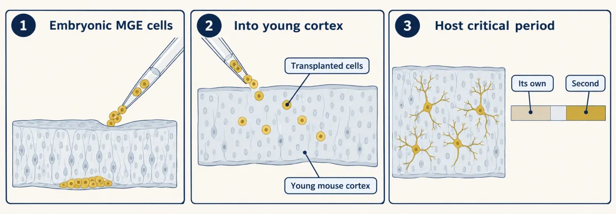
This is the most direct evidence that the window follows the inhibitory cells: an animal past its own window got a second one from a young cohort of inhibitory cells. It does not single out parvalbumin cells, because the donor cells became several kinds of interneuron.
What sets the pace of the basket cells
If the maturation of these inhibitory circuits helps set the timing, changing when they mature should move the window. Three findings fit.
- Brain-derived neurotrophic factor (BDNF). In mice built by Josh Huang, Susumu Tonegawa, Alfredo Kirkwood, Lamberto Maffei and colleagues (1999), cortical neurons make extra BDNF, a neurotrophin that among other effects promotes inhibitory synapses. Their inhibition matured early, and their critical period ended early.
- Darkness. Rearing animals in darkness from birth, which slows the maturation of inhibition, delays the window in rodents and in cats.
- Otx2. Otx2 is a homeoprotein, a transcription factor with a homeodomain. In 2008, Sayaka Sugiyama, Hensch, Alain Prochiantz and colleagues reported that Otx2 protein builds up in parvalbumin cells of mouse visual cortex although these cells make little Otx2 RNA. Its build-up depended on visual experience, and infusing Otx2 early opened plasticity early. Its source is uncertain.
Closing the window: brakes, not decay
Read the transcript of “Brakes, and Borrowed Cells”
Film transcript: Brakes, and Borrowed Cells
If maturing inhibition opens the window, what closes it? Does the machinery for change simply wear out? If so, removing one molecule in an adult should not bring the window back. As the window ends, a lattice of matrix called a perineuronal net condenses around many parvalbumin cells. Pause and predict. Digest that net in an adult rat, then close one eye. Does the cortex shift?
It does. Chondroitinase, an enzyme that cuts the net's sugar chains, let deprivation shift adult rat cortex. Myelin carries proteins such as Nogo. Mice lacking the Nogo receptor kept plasticity past the usual closing age. Lynx1 is a small membrane protein that damps nicotinic receptors. Adult mice without it kept strong plasticity. Blocking nicotinic receptors prevented that plasticity. Inhibition itself has a second job. Past the threshold, it keeps maturing and becomes a brake. In adult rats, reducing GABA transmission partly restored plasticity. In adult rats, weeks of the antidepressant fluoxetine lowered inhibition and restored plasticity.
Diazepam blocked that. In the knockout, diazepam opened the window. In the adult, it put the brake back. Now a direct test of whether the window follows the cells. Transplant embryonic inhibitory neuron precursors into a young mouse's cortex. They mature into interneurons, including parvalbumin cells. The host's own window closes.
Pause and predict. Will a second window come at the host's age, or at the cells' age? The cells' age. When they reached the age of native interneurons in the normal window, the host shifted again. Why does this matter? In amblyopia, one eye sees poorly because its input to the cortex was degraded in childhood. Patching the good eye works best in young children, and much less reliably in adults. These rodent results show mechanism, not treatment. Reopening plasticity can also reopen vulnerability. Takeaway: in rodent visual cortex, this window is set by the state of a circuit, not simply by age. Inhibition opens it, and added brakes, including more mature inhibition, close it.
From the film
- In rodents, removing a brake such as the perineuronal net, the Nogo receptor or Lynx1 let plasticity persist or return, so the window is actively held shut rather than decaying.
- Transplanted inhibitory neuron precursors gave the young host a second window when the donor cells reached about the age of native interneurons, so the window follows the cells.
- These rodent results show mechanism, not an established treatment for amblyopia in people.
If the window simply faded as the machinery for plasticity was lost, removing one molecule in an adult should not bring it back. Experiments in adult rodents suggest instead that it is actively held shut.
Predict. As the critical period ends, a perineuronal net, a dense lattice of extracellular matrix rich in chondroitin sulfate proteoglycans, condenses around many parvalbumin cells. Dark rearing delays it. Inject an enzyme that cuts chondroitin sulfate chains into the visual cortex of an adult rat, then close one eye. What does “decay” predict? What does “brake” predict?
Check your prediction
Decay predicts no shift. A brake predicts a shift once it is removed. In 2002, Tommaso Pizzorusso, Maffei and colleagues in Pisa did the experiment with chondroitinase ABC, a bacterial enzyme. Monocular deprivation then shifted ocular dominance, which it did not do in untreated adults. The nets help hold the window shut.
Two more brakes have been found by deleting a gene and finding plasticity that lasts past the usual closing age.
Myelin signals. Myelin, the insulating wrap on axons (its own lesson), carries proteins such as Nogo that inhibit axon growth through the Nogo receptor. In 2005, Aaron McGee, Stephen Strittmatter and colleagues reported that mice lacking the Nogo receptor kept ocular dominance plasticity after the usual closing age.
Lynx1. Lynx1 is a small membrane protein that binds nicotinic acetylcholine receptors and damps their response. Its level in mouse visual cortex rises in adulthood. In 2010, Hirofumi Morishita, Hensch and colleagues reported that adult Lynx1 knockouts kept strong plasticity, and that blocking nicotinic receptors prevented it.
Inhibition itself: two jobs at two times. Here a second puzzle appears. Inhibition opened the window, so how can it also help close it? Reaching a threshold starts the window. Inhibition that goes on maturing after that becomes one of the brakes. In GAD65 knockouts given diazepam, plasticity was eventually lost after a long delay, and the loss came with improved function of their own inhibitory circuits (Iwai and colleagues, 2003). In adult rats, the Pisa group partly restored ocular dominance plasticity by reducing GABA transmission in visual cortex (Harauzov and colleagues, 2010). That is why some of the reopening treatments below work partly by lowering inhibition.
Reopening the window, and what it promises for amblyopia
The reason to reopen it is amblyopia, in which one eye (occasionally both) sees poorly because its input to the cortex was degraded in childhood, for example by a misaligned or blurred eye. Patching the good eye works much less reliably in adults than in young children. A drug that reopened the window could, in principle, give adults another chance.
Predict. In 2008, José Fernando Maya Vetencourt, Maffei, Eero Castrén and colleagues gave adult rats weeks of fluoxetine, an antidepressant. It restored ocular dominance plasticity and lowered intracortical inhibition. What should diazepam, given along with the fluoxetine, do? Compare it with what diazepam did in the GAD65 knockout.
Check your prediction
Diazepam blocked these effects, the mirror image of the knockout rescue. In the knockout, diazepam raised inhibition that was too weak, and the window opened. In the adult, fluoxetine lowered inhibition that had become a brake, and diazepam put the brake back. The same drug had opposite effects because the circuit was in a different state.
Paired with visual experience through the weak eye, fluoxetine also allowed recovery of vision in rats made amblyopic by long deprivation in early life. Elizabeth Quinlan’s laboratory reported that a period of complete darkness restored plasticity in adult rats (He and colleagues, 2006) and, in a later study, allowed recovery of vision after long deprivation (He and colleagues, 2007). With chondroitinase ABC and the interneuron transplant, that makes several routes back to a plastic cortex in rodents.
Read these as evidence about mechanism, not as treatments. Each result is in rats or mice, mostly in visual cortex, with deprivations that differ from human amblyopia. Reopening plasticity can also reopen vulnerability, since a plastic cortex can be changed for the worse. Small trials of drugs in adults with amblyopia have so far given mixed results, and none of these approaches is an established therapy. Song learning and language have their own windows, taken up in the lesson on sensitive periods.
Putting it together
| Experiment | Animal and age | Observed result | What it supports | What it does not establish |
|---|---|---|---|---|
| Close one eye in GAD65 knockouts | mouse, P28 | no shift (1998) | the window needs enough inhibition | which inhibitory cells |
| Diazepam during deprivation | GAD65 knockout, P28 | shift restored (1998) | raising inhibition is enough to open it | how inhibition enables plasticity |
| Diazepam in older knockouts | GAD65 knockout, P60 | shift (2000) | age did not close the window | that age plays no part in normal mice |
| Early diazepam | normal mouse, about P15 | early shift (2000) | the window can open early | — |
| Early diazepam, tested at the usual peak | normal mouse, P28 | much less shift (2003) | a window triggered early also closes early | what sets its length |
| Diazepam-insensitive alpha1 or alpha2 | mouse | alpha1 change blocks early opening; alpha2 change does not (2004) | the drug acts through alpha1 receptors | that basket cells alone carry the effect |
| MGE transplant | young mouse host | a second window when donor cells reach critical-period age (2010) | the window follows the inhibitory cells | that these cells are the only clock, or that parvalbumin cells alone carry it |
| Extra BDNF | mouse | inhibition matures early; window ends early (1999) | fits the idea that the pace of inhibition sets the timing | that BDNF acts only through inhibition |
| Dark rearing from birth | rodents, cats | window delayed | experience paces maturation | — |
| Chondroitinase ABC | adult rat | shift after deprivation (2002) | perineuronal nets help hold the window shut | how the brakes interact |
| Nogo receptor knockout | mouse | plasticity past the usual closing age (2005) | myelin signals act as a brake | how the brakes interact |
| Lynx1 knockout | adult mouse | strong plasticity; a nicotinic blocker prevents it (2010) | Lynx1 acts as a brake | how the brakes interact |
| Reduced GABA transmission | adult rat | plasticity partly restored (2010) | mature inhibition acts as a brake | — |
| Fluoxetine, with and without diazepam | adult rat | plasticity restored; diazepam blocks it (2008) | lowering inhibition can reopen the window | a treatment for people |
| Dark exposure | adult rat | plasticity restored (2006) | a change in visual experience can reopen the window | a treatment for people |
The evidence points the same way across genes, drugs, rearing and transplanted cells. In mouse and rat visual cortex, the ocular dominance window tracks the maturation state of inhibitory circuits: a threshold starts it, further maturation and added brakes end it, and both steps can be moved. It is set by the state of a circuit, not simply by age.
Going deeper
Critical period or sensitive period
A sensitive period is a limited time when experience has unusually strong effects on a circuit. A critical period is the stricter case, in which the experience is needed for normal development and its effects last. Usage varies between fields. The mouse ocular dominance window is usually called a critical period, though its edges are softer than the name suggests.
Where cortical Otx2 comes from
The source of the Otx2 that builds up in parvalbumin cells was first proposed to be the retina; later work pointed to the choroid plexus, the tissue that makes cerebrospinal fluid, as a source as well. The full answer is still debated, so hold this part loosely. Otx2 uptake into parvalbumin cells has been reported to depend on perineuronal net sugars, which, if it holds, would link the opening and closing steps.
The adult form of ocular dominance plasticity
Adult mouse visual cortex is not completely fixed. Sawtell and colleagues (2003) found an adult form of ocular dominance plasticity in mice, mostly a strengthening of the open eye’s responses, which differs from the juvenile weakening of the closed eye’s responses. It depends on N-methyl-D-aspartate (NMDA) receptors. So “closed” means reduced and changed, not absent.
How the brakes might overlap
The Nogo receptor, Lynx1, the extracellular matrix and mature inhibition each restrain plasticity: in rodents, removing or reducing any one of them lets some plasticity persist or return. It is not settled how they interact, or whether they are partly redundant.
Why the drug’s lack of effect on the index is a finding
It is tempting to say that boosting inhibition could never move the index, because it acts on both eyes’ inputs. That holds only for one kind of effect. If inhibition scaled both responses by the same factor, the index would not change: 30 and 10 spikes per second becoming 18 and 6 still give 0.50. If it subtracted the same amount from both, the index would change: 30 and 10 becoming 25 and 5 give about 0.67. So the statement that diazepam alone did not shift ocular dominance is an experimental result, not a consequence of the arithmetic.
Your notes
Select any text in the lesson to highlight it or add a note to it. Your highlights and notes are saved in this browser and collected in your notebook.
This browser is not letting the site save anything (a private window or strict privacy settings), so highlights and notes will be lost when you leave the page.
How we measure it
Monocular deprivation with an ocular dominance readout
One eyelid is sutured shut for a few days, then responses of visual cortex to each eye are measured with single-unit recording, visual evoked potentials or optical imaging. A shift toward the open eye shows that the cortex was plastic during the deprivation.
Knockout and knock-in mice
A knockout mouse lacks a working copy of one gene; a knock-in carries a chosen change in it. GAD65 knockouts tested whether activity-dependent GABA release was needed for the window, and knock-ins that make one GABA-A receptor subunit insensitive to diazepam tested which subunit carries the drug's effect.
Local drug and enzyme infusion
A drug or enzyme is delivered into one region of cortex through a minipump or an injection. Diazepam infusion tested whether boosting inhibition is enough to open the window, and chondroitinase ABC injection tested whether the extracellular matrix holds it closed.
Interneuron transplantation
Inhibitory neuron precursors are taken from the embryonic medial ganglionic eminence and injected into the cortex of a young host, where they migrate and mature. Testing the host later asks whether the age of the inhibitory cells, rather than the age of the animal, sets the window.
Practice questions on this lesson (3) → MCAT-style, original, each with an explanation
Where this lesson fits
Before thisReview first
- Ocular Dominance and the Critical PeriodThis course · Lesson 39
This lesson takes the monocular deprivation shift and the ocular dominance index from there and asks what sets the window.
- Tangential Migration: Interneurons and the Long Way RoundThis course · Lesson 19
The inhibitory basket cells and MGE transplants in this lesson depend on interneurons being born outside cortex and migrating in.
After thisStudy next
- Microglia and Complement Prune SynapsesThis course · Lesson 41
It asks how non-neuronal cells prune synapses during the same experience-driven refinement, which this lesson's window regulation sets the timing for.
- Sensitive Periods in Behavior: Song, Language and SightThis course · Lesson 42
It extends the idea of a regulated window from visual cortex to song and language, which this lesson explicitly leaves for there.
- Disorders of the Synapse: Built, Then KeptThis course · Lesson 49
It uses the idea that a silenced state can be reversed in adults, as reopening plasticity is here.
Elsewhere on the siteFilms, simulations and method films in other courses
- Shunting inhibitionSimulation · Neuroscience
Shows how inhibition changes a cell's response, the property that opens the window here.
- Depletion and the paired-pulse ratioSimulation · Neuroscience
Shows how activity strengthens or weakens synapses, the plasticity the critical period gates.
Suggested by an AI model from the lessons themselves, then checked. How we use AI
Finish line
Test yourself
8 questions
Practice and MCAT-style, with explained answers.
Next · Lesson 41
Microglia and Complement Prune Synapses
Why can a developing neuron make the first protein of an immune cascade that marks bacteria for destruction?
Continue →