Developmental Neuroscience · Skim version
Microglia and Complement Prune Synapses
Read the full lesson → Skim the whole course →
The guiding idea
Why can a developing neuron make the first protein of an immune cascade that marks bacteria for destruction?
In the young mouse visual thalamus, relative activity biases which retinal inputs lose; microglia take up relatively more material from the less active eye, and complement helps them take up part of it.
From the film: Which Inputs Lose?
- In the young mouse visual thalamus, the two eyes' inputs start out overlapped and sort into separate territories over roughly the first week and a half.
- Relative activity biases which retinal inputs lose, and microglia take relatively more material from the quieter eye.
- Complement helps microglia take up part of that material, and the evidence does not show that it carries the activity information.
From the film: A Tag and a Reader
- Complement marks a target for eating in three steps: C1q binds a surface, C3 is cut and its large piece fixes to that surface, and complement receptor 3 on an eating cell binds the tag.
- In mice lacking C1q or C3, the two eyes' territories failed to refine normally and still overlapped more than in normal littermates.
- These knockouts show that complement is needed for normal refinement in this circuit, but not which synapse got a tag, what read it, or which cell carried the input away.
From the film: Caught in the Act
- Microglia in the young mouse visual thalamus held retinal material inside their lysosomes, and this uptake was high at postnatal day five and much lower by day nine.
- Without complement receptor 3 or C3, microglia took up about half as much, so complement is one route of several.
- With one eye silenced, microglia held relatively more of that eye's material, and with one eye boosted, relatively more of the untreated eye's, so the comparison must be made against how much of each eye's input is there.
- This compares whole eyes, and how activity and complement connect is still open.
-
Two eyes, one relay: which inputs lose?
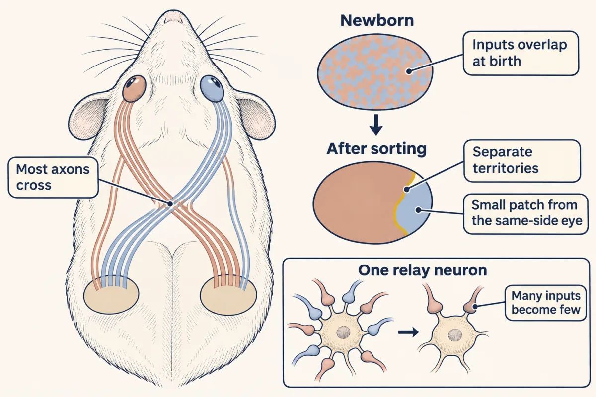
Over the first postnatal week and a half, inputs from the two eyes sort into separate territories in the relay, each nucleus ending up mostly the territory of the opposite eye with a small patch for the eye on the same side. A single relay neuron also goes from many retinal inputs to a few. AI-generated illustration -
A tag from the immune system
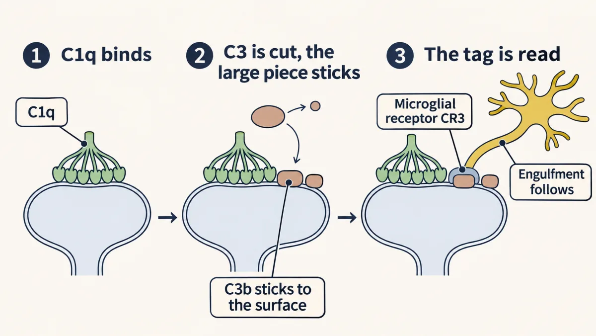
The three steps of the classical pathway that matter here: C1q binds a surface, C3 is cut and its large piece fixes to that surface, and a phagocyte carrying complement receptor 3 binds the tag, which promotes engulfment. AI-generated illustration -
Caught in the act: microglia take the quieter eye
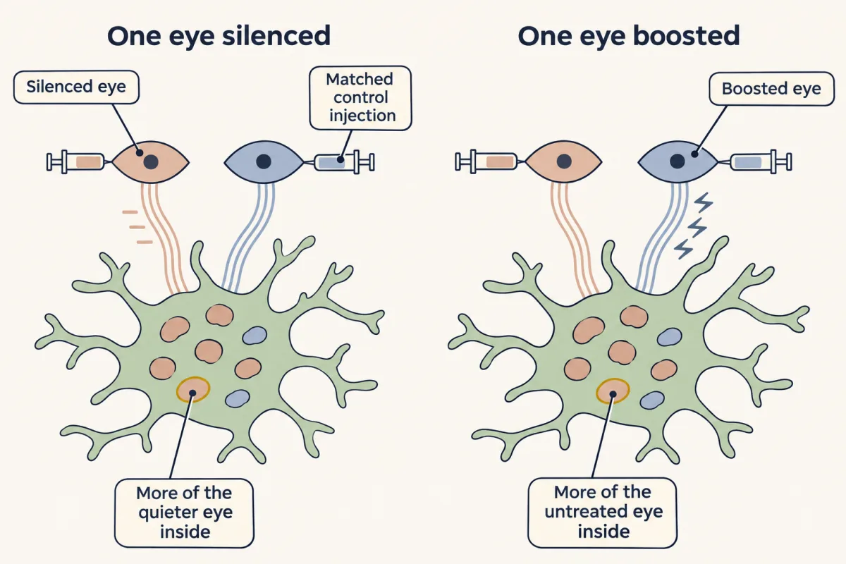
The two drugs push in opposite directions: with one eye silenced, microglia held relatively more of that eye's material, and with one eye boosted, relatively more of the untreated eye's. The comparison has to be made against how much of each eye's input is there, not as a raw share. AI-generated illustration -
Find me, eat me
-
When pruning goes wrong: links, not causes
-
Putting it together
-
Going deeper
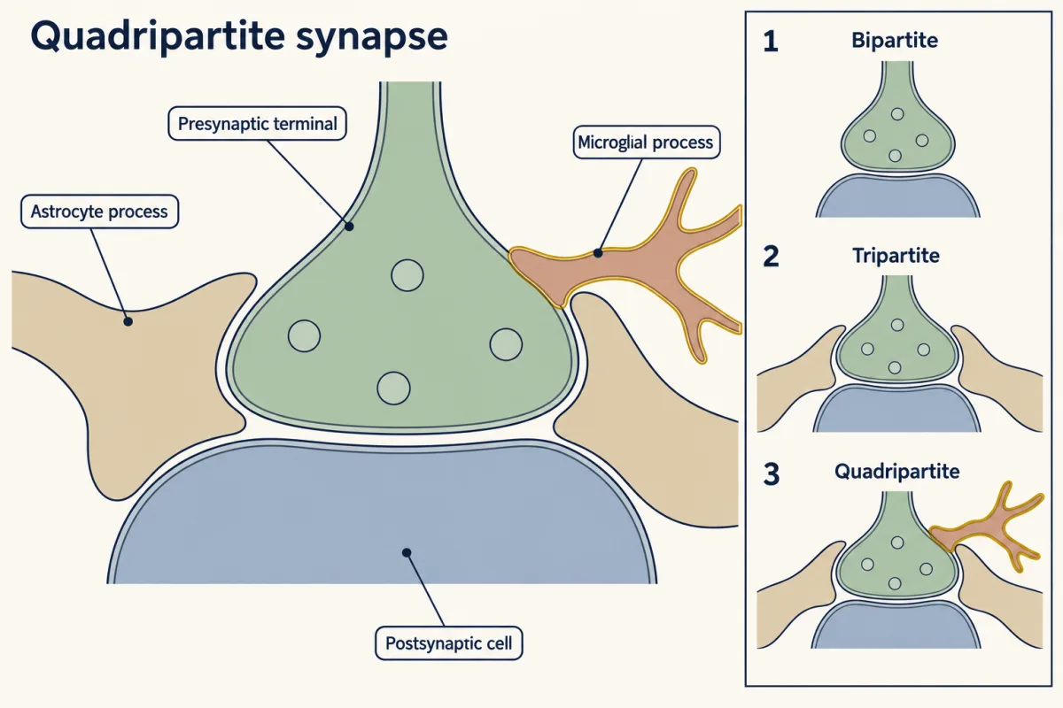
Bipartite, then tripartite with the astrocyte, then quadripartite with the microglial process. AI-generated illustration
Where this lesson fits
Before thisReview first
- Activity Before Experience: Spontaneous Retinal WavesThis course · Lesson 38
Retinal waves drive the activity differences between inputs that this lesson says microglia and complement act on.
- Ocular Dominance and the Critical PeriodThis course · Lesson 39
You need its idea that the more active eye's inputs win and the quieter eye's lose before asking what removes them.
- Too Many Neurons: Naturally Occurring Cell DeathThis course · Lesson 28
It sets up the idea that a nervous system makes too much and then removes some of it.
After thisStudy next
- Disorders of the Synapse: Built, Then KeptThis course · Lesson 49
It takes up the proposed links between altered synapse number and autism or schizophrenia that this lesson only introduces.
- Sensitive Periods in Behavior: Song, Language and SightThis course · Lesson 42
It uses the idea that experience-dependent removal of connections closes off circuit change as development goes on.
Elsewhere on the siteFilms, simulations and method films in other courses
- Cell-free extracts and reconstitutionMethod film
It shows how a cell process is rebuilt in a tube to test which parts are needed, as for the C1q, C3 and CR3 tagging steps.
- A Cell Keeps Its Answer ReadyFilm · Cell Signaling
It shows how innate immune cells recognize targets they have never met, the system whose tagging proteins neurons borrow here.
Suggested by an AI model from the lessons themselves, then checked. How we use AI