Developmental Neuroscience · Unit 9: Experience refines the wiring
Ocular Dominance and the Critical Period
Rewritten on : new text, figures, flashcards and film. Your notes and highlights stay, but the text around them may have moved.
Why does closing one eye of a kitten for a week rewire its cortex, when closing both eyes shifts the balance much less?
Pause when the reading reaches a film, and offer to play it. Saved on this device.
Watch the film now, or keep listening
Builds on:Topographic Maps: Sperry's Eye and Eph GradientsSynapse Elimination: One Muscle Fiber, One AxonWhy these?
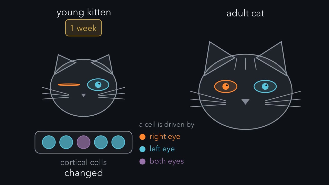 ▶ Watch One Cell, Two Eyes · 3:03
Read the transcript of “One Cell, Two Eyes”
Film transcript: One Cell, Two Eyes
Close one eye of a young kitten for a week, and its visual cortex changes. Close an adult cat's eye for months, and the cortex looks normal. Why? To see what changed, follow the signal from the eyes to the cortex. Each eye sends its axons to a relay station in the thalamus, the lateral geniculate nucleus. We will call it the LGN. There the two eyes' axons end in separate layers. So most LGN cells are driven by one eye. LGN axons carry the signal on to the primary visual cortex, at the back of the brain. They end mainly in layer 4. There the two eyes' endings still lie apart, in alternating stripes. In macaque monkeys these stripes, called ocular dominance columns, are sharp.
In cats the pattern is less regular. In macaque layer 4C, most cells are driven mainly by one eye. Cat layer 4 has more cells that hear both. Above and below layer 4, cortical neurons combine the two streams. Many cells there can be driven by both eyes. In cats and monkeys, this is the first place where many single cells are driven by both eyes. The eyes compete for territory in layer 4, and for those shared cells. Which eye is which depends on where you record. We will always record from the left hemisphere. The eye on the opposite side, the right eye, is contralateral, in orange. The left eye, on the same side, is ipsilateral, in blue. David Hubel and Torsten Wiesel measured the balance one cell at a time.
A fine metal electrode sits next to one neuron and records its spikes from outside the cell. With the animal anaesthetized, they showed a bar of light to one eye, with the other covered. Then they switched. Each cell went into one of seven ocular dominance groups. Group 1 answers only to the contralateral eye. Group 7 answers only to the ipsilateral eye. Group 4 answers about equally to both. The groups between favor one eye, more or less. One worked cell fires to the right eye and not at all to the left. That puts it in group 1. A cell that fires only to the left eye goes in group 7. A cell no eye can drive is counted as unresponsive. Count the cells in each group, and you get an ocular dominance histogram.
In a normal adult cat it is a hump. Most cells sit in the middle groups, leaning toward the contralateral side. Takeaway: the eyes' paths stay apart until the cortex, where shared cells can be driven by both. So what sets each cell's group? The next chapter closes one eye and watches the balance move.
From the film
- The two eyes' pathways stay apart through the LGN and the layer 4 stripes of primary visual cortex.
- Above and below layer 4, many cells in cats and monkeys can be driven by both eyes, and the eyes compete for these shared cells.
- An ocular-dominance histogram sorts cells into seven groups, and in a normal adult cat it is a hump leaning toward the contralateral side.
Sew one eyelid of a kitten shut for about a week when it is a month old, and most cells in its visual cortex stop answering to that eye. Close an adult cat’s eye for months, and the cortex looks normal.
The loss is not explained by damage to the eye. When it is reopened, its retina still responds to light. What changed is in the brain, and it changed because of what the animal saw, at an age when the wiring could still be rewritten. This lesson asks how that change is measured, what the closed eye lost, and why it happens mainly in the young. The same logic explains why a child with a weak eye is treated by patching the good one.
Three puzzles run through the lesson.
- Why does one closed eye lose the cortex, when its retina still works?
- Why do two closed eyes lose so much less, when each is as unused as the single closed eye?
- Why do misaligned eyes, each seeing normally, lose the cells they shared? Neither eye is short of light.
The layout
Every experiment in this lesson is read on one map, which the opening film draws.
- Each eye sends its axons to the lateral geniculate nucleus (LGN), a relay station in the thalamus.
- In the LGN the two eyes’ axons end in separate layers, so most LGN cells are driven by one eye.
- LGN axons carry the signal on to the primary visual cortex, at the back of the brain, and end mainly in layer 4. There the two eyes’ endings still lie apart, in alternating stripes, one set for each eye. In macaque monkeys these ocular dominance columns are sharp in layer 4C, and most cells there are driven mainly by one eye. In cats the stripes are less regular, and more layer 4 cells hear both eyes.
- Above and below layer 4, cortical neurons combine the two streams. In cats and monkeys this is the first place where many single cells are driven by both eyes. The eyes compete for territory in layer 4 and for these shared, binocular cells.
- Which eye is which depends on the side you record from. The contralateral eye is on the opposite side from the recorded hemisphere, and the ipsilateral eye is on the same side. Every example here records from the left hemisphere, so the right eye is contralateral and the left eye is ipsilateral.
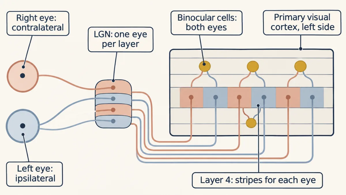
The one idea to carry through this lesson. While the cortex develops, the two eyes compete for the cells they share, and each cell keeps the eye that helps it fire, and keeps both when both do. So what counts is an eye’s activity compared with the other eye’s, and in step with the cell, not its use alone. The contest is decisive mainly while the window is open.
What the rule covers. It is a qualitative guide to how the balance between the eyes shifts in the binocular part of primary visual cortex, in cats, monkeys and mice, after closing one eye or both, misaligning the eyes, or silencing one eye with a toxin. “Helps it fire” is shorthand for a learning rule built in the section Out of step. The rule says which way the balance tips, not where it settles, and it concerns the balance between the eyes, not the health of their responses. On its own it does not explain what opens and closes the window, how each eye’s axons sort into stripes, how a weak eye recovers, or every part of the change in mice. Those need further mechanisms, noted where they arise.
Measuring ocular dominance
David Hubel and Torsten Wiesel measured each cell’s eye preference one cell at a time. In extracellular single-unit recording, a fine metal electrode sits next to one neuron and records its spikes from outside. With the animal anaesthetized, they showed a bar of light to one eye while the other was covered, then switched.
Each cell was placed on a scale of seven ocular dominance groups.
- Group 1 cells answer only to the contralateral eye.
- Group 7 cells answer only to the ipsilateral eye.
- Group 4 cells answer about equally to both.
- Groups 2 and 3 favor the contralateral eye, and groups 5 and 6 the ipsilateral eye, to a lesser or greater degree.
A worked example, in the left hemisphere: a cell driven only by the right eye is group 1, one driven about equally by both is group 4, and one driven only by the left eye is group 7. A cell that no eye can drive is left off the scale and counted as unresponsive. If the right eye is closed and loses the contest, cells pile up at the group 7 end.
Count the cells in each group and you get an ocular-dominance histogram, drawn with group 1 on the left and group 7 on the right. In a normal adult cat it is a hump: most cells sit in the middle groups, and the hump leans toward the contralateral side.
One closed eye
Read the transcript of “Six Days at Four Weeks”
Film transcript: Six Days at Four Weeks
If you sew one eye shut, what happens to the cortical cells it used to drive? In 1963, Wiesel and Hubel reported what happens when a kitten grows up with one eye closed. They sutured the lids of one eye at or near birth. They recorded after about three months of closure. Very few cortical cells could be driven by the closed eye. Yet its retina still responded. So did its LGN cells, though those serving the closed eye looked smaller. So the eye was not broken. What changed was in the brain. An adult cat with one eye closed for months showed no such change. In 1970 they asked when. They closed one eye in kittens of different ages, for different lengths of time.
Predict. That closure from birth lasted months, so it spanned many ages. Compare brief closures begun at different ages. Is there one switch-off point, or a rise and a fall? A rise and a fall. In kittens, susceptibility began around the start of the fourth week. It stayed high until somewhere between six and eight weeks. Then it declined through the third month. So a closure from birth lasting 3 months overlaps those weeks, which fits its large effect.
At the peak, the effect was fast. Some kittens had the right eye closed for only 6 days, in the fourth or fifth week. Recording in the left hemisphere, the closed right eye is the group 1 side. The number of cells it could drive fell to a fraction of normal. Most of the histogram moved toward group 7, the open eye. Six days of unequal vision had been enough. The anatomy follows. Hubel, Wiesel and Simon LeVay closed one eye in infant macaque monkeys. Later they traced the stripes in layer 4C. The open eye's stripes had widened, and the closed eye's had narrowed. In kittens, single thalamic axons serving a deprived eye were later seen to lose branches within about a week.
So a closure in this window changes the territory each eye's axons hold, not only how cells respond. Takeaway: in the young, a few days of unequal vision move the balance. In the adult, months do not. That leaves a question. Did the closed eye lose because it was unused, or because the other eye was not?
From the film
- In kittens, closing one eye for a few days in the fourth or fifth week moves most of the histogram toward the open eye.
- Susceptibility in kittens rises from about the fourth week and declines through the third month, while months of closure in an adult cat leave the cortex looking normal.
- The closed eye's retina still responds, so the loss is in the brain, not the eye.
- Closure also changes the territory each eye's axons hold, not only how cells respond.
In 1963 Wiesel and Hubel reported what happens when a kitten grows up with one eye closed. They sutured the lids of one eye at or near birth, so that eye got only diffuse light, and recorded after about three months of closure. (Closure in these experiments means lid suture.) Very few cortical cells could be driven by the closed eye, though its retina and its lateral geniculate nucleus (LGN) cells still responded. The LGN cells serving the closed eye did look smaller. An adult cat with one eye closed for months showed no such change.
Predict. That closure lasted months, so it spanned many ages. In 1970 they asked when closure matters, by closing one eye in kittens of different ages for different lengths of time. Compare brief closures begun at different ages and sketch how the size of the effect should change with age. Is there one switch-off point, or a rise and a fall?
Check your prediction
A rise and a fall. In this kitten series, susceptibility began around the start of the fourth week, stayed high until somewhere between six and eight weeks, and declined through the third month. A closure from birth lasting three months overlaps those weeks, which fits its large effect. At the peak the effect was fast. In kittens whose right eye was closed for only six days during the fourth or fifth week, the number of cells the closed eye could drive fell to a fraction of normal. Six days of unequal vision had moved most of the histogram toward the open eye.
The closed eye loses cortical cells mainly to the open eye's advantage and, in the lesson's data, little once the window has passed; closing both eyes removes that advantage. Bar heights are relative units, a sketch of the logic rather than measured counts.
The anatomy follows the physiology. Hubel, Wiesel and Simon LeVay closed one eye in infant macaque monkeys and later traced the stripes in layer 4C. The open eye’s stripes had widened and the closed eye’s had narrowed. In kittens, single thalamic axons serving a deprived eye were later seen to lose branches within about a week of closure. So a closure in this window changes the territory each eye’s axons hold, not only how cells respond.
Disuse or competition?
Read the transcript of “Two Closed Eyes”
Film transcript: Two Closed Eyes
The closed eye lost the cortex, though its retina still worked. Was that disuse, or competition? Disuse says connections that carry little signal wither. On this reading, an eye's fate depends on its own use. Competition says the two eyes compete for influence over each shared cell. One eye's advantage costs the other its share. On this reading, an eye's fate depends on its use compared with the other eye's. For one closed eye, both ideas predict the same loss. So that experiment cannot tell them apart. They part company when both eyes are closed. Some toy bookkeeping makes this concrete. The numbers are illustrative, not measured. Give an open eye 10 units of useful activity, and a sutured eye 1 unit.
With both eyes open, disuse says each eye keeps all of its own connections. Competition gives each eye half the influence. Close one eye. Under disuse, it keeps only what its own use supports, a tenth. Under competition, its share shrinks to a sliver. Now close both. Under disuse, each eye is as unused as the single closed eye was. Under competition, neither eye has an advantage. Predict. Close both eyes of young kittens for the same weeks. What does each idea expect the histogram to show?
Disuse expects weak responses to both eyes, and perhaps many silent cells. Competition expects the balance to hold. Wiesel and Hubel did this experiment in 1965. They scored many cells on the seven-group scale. They also counted cells that responded abnormally, or not at all. Many cells still responded to each eye. The balance moved far less than after one-eye closure.
A share of cells did respond abnormally or not at all. So going without vision is not harmless. But the large loss that disuse predicts was absent. What cost the single closed eye its cells was mainly the open eye's advantage. That is the first part of the rule. The two eyes compete for the cells they share. One warning about the table. It is only bookkeeping. Models of competition add feedback. An eye that gets ahead is strengthened further. So inside the window, a strong imbalance can move most cells, not a fixed fraction of them. The rule gives the direction of change. It does not say where the balance finally settles. Takeaway: closing both eyes shifts the balance far less.
So what hurt one closed eye was mainly the open eye's advantage. But is the amount of activity all that counts? The next chapter keeps the amounts equal.
From the film
- Closing both eyes shifts the balance between the eyes far less than closing one.
- What cost the single closed eye its cells was mainly the open eye's advantage, so the eyes compete for the cells they share.
- Going without vision is not harmless, since some cells respond abnormally or not at all.
There are two ways to read the closed eye’s loss.
- Disuse. Connections that carry little patterned signal wither. On this reading, an eye’s fate depends on its own use.
- Competition. The two eyes compete for influence over each shared cell, so one eye’s advantage costs the other its share. On this reading, an eye’s fate depends on its use compared with the other eye’s.
For one closed eye, both readings predict the same loss. They part company when both eyes are closed.
Predict. Take kittens at the age when one-eye closure moves nearly every cell to the open eye. Close both eyes for the same weeks. Under disuse, what should the histogram look like? Under competition?
Check your prediction
Some toy bookkeeping makes the two predictions concrete. Give an open eye 10 units of useful activity and a sutured eye 1 unit. The numbers are illustrative, not measured.
| Rearing | Eye A | Eye B | Disuse: eye B keeps this much of its own normal connections | Competition: eye B’s share of influence, B ÷ (A + B) |
|---|---|---|---|---|
| Normal | 10 | 10 | 100% | 50% |
| One eye closed | 10 | 1 | 10% | 1 ÷ 11, about 9% |
| Both eyes closed | 1 | 1 | 10% | 50% |
The two columns measure different things: disuse tracks each eye’s own strength, and competition tracks the split between the eyes. Under disuse, closing both eyes costs each eye as much as closing one did, so responses to both eyes should be weak, and many cells might not respond at all. Under competition, neither eye has an advantage, so the balance between them should hold.
Wiesel and Hubel did this experiment in 1965. They scored many cells on the seven-group scale and also counted those that responded abnormally (for example weakly, or without a clear preferred orientation) or not at all. Many cells still responded to each eye, and the balance between the eyes moved far less than after one-eye closure. Some cells were abnormal or unresponsive, so going without vision is not harmless. But the large loss that disuse predicts was absent. What cost the single closed eye its cells was mainly the open eye’s advantage.
The table is only bookkeeping. Models of competition add feedback, in which an eye that gets ahead is strengthened further, so inside the window a strong imbalance can move most cells to the open eye, not a fixed share of them.
Out of step
Read the transcript of “Out of Step”
Film transcript: Out of Step
If two eyes send equal amounts of activity, should they always share a cell? The bookkeeping counts only amount. Strabismus tests whether amount is enough. In strabismus the eyes are misaligned. Each points at a different part of the scene. Hubel and Wiesel produced it in young kittens by cutting one eye muscle. Each eye still received a normal amount of patterned light. Predict. Each eye sends about as much activity as in a normal kitten. If amount were all that mattered, what should the histogram look like?
If amount alone set each cell's split, the histogram would look like a normal one. It did not. Most cells answered to one eye or the other, and few to both. The histogram had piles at groups 1 and 7, and a hollow middle.
Neither eye took over. The cells the two eyes had shared went to one eye or the other. Equal amounts were not enough. What changed was not the amount, but the timing between the eyes. When the eyes are aligned, they look at the same point. An edge crossing that point excites both eyes' inputs to a shared cell at nearly the same moments. Their signals are in step, or correlated. When the eyes are misaligned, each eye's input arrives while the other is doing something unrelated. A rule that explains this starts with Donald Hebb, in 1949. Hebb proposed that an input that repeatedly takes part in firing a cell is strengthened. Later models added a weakening half.
In the BCM model, an active input weakens when the cell responds, but only weakly. An active input strengthens when the cell responds strongly. A silent input changes little. The line between strong and weak slides with the cell's recent activity. Two eyes in step arrive together. The cell responds strongly when both are active, so both are kept. Out of step, each eye's input often arrives alone. Suppose one eye starts slightly stronger at this cell. Alone, it drives a strong response and is strengthened. Alone, the other drives only a weak response, and is weakened. The lead grows until the cell answers mainly to one eye. Which eye leads differs from cell to cell.
One molecular candidate for detecting together is the NMDA receptor. It passes much current only with glutamate and a co-agonist bound and the cell depolarized enough to clear magnesium from its pore. Now we have both halves of the rule. The cell keeps the eye that helps it fire. Takeaway: amount matters, but only activity in step with the cell's firing helps an eye keep it. Next, a sharper test: an eye that is silent, against an eye that is noisy.
From the film
- In strabismic kittens each eye sends a normal amount of activity, yet most cells answer to one eye or the other and few to both.
- Amount matters, but only activity in step with the cell's strong responses helps an eye keep it.
- The cell keeps the eye, or both eyes, that help it fire, and which eye leads differs from cell to cell.
The bookkeeping counts only amount. Strabismus tests whether amount is enough. In strabismus the eyes are misaligned, so each points at a different part of the scene. Hubel and Wiesel produced it in young kittens by cutting one eye muscle. Each eye still received a normal amount of patterned light.
Predict. If amount were all that mattered, what should the histogram of a strabismic kitten look like?
Check your prediction
Like a normal one, since the amounts have not changed. It did not. Most cells answered to one eye or the other, and few to both: the histogram had piles at groups 1 and 7 and a hollow middle. Neither eye took over, so equal amounts are not enough to keep a cell binocular.
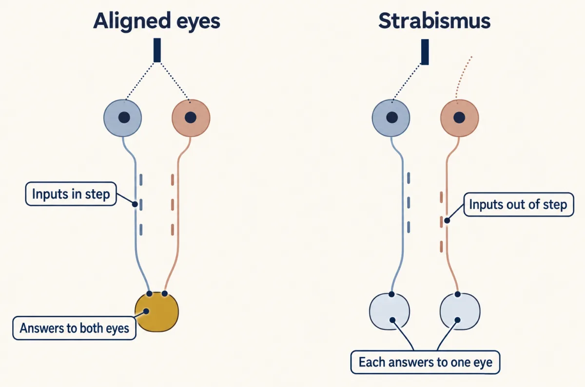
What changed was not the amount but the timing between the eyes. When the eyes are aligned they look at the same point, so an edge crossing it excites both eyes’ inputs to a shared cell at nearly the same moments. Their signals are in step, or correlated. When the eyes are misaligned, each eye’s input arrives while the other is doing something unrelated.
A rule that explains both results starts with Donald Hebb, who proposed in 1949 that an input that repeatedly takes part in firing a cell is strengthened. Later models added a weakening half. The version used here comes from the BCM model (Bienenstock, Cooper and Munro, 1982).
- An input that is active while the cell responds strongly is strengthened.
- An input that is active while the cell responds, but only weakly, is weakened.
- An input that is silent changes little in this model.
- The line between strong and weak slides with the cell’s recent activity.
That is what “helps it fire” means. Walk one shared cell through both cases.
- In step, the two eyes’ inputs arrive together. The cell’s strong responses come when both are active, so both inputs are kept.
- Out of step, each eye’s input often arrives alone. Suppose one eye’s synapses on this cell start slightly stronger. Alone, that eye drives a strong response and is strengthened. Alone, the other drives only a weak response and is weakened. The lead grows until the cell answers mainly to one eye. Which eye starts ahead differs from cell to cell, so neither eye takes over the cortex.
This walk-through holds in models with such a threshold, and it describes average activity, not a rule applied spike by spike. Amount matters because an eye that sends more activity can drive more strong responses, but only activity that arrives with the cell’s strong responses helps. One molecular candidate for detecting “together” is the N-methyl-D-aspartate (NMDA) receptor, which passes substantial current only when glutamate and a co-agonist (glycine or D-serine) are bound and the cell is depolarized enough to clear magnesium from its pore (see that lesson).
Silence is not the same as noise
Read the transcript of “Silence Is Not Noise”
Film transcript: Silence Is Not Noise
Which loses more of the cortex: an eye that sends nothing, or an eye that sends noise? Lid suture blurs an eye's image. But its retina keeps firing under the lid. In 1986, Michael Stryker's laboratory asked whether electrical activity itself is what counts. They silenced one eye of kittens with tetrodotoxin, a toxin that blocks sodium channels. It silences the retinal ganglion cells, the eye's output neurons. Cortical responses shifted toward the eye that kept firing. So an imbalance of electrical activity between the eyes is enough to cause a shift. In the same year, Stryker and William Harris silenced both eyes during development. Then the two eyes' axons failed to separate into stripes.
Now there are two ways to take an eye out of the contest. A sutured eye, whose retina still fires. And a silenced eye, which sends nothing at all. Predict. If the more active eye simply wins, which closed eye should lose more? What does our rule predict?
By amount, the silenced eye is quieter, so it should lose more. It loses less. In kittens, lid suture weakens the closed eye's responses more than tetrodotoxin does. Why? The sutured eye's retina is still active under the lid. That leftover activity is unpatterned. It is out of step with the cells it drives. And it seems to weaken the eye's own synapses. A silent eye is never active at the wrong moments. So it loses less. So the rule that the more active eye wins fails this test. So does simple disuse. The rule in the box can account for the difference. The sutured eye's input is the kind its weakening half acts on; a silent input is left nearly alone.
The open eye drives the cell's strong responses. On its own, the sutured eye evokes only weak ones. Takeaway: silence is not the same as noise. An eye whose activity is out of step with the cell loses more than an eye that is simply silent. One question remains. Why does all of this work mainly in the young?
From the film
- Silencing one eye of kittens with tetrodotoxin shifts cortical responses toward the eye that kept firing, so an imbalance of activity is enough to cause a shift.
- In kittens, lid suture weakens the closed eye's responses more than tetrodotoxin does.
- A sutured eye's leftover activity is out of step with the cells it drives, while a silent eye is not active at the wrong moments, so silence is not the same as noise.
Lid suture blurs an eye’s image but leaves its retina firing. To ask whether electrical activity itself is what counts, Michael Stryker’s laboratory silenced one eye of kittens with tetrodotoxin, a toxin that blocks sodium channels and silences the retinal ganglion cells, the eye’s output neurons (Chapman and colleagues, 1986). Cortical responses shifted toward the eye that kept firing. So an imbalance of electrical activity between the eyes is enough to cause a shift. In the same year, Stryker and William Harris reported that silencing both eyes during development kept the two eyes’ axons from separating into stripes.
That makes “activity” literal. It also allows a sharper test, because there are now two ways to take an eye out of the contest: a sutured eye, whose retina still fires, and a silenced eye, which sends nothing.
Predict. If the rule were simply “the more active eye wins”, which closed eye should lose more of the cortex, the sutured one or the silenced one? What does the rule in the box predict?
Check your prediction
By amount, the silenced eye is the quieter one, so it should lose more. It loses less. In kittens, lid suture weakens the closed eye’s responses more than tetrodotoxin does (Rittenhouse and colleagues, 1999). The sutured eye’s leftover, unpatterned activity is out of step with the cells it drives, and it seems to weaken its own synapses. A silent eye is not active at the wrong moments.
“The more active eye wins” fails this test, as do the table’s amount-only competition and plain disuse, which predicts that the eye used least should lose most; here it lost less. The rule in the box can account for the difference. The open eye drives the cell’s strong responses, so the sutured eye’s input, on its own, evokes only weak ones and is weakened, while a silent input changes little in this model. Real cortex also weakens a silent input somewhat when a competitor is active, which the rule does not capture: the silenced eye does lose ground, only less than the sutured eye.
The window and the patch
Read the transcript of “The Window and the Patch”
Film transcript: The Window and the Patch
Why does a week of closure rewire a kitten's cortex, while months do nothing visible in an adult? Every result so far depends on age. Two terms help. A sensitive period is a window in which experience has an unusually strong effect. A critical period, in Eric Knudsen's definition, is a sensitive period with two extra features. The experience is essential for normal development. And its effects last. Real windows often fall between the two. How sharp one looks depends on the species, the manipulation and the measure. An adult cat's histogram may not move after a closure. That does not mean the adult cortex cannot change at all. Name the species with every number, because the window differs.
In kittens, susceptibility begins around the start of the fourth week. It stays high to about six to eight weeks, then declines through the third month. In macaques, early closure has its strongest effect in the first weeks after birth. In mice, Joshua Gordon and Michael Stryker found the strongest shifts near postnatal day 28. The same few days of closure in adult mice did little. Mice have no ocular dominance columns. Their value is genetics, used to ask what opens and closes the window. Now the clinic. Amblyopia is reduced vision, usually in one eye, that glasses do not immediately restore. No structural disease of the eye explains it. It usually arises when the two eyes give the young brain unequal input.
Common causes are strabismus, a large difference in focus between the eyes, and anything that blocks one eye's image. Strabismus affects a few percent of children. Predict. A young child's left eye sees poorly after early strabismus. Glasses correct the optics. Which eye do you patch?
The good eye. A patch covers it for part of the day, or atropine drops blur it. Then the weak eye carries the useful visual input. For those hours the weak eye is the one helping the shared cells fire, so the contest tilts its way. That can work because the weak eye keeps pathways that can still be strengthened, even if some connections were lost. Treatment generally works best in young children, consistent with a sensitive period. Trials have found some older children still improve. A cataract that blocks one eye from birth is usually removed early in infancy, for the same reason. Takeaway: the cell keeps the eye that helps it fire. That contest counts most while the window is open.
What opens and closes that window is the next lesson's question.
From the film
- A sensitive period is a window of unusually strong effect of experience, and a critical period is one in which the experience is essential and its effects last.
- The window differs between species, so name the species with every number.
- Amblyopia usually arises when the two eyes give the young brain unequal input, and patching the good eye tilts the contest toward the weak eye.
- Treatment generally works best in young children, though trials have found that some older children still improve.
Every result so far depends on age. A sensitive period is a window in which experience has an unusually strong effect. A critical period, in Eric Knudsen’s definition, is a sensitive period in which the experience is essential for normal development and its effects last. Real windows often fall between the two, and how sharp one looks depends on the species, the manipulation and the measure. This lesson follows common usage in calling the ocular dominance window a critical period. A closure that does nothing detectable to an adult cat’s ocular dominance histogram does not mean the adult cortex cannot change in any way.
The window differs between species.
- Kittens: the rise and fall traced above, from the fourth week through the third month.
- Macaques: the effect of early closure is strongest in the first weeks after birth.
- Mice: in 1996 Joshua Gordon and Michael Stryker found that a few days of closure shifted responses most strongly near postnatal day 28, with little effect from the same closure in adults.
Mice have no ocular dominance columns. Their binocular cortex is a small zone dominated by the contralateral eye, so plasticity is read from single cells, summarized as a contralateral bias index (1 when every cell answers only to the contralateral eye, 0 when every cell answers only to the ipsilateral eye), or from evoked potentials and imaging. The mouse’s value is its genetics, which later work used to ask what opens and closes the window (that lesson).
Amblyopia and patching
The clinical version of this story is amblyopia: reduced vision, usually in one eye, that glasses do not immediately restore, with no structural disease of the eye to explain it. It usually arises when the two eyes give the young brain unequal input, though poor input to both eyes, such as a large focusing error in both, can cause it too. Common causes are strabismus, a large difference in focus between the eyes, and anything that blocks one eye’s image, such as a congenital cataract or a drooping lid. Strabismus affects a few percent of children. It causes amblyopia mainly when one eye is habitually used and the other suppressed; the strabismic kittens above kept roughly balanced eyes.
Predict. A young child’s left eye sees poorly after early strabismus, though the eye itself is healthy. Glasses correct the optics. Which eye would you cover with a patch, and what does the rule say the patch is doing?
Check your prediction
Cover the good eye, with a patch for part of the day or by blurring its focus with atropine drops, so the weak eye carries the useful visual input. For those hours the weak eye is the one helping the shared cells fire, so the rule says the patch tilts the contest toward it. Whether and how far the weak eye recovers depends on more than the rule: it needs pathways that can still be strengthened while the system is plastic, even if some of its connections were lost or remodeled. Treatment generally works best in young children, consistent with a sensitive period, although trials have found that some older children still improve.
A cataract that blocks one eye from birth is usually removed early in infancy for the same reason.
Putting it together
The three puzzles share one answer: the cell keeps the eye, or both eyes, that help it fire.
- One closed eye loses because the open eye helps the shared cells fire, while the closed eye’s leftover activity, which appears to be out of step with them, seems to weaken its own synapses.
- Two closed eyes lose much less, because neither has a systematic advantage. Going without vision still leaves a share of cells abnormal or unresponsive.
- Misaligned eyes lose their shared cells although the amounts are normal. Out of step, a small lead for one eye at a cell grows, and which eye leads differs from cell to cell.
What opens and closes the window in which the contest is decisive is the subject of the next lesson. Before the eyes open, structured activity of a different kind comes from the retina itself (that lesson).
Going deeper
The two halves of the rule
Hebb’s 1949 proposal was only the strengthening half, and later proposals supplied different weakening conditions. Stent (1973) proposed that an input weakens when it fails to help excite a cell that other inputs are firing. Covariance rules weaken an input when its activity and the cell’s depart from their averages in opposite directions. The BCM model, used in this lesson, calls the sliding line between strong and weak responses the modification threshold.
A simple Hebbian model shows the limit stated under What the rule covers. If the total strength on each cell is held fixed by subtracting the same amount from both inputs, and the two eyes’ inputs are only partly correlated, the weight difference between the eyes grows and cells drift toward one eye. With multiplicative normalization the binocular state is stable instead, so the outcome depends on the constraint as well as the rule. Either way, the rule gives the direction of change, not where the balance settles.
BDNF and TrkB
Activity also raises the cortex’s production of brain-derived neurotrophic factor (BDNF), a neurotrophin that acts through the receptor kinase TrkB (that lesson), and it was proposed as one of the signals that rewards the winning input. In mice, though, blocking TrkB did not prevent the shift; it impaired recovery after the closed eye was reopened.
Two phases in the mouse
In mice, four days of closure near postnatal day 28 lowers the contralateral bias index in two phases: depression of the deprived eye’s responses, then potentiation of the open eye’s. Silencing the deprived eye with tetrodotoxin prevents the early loss of its responses. The later rise of the open eye’s responses is thought to involve, at least in part, homeostatic mechanisms that raise a cell’s responsiveness after it loses input, so “helps it fire” does not fully describe it.
When do monkey columns separate?
Their 1980 study proposed that in newborn monkeys the two eyes’ territories in layer 4C still overlap and separate over the first postnatal weeks, so a closure tilts the separation toward the open eye. Later tracing in newborn monkeys with no visual experience found the stripes already well separated at birth, so an early closure may instead move borders that already exist.
Tracing eye-specific columns
Inject a radioactive amino acid such as tritiated proline into one eye. It is built into proteins, carried to the lateral geniculate nucleus (LGN), partly passed across to the LGN cells, and carried on to their endings in layer 4. Expose film to slices of cortex and the injected eye’s endings appear as stripes, alternating with unlabeled stripes that belong to the other eye.
Other definitions, and an older wording of the rule
Some accounts define a critical period as a time when a developing system is especially susceptible to, and requires, specific experience to develop normally. Some call such windows “windows of opportunity and vulnerability”, and draw a different distinction from Knudsen’s: critical periods begin and end abruptly, while sensitive periods rise and fall gradually.
An older statement of the competition hypothesis is that differences in the amount of activity determine the strength of the projection (the set of axons one eye sends to the cortex). The strabismus and tetrodotoxin results above show why amount alone is incomplete.
Some teaching versions collapse the seven ocular dominance groups into fewer bins. This lesson uses the original seven.
Your notes
Select any text in the lesson to highlight it or add a note to it. Your highlights and notes are saved in this browser and collected in your notebook.
This browser is not letting the site save anything (a private window or strict privacy settings), so highlights and notes will be lost when you leave the page.
How we measure it
Extracellular single-unit recording
A fine metal electrode is placed next to one neuron in an anaesthetized animal and records its spikes from outside the cell. Stimuli are shown to each eye in turn, with the other eye covered, so the strength of each eye's drive on that one cell can be compared.
Monocular deprivation by lid suture
The eyelids of one eye are sewn shut for a chosen period at a chosen age, so that eye gets only diffuse light while the other sees normally. Reopening the eye and recording from the cortex shows how the balance between the eyes has changed.
Transneuronal autoradiography
A radioactive amino acid such as tritiated proline is injected into one eye. It is built into proteins, carried to the thalamus, partly passed to the thalamic neurons, and carried on to their endings in layer 4 of the cortex, where film exposed to brain sections shows that eye's stripes.
Ocular dominance in the mouse
Mouse visual cortex has a small binocular zone and no ocular dominance columns. Plasticity is read from single units scored for eye preference and summarized as a contralateral bias index, or from visually evoked potentials and imaging, before and after closing one eye.
Practice questions on this lesson (3) → MCAT-style, original, each with an explanation
Where this lesson fits
Before thisReview first
- Topographic Maps: Sperry's Eye and Eph GradientsThis course · Lesson 26
You need the eye-specific maps and activity-dependent sorting of retinal axons from there to follow how the two eyes' inputs are arranged.
- Synapse Elimination: One Muscle Fiber, One AxonThis course · Lesson 37
It teaches competition between inputs for one target, which is the core logic of the eyes competing for cortical cells.
After thisStudy next
- Opening and Closing a Critical PeriodThis course · Lesson 40
It explains what opens and closes the window, which this lesson leaves open.
- Microglia and Complement Prune SynapsesThis course · Lesson 41
It shows how synapses of the losing input are pruned during experience-dependent refinement.
- Sensitive Periods in Behavior: Song, Language and SightThis course · Lesson 42
It takes the sensitive-period idea from the eye to song, language and sight.
Elsewhere on the siteFilms, simulations and method films in other courses
- A Synapse That Could LearnFilm · Neuroscience
It shows Hebb's rule, that a synapse strengthens when its input and the target cell fire together, which this lesson's competition rests on.
- Adding Up the LightFilm · Neuroscience
It shows how a cortical cell sums input from its patch of the visual world, as in the cells tested for eye preference.
Suggested by an AI model from the lessons themselves, then checked. How we use AI
Finish line
Test yourself
8 questions
Practice and MCAT-style, with explained answers.
Next · Lesson 40
Opening and Closing a Critical Period
A mouse that lacks one enzyme for making GABA keeps its critical period waiting for months. One drug starts it.
Continue →← Lesson 38 · Activity Before Experience: Spontaneous Retinal Waves