Biology · Unit 3: Bacteria and viruses
How Microbes Multiply
Rewritten on : new text, flashcards and film. Your notes and highlights stay, but the text around them may have moved.
One bacterium can become millions of cells in an afternoon, yet a virus never divides at all. How, then, can a cold sore heal and come back in about the same spot?
Pause when the reading reaches a film, and offer to play it. Saved on this device.
Watch the film now, or keep listening
Builds on:Living or Not? Viruses and BacteriaThe Cell Cycle and MitosisWhy these?
Where this lesson sits
- Virginia SOL BIO.4b
- Practices BIO.1c, BIO.1d, BIO.1e, BIO.1f
Essential question: How do bacteria and viruses make more of themselves, and why do some infections come back years later?
-
Life Science (grade 7) LS.10 b
Review it at grade 7 level
In Life Science you learned that some living things reproduce asexually. One parent makes offspring, and the offspring are genetically identical to the parent. Other living things reproduce sexually, usually with two parents, and the offspring are different from each parent. You may have seen a picture of one cell becoming two. Biology adds the names and steps: bacteria split by binary fission or grow buds, can trade DNA by conjugation, and viruses are copied only inside a host cell. Phages, the viruses of bacteria, can follow the lytic cycle, and some can also follow the lysogenic cycle.
-
Biology BIO.4b You are here
This lesson
-
Biology II Biology II: Advanced Survey of Biology Topics, BIOII.2 d; Genetics, GEN.4 d
Preview where it goes next
Virginia’s Advanced Survey of Biology course compares ways of reproducing asexually across life: binary fission in prokaryotes, budding, fragmentation, vegetative propagation in plants and spores. The Genetics course asks how bacteria vary if their offspring are clones. It names three ways bacteria gain genes from outside: conjugation (DNA passed from cell to cell), transformation (DNA taken up from the surroundings) and transduction (DNA carried by a virus). It also explains that bacteria usually have one circular chromosome and may carry plasmids, small extra circles of DNA that often move during conjugation. Mutation adds variation too, even in a clone.
-
College
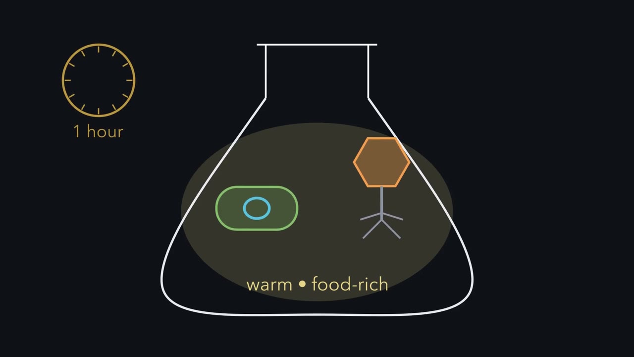 ▶ Watch Copy Yourself, or Be Copied · 3:05
Read the transcript of “Copy Yourself, or Be Copied”
Film transcript: Copy Yourself, or Be Copied
One bacterium and one virus land in a warm flask full of food. An hour later, which has made more of itself? And how did it do it? Start with the bacterium. It is a single cell, with its DNA in one loop called the chromosome. There is no nucleus around it. First the cell copies its chromosome. Then it grows longer, with one copy at each end. Then it pinches in the middle and splits in two. This is binary fission. Each new cell gets a complete copy of the chromosome. So the two cells match, except for rare copying mistakes. Now each new cell does the same thing. One becomes two. Two become four, then eight, then sixteen. Every round doubles the count.
The time for one round is called the doubling time. In the best lab conditions, an E. coli cell can double about every 20 minutes. Keep that pace for seven hours, with no cells dying. Will one cell become closer to a thousand cells, or closer to a million?
More than a million. The counter reaches 21 doublings, about two million cells. That is a calculated number, not a count of real cells. But a real flask is closed. As the cells multiply, they use up the food. Their wastes build up in the liquid around them. So the climb slows, and the count levels off. Doubling makes numbers huge, but only while food lasts and wastes stay low. Now the virus. This one is a phage, a virus that infects bacteria. Left alone in all that food, what does it do? Nothing. It does not grow. It does not split. It cannot make anything on its own. Its counter stays at one. Then it lands on a bacterium and injects its DNA.
Soon, orange phage parts start to appear inside the cell. The phage is not making them. The cell is. The cell reads the phage genes as if they were its own, and builds phage parts. So the two multiply in opposite ways. The bacterium grows and splits on its own. The virus never divides. A host cell builds every new virus. Here is the one rule to keep from this film. A bacterium copies itself. A virus is copied by a host cell, never by itself.
From the film
- Most bacteria reproduce by binary fission: a cell copies its DNA, grows and splits into two.
- Each doubling multiplies the count by 2: at the best-case pace, 21 doublings turn one cell into about two million, a calculated number.
- In a closed flask, food runs low and wastes build up, so growth levels off.
- A virus cannot split, grow or make anything on its own; a host cell builds every new virus.
Under the best laboratory conditions, one E. coli cell can split into two about every 20 minutes. A cold sore can heal, then come back later in about the same spot. The herpes virus behind cold sores cannot copy itself, yet it can stay in your body for years. How can something that never divides keep coming back, and what stops a bacterium from doubling forever?
The one idea. A bacterium copies itself; a virus is copied by a host cell. A bacterium grows and splits in two, so with food its numbers double every generation, until food runs low or wastes build up. A virus never divides: a host cell, the living cell a virus infects, does the copying. In the lytic cycle it builds new viruses right away. In the lysogenic cycle it copies only the viral DNA, along with its own, each time it divides; new viruses are built only if that DNA switches to the lytic cycle, which may never happen.
Scope: bacteria and the viruses that infect them, with phage lambda as the model for the two paths. Some other phages have different cycles; a few release new phages without bursting the cell. Budding still makes a new cell; conjugation adds genes, not cells. Herpes is a comparison, not the same mechanism.
What do you think? Decide before you read.
-
Scientists disagree: There is no mother, father or egg: a bacterium copies its DNA and splits into two cells (binary fission). A few kinds of bacteria grow a bud instead, which researchers have filmed with time-lapse microscopes.
What scientists think
Scientists disagree: There is no mother, father or egg: a bacterium copies its DNA and splits into two cells (binary fission). A few kinds of bacteria grow a bud instead, which researchers have filmed with time-lapse microscopes.
-
Scientists disagree: A virus cannot make copies of itself without a host cell. The cell’s own machinery builds viral parts, and new viruses are put together from those parts. The second film shows the experiment behind this.
What scientists think
Scientists disagree: A virus cannot make copies of itself without a host cell. The cell’s own machinery builds viral parts, and new viruses are put together from those parts. The second film shows the experiment behind this.
-
Scientists disagree: In a closed flask, scientists measure the culture growing cloudier, then holding steady. The 20-minute doubling time is a best-case lab number for one kind of bacterium, not a rule that lasts.
What scientists think
Scientists disagree: In a closed flask, scientists measure the culture growing cloudier, then holding steady. The 20-minute doubling time is a best-case lab number for one kind of bacterium, not a rule that lasts.
Explore: grow a population and follow a virus
In the simulation’s first panel, you pick a doubling time and how much food is in the dish. Its second panel, a phage infection, returns in the virus section.
Panel 1. Double a bacterial population
- Doublings finished
- 6
- Cells with endless food
- 64
- Cells with the food ceiling
- 64
In this sketch: one starting cell, no waiting before growth starts, no cells dying, and growth runs at full speed until it stops at a ceiling set by food alone (a real culture slows first, and wastes build up). The ceiling (4,096 cells at full food) and doubling times other than 20 minutes are made-up sketch values, not measurements.
Panel 2. Follow a phage
- Cells carrying the prophage
- –
- Host divisions
- –
- Cells that burst
- –
In this sketch: you pick the path, the only cell that dies is one that bursts, and the picture shows at most 8 cells.
In this sketch, a population doubles once each doubling time until food runs low and wastes build up, and a prophage is copied into both daughter cells each time its host divides.
Try this.
- Predict how many cells one cell becomes after 2 hours with a 20-minute doubling time. Then run it.
- Keep the doubling time the same and halve the food. Predict what changes about the curve, then test.
Explain: how bacteria make more bacteria
- asexual reproduction
- binary fission
- budding
- conjugation
- plasmid
- bacteriophage
- host cell
- lytic cycle
- lysogenic cycle
- prophage
- doubling time
Asexual reproduction is reproduction with one parent, where the offspring get copies of that parent’s DNA. Most bacteria reproduce this way, mostly by binary fission, the copy, grow and split of the first film. It looks like mitosis from the cell cycle lesson, but it is simpler: a bacterium has no nucleus, and each new cell just gets a complete copy of the chromosome, the cell’s main DNA.
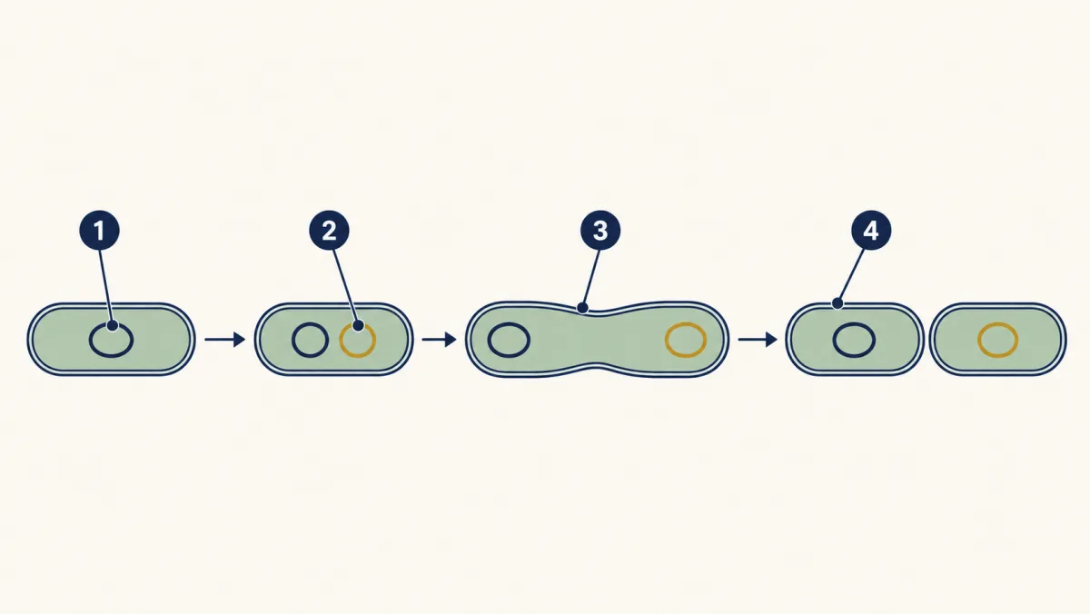
- chromosome
- copy of the chromosome
- grows longer
- daughter cells
- chromosome
- blank
- blank
- daughter cells
Word bank: copy of the chromosome, grows longer
- blank
- blank
- blank
- blank
Check your labels
- chromosome
- copy of the chromosome
- grows longer
- daughter cells
Some bacteria use budding: a small new cell grows out of the parent and then breaks off, as in the ocean bacterium below.
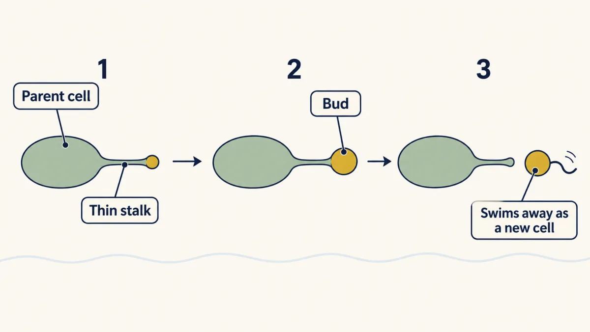
The daughter cells are usually genetically identical. Copying DNA sometimes makes mistakes: sequencing studies of lab E. coli, which read the cells’ DNA, found mutations (changes in the DNA) building up over many generations.
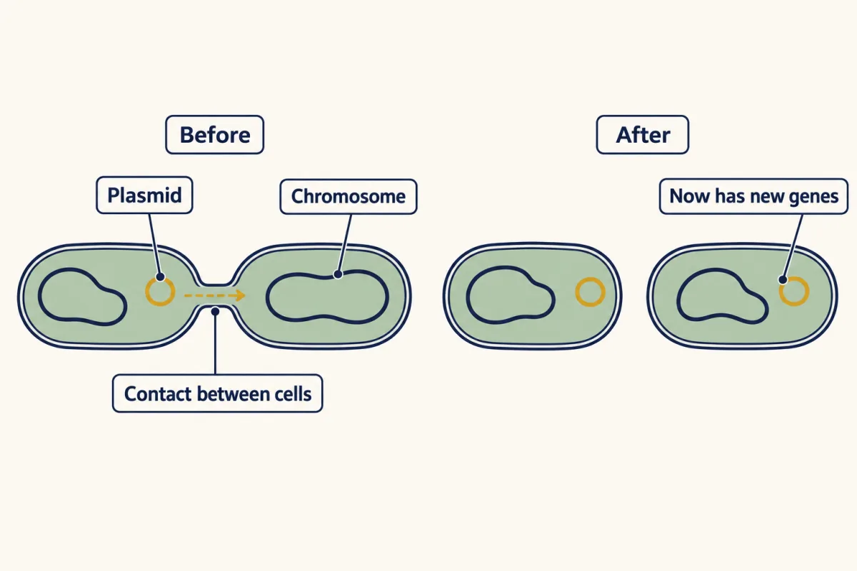
Bacteria have one more way to change their genes. In conjugation, one bacterial cell passes DNA directly to another through contact. The DNA is often a plasmid, a small extra circle of DNA separate from the chromosome. Virginia’s science standards (the SOL) call it a form of sexual reproduction, because genes from two cells combine. Many biologists call it gene transfer, because no new cell is made.
Explain: doubling, and why it stops
Doubling time is the time a population takes to double. If it stays the same, you can predict the count:
number of cells = starting number × 2 × 2 × 2 … (one × 2 for each doubling)
Worked example: a model population starts with 100 cells and doubles every 20 minutes. How many cells after 2 hours?
- 2 hours is 120 minutes, and 120 ÷ 20 = 6 doublings.
- 100 → 200 → 400 → 800 → 1,600 → 3,200 → 6,400 cells.
The first film’s two million is the same sum: one cell after 21 doublings (7 hours) gives 2,097,152. These are calculated numbers, not counts. The model assumes no cells die, no waiting before growth starts and endless food.
The flat start of the film’s curve is a lag, where cells are active but the count barely rises. If the culture is left long enough, the number of living cells can start to fall. Every population meets limits; which ones matter depends on the organism and where it lives, as the carrying capacity lesson shows.
Elaborate: a real growth curve
Scientists often follow a liquid culture by its cloudiness, called optical density at 600 nanometers (OD600): a machine shines light through the liquid, and more cells scatter more light. It is not a count of cells, and even liquid with no cells reads a little cloudy.
This E. coli culture was started by diluting cells 1,000-fold into fresh medium (liquid food) in one small well of a lab plate (sources in Going deeper). The first reading was 0.078. After that dilution the cells likely added very little to it, but the dataset has no cell-free well, so we cannot say exactly how much is background. The third column subtracts the first reading: it shows the change since 0.5 hours, a rough guide to growth, not a reading of the cells alone.
| Time (hours) | OD600 reading (no units) | Rise since the first reading |
|---|---|---|
| 0.5 | 0.078 | 0.000 |
| 2 | 0.079 | 0.001 |
| 4 | 0.080 | 0.002 |
| 6 | 0.084 | 0.006 |
| 8 | 0.134 | 0.056 |
| 10 | 0.198 | ___ |
| 12 | 0.313 | ___ |
| 14 | 0.322 | 0.244 |
| 16 | 0.318 | 0.240 |
| 18 | 0.315 | 0.237 |
Step 1. Fill in the two blanks. When does the rise barely change, and when does it stop?
Step 2. Doubling means the number is multiplied by 2, so compare by dividing, not subtracting. From 8 to 10 hours, did the rise roughly double? From 10 to 12 hours?
Predict. The best-case E. coli doubling time is 20 minutes. If these cells had kept that pace from 8 to 12 hours, how many doublings would that be, and what would the doubling arithmetic give for the rise at 12 hours?
Check your prediction
Four hours at 20 minutes is 12 doublings. On paper, 0.056 × 212 = 0.056 × 4,096, about 230. No machine would read that: long before then, cloudiness stops rising in step with cell number. The arithmetic shows only that this culture did not keep up a 20-minute pace from 8 to 12 hours. The real rise went 0.056, 0.120, 0.235, roughly doubling every 2 hours.
Predict. For the first 6 hours the rise hardly moved. Does that prove the cells were not dividing?
Check your prediction
No. Diluted 1,000-fold, the cells started far below what the machine can see. If they came from a culture about as cloudy as this one’s plateau, climbing back takes about 10 doublings (210 = 1,024), and the first several are too faint to read. A flat start can be a true lag or just too few cells to see.
Claim, evidence, reasoning
Make a claim that answers this question: can a bacterial population keep doubling forever? Use the growth table.
Start with: The data show that the population blank.
Start with: From 6 to 12 hours the reading rose from blank to blank, but from 14 to 18 hours it blank.
Start with: This happens because blank, so doubling stops when blank.
Compare with a model answer
A bacterial population cannot keep doubling forever. In this E. coli culture the reading barely moved for the first 6 hours, from 0.078 to 0.084, perhaps because the 1,000-fold diluted start was too faint to read. It then rose steeply to 0.313 by 12 hours. From 14 to 18 hours it stayed near 0.32, so growth had levelled off. In a closed well, food running low or wastes building up could explain this, but this curve alone cannot tell us which, or whether every cell stopped dividing. One limit: this is one well of one strain in one medium, and cloudiness is not a cell count.
Saved in this browser only.
Design problem
Baker’s yeast is a single-celled fungus, not a bacterium, but it also reproduces by budding and is safe for a classroom. Plan an investigation to test whether the amount of sugar changes how high a yeast population grows before it levels off.
Saved in this browser only.
One way to do it
- Independent variable: sugar added to the same nutrient broth in every tube, for example 0, 1, 2 and 4 grams per 100 mL. The broth must also supply nitrogen and minerals (for example a yeast nutrient from your teacher); in sugar water alone, yeast soon runs short of them and stops growing.
- Dependent variable: cloudiness every hour, using a turbidity probe or a printed card behind the tube, until the readings stop rising. Mix each tube gently first, because yeast settles and gas bubbles add cloudiness. To check that cloudiness tracks cell number, count cells in a drop under a microscope a few times.
- Constants: the same yeast packet and amount, the same water volume and temperature, the same tubes.
- Control: the tube with no sugar.
- Trials and sample size: three tubes per sugar level, then average them.
- Safety: use only yeast from the store, as your teacher directs. Never grow microbes from skin, soil or surfaces, because you cannot know what will grow.
If the high-sugar tubes level off higher, sugar was limiting how far the population grew. If all tubes level off at the same height, something else, such as another nutrient running out or wastes building up, may set the limit first.
Explain: how viruses make more viruses
The cells in that well used food to multiply until the reading stopped rising. A free phage, a virus that infects bacteria, takes in no food and does nothing on its own. Inside a bacterium, though, the host’s food, energy and machinery build new phages, and one infected cell can release many. The second film follows one phage into one cell.
Read the transcript of “Built Now, or Carried Along”
Film transcript: Built Now, or Carried Along
The first film ended with phage DNA inside a bacterium, and the cell building phage parts. So how does one phage, which never divides, become many? Scientists tested this. They infected bacteria with phages, then broke some cells open at different times. Each time, they looked for whole phages. Early in the infection, the cells already hold plenty of phage material. How many whole phages will they find inside?
None. There are phage parts, but no whole phages. So a phage is not split from a parent. It is built from parts. Later, the parts are put together into many new phages. Then the cell bursts open. The new phages escape, and the host cell dies. This path is the lytic cycle. The phage takes over now. The host builds new phages, and then the cell bursts to release them. Some phages have a second path. The best known is phage lambda, which infects E. coli. Its DNA does not have to take over right away. Lambda's orange DNA slips into the blue chromosome and becomes part of it. Stored viral DNA like this is called a prophage. The cell stays alive and keeps dividing by binary fission.
Each time, it copies its chromosome, prophage and all. Both new cells get a copy. Here is the counter from the first film. One cell, two, four, eight, sixteen, each one carrying the orange DNA. Now look at the second counter. New phages: zero. The viral DNA multiplies by riding along with the host's doubling. This path is the lysogenic cycle. The prophage can stay for many generations. But something such as damage to the cell's DNA can switch it into the lytic cycle. In that one cell, the host now builds new phages. The cell bursts and dies. The other cells carry on, prophage and all. So the two paths do not have to end the same way. The lytic path builds new phages now.
The lysogenic path copies only the phage DNA, maybe for many generations. New phages come only if it switches to lytic, which may never happen. Here is the rule to keep. A virus never builds itself. For lambda's two paths, a host cell builds new phages only on the lytic path.
From the film
- Cells broken open early in an infection hold viral parts but no whole viruses: viruses are built from parts, not divided.
- In the lytic cycle the host builds new phages right away, then bursts and dies.
- In the lysogenic cycle of phage lambda, the viral DNA joins the host chromosome as a prophage and is copied each time the host divides; no new phages are built.
- Something such as damage to the cell's DNA can switch a prophage into the lytic cycle; only then are new phages built.
What the second film left out: in a real infection, nobody picks the path. In phage lambda the outcome depends on the state of the host cell and on how many phages infect it at once. It is also partly a matter of chance. Why a virus needs a host at all is the subject of the living-or-not lesson.
In the lytic cycle, the build-now-then-burst path, the host’s own machinery does the copying. Some proteins made from phage genes help by steering the cell to copy phage DNA.
In the lysogenic cycle, the carried-along path, the stored viral DNA is called the prophage. Lambda’s prophage joins the chromosome, but some other phages keep theirs as a separate piece inside the cell. Either way, both daughter cells get a copy at each division. A bacterium carrying a prophage is a lysogen.
Try this. In the simulation’s second panel, take the lysogenic path and let the cells divide for three rounds. Then switch one cell to lytic and step it through. How many cells still carry the prophage?
Check your prediction
Seven: three rounds of division make 1 × 23 = 8 carriers, and after the switched cell bursts, 8 − 1 = 7 remain.
Predict. A culture of lysogens grows in a closed well until its cloudiness levels off. What happens to the number of cells carrying the prophage?
Check your prediction
Roughly, it levels off too. The prophage is copied only along with its host’s DNA, so while cells keep it and do not switch, it meets its host’s limits. New phages come only after a switch to lytic, for example after damage to a cell’s DNA.
| Binary fission (bacteria) | Lytic cycle (virus) | Lysogenic cycle (virus) | |
|---|---|---|---|
| Who does the copying | The bacterium itself | The host cell | The host cell, each time it divides |
| What is made | Two new cells | Many new virus particles | Host cells that carry viral DNA, no new viruses |
| Host cell afterward | No host needed for most bacteria | Dies, for a phage (the cell bursts) | Alive, unless it later switches to lytic |
Now the cold sore. Herpes simplex virus infects people, not bacteria, but it hides in a similar way. It can stay in sensory nerve cells, the cells that carry feeling from the skin. Studies of infected human and mouse tissue suggest its DNA stays as a separate circle in the nucleus. It does not join the chromosome the way lambda does. From time to time the virus can become active again and travel back to the skin. Sometimes a new sore appears, but sometimes it does not.
Evaluate: check your thinking
A bacterium copies itself; a virus is copied by a host cell. In lambda’s two paths, the host builds new phages only on the lytic path; on the lysogenic path it copies just the prophage DNA. Doubling cannot go on forever: in the well above, the rise roughly doubled every 2 hours, then levelled off. A cold sore comes back because herpes can hide in nerve cells and become active again, like, but not the same as, a lysogenic phage.
Science practices you used: predicting and testing with a model, calculating with doublings, reading a growth table, naming the limits of one data set and planning a fair test with a control and repeated trials.
Going deeper
Where the growth data come from. The table shows raw readings from one well, curve 15, of E. coli strain BW25113 grown in a defined lab medium, from the dataset of Honoka Aida and Bei-Wen Ying (deposited on figshare in 2025). The readings were taken every 30 minutes; the table shows ten of them.
How common is conjugation? The Virginia SOL describes conjugation as rare. How often it really happens depends on the kind of bacteria, the DNA they carry and their surroundings.
Your notes
Select any text in the lesson to highlight it or add a note to it. Your highlights and notes are saved in this browser and collected in your notebook.
This browser is not letting the site save anything (a private window or strict privacy settings), so highlights and notes will be lost when you leave the page.
How we measure it
Optical density (cloudiness) readings
A machine shines light of one color (600 nanometers) through a liquid culture and measures how much gets scattered by cells. Readings taken every 30 minutes trace a growth curve, but turning cloudiness into a number of cells needs a separate calibration.
Viable plate count
A diluted sample is spread on a dish of food gel, and each live cell, or small clump of cells, that can grow there makes a visible colony. Counting colonies estimates live cells, reported as colony-forming units.
Time-lapse fluorescence microscopy
A glowing tag marks one part of the chromosome, and a microscope takes pictures over hours. In a budding bacterium this showed when the copied chromosome moved into the bud.
Where this lesson fits
Before thisReview first
- Living or Not? Viruses and BacteriaThis course · Lesson 12
It explains why a virus needs a host cell, which this lesson builds on when viruses are copied by hosts.
- The Cell Cycle and MitosisThis course · Lesson 10
Binary fission is compared with mitosis, so you need to know how a eukaryotic cell copies and divides.
After thisStudy next
- Populations and Carrying CapacityThis course · Lesson 29
It takes the idea that growth levels off when food runs low and shows what limits real populations.
- Germs, Vaccines and AntibioticsThis course · Lesson 15
It uses fast bacterial doubling to explain how infections spread and how resistance can arise.
- Engineering GenesThis course · Lesson 22
It uses bacteria multiplying and plasmids passing between cells to make human insulin.
Elsewhere on the siteFilms, simulations and method films in other courses
- Bacterial and phage geneticsMethod film
This shows how conjugation and phage transfer of DNA are used to map genes in bacteria.
Suggested by an AI model from the lessons themselves, then checked. How we use AI
Finish line
Test yourself
8 questions
Practice and MCAT-style, with explained answers.
Next · Lesson 14
Microbes in Bodies and Ecosystems
Why do the roots of a healthy soybean plant carry small bumps full of bacteria, and what would the plant lose without them?
Continue →