Biochemistry · Cell Signaling
Signal-Transduction Pathways: How Cells Compute With Molecular Switches
Rewritten on : new text, figures and flashcards. Your notes and highlights stay, but the text around them may have moved.
One epinephrine molecule can mobilize millions of glucose units, and the same relay has to stop on time.
Pause when the reading reaches a film, and offer to play it. Saved on this device.
Watch the film now, or keep listening
Builds on:Enzyme Kinetics: The Michaelis–Menten ModelProtein Binding and Allostery: How Hemoglobin Loads and Delivers OxygenMembranes: The Impermeable Sheet That Builds ItselfWhy these?
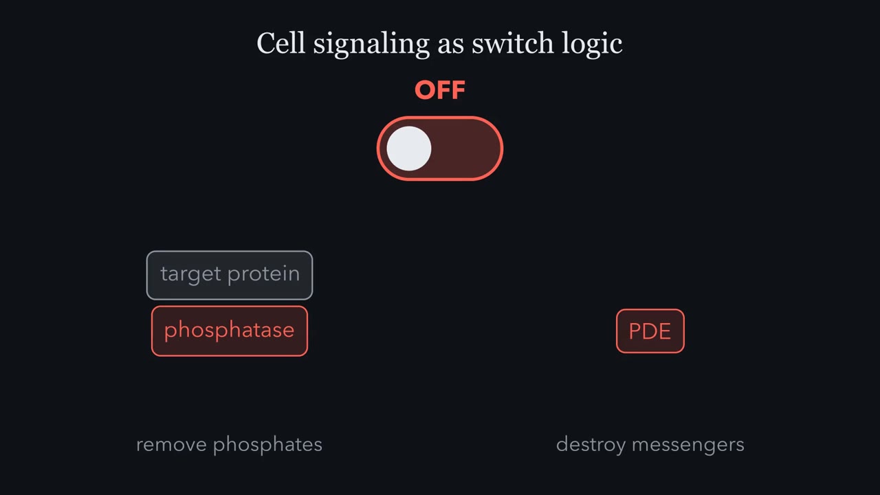 ▶ Watch Cells Compute With Switches · 2:42
Read the transcript of “Cells Compute With Switches”
Film transcript: Cells Compute With Switches
Think of cell signaling as switch logic. When a ligand binds its receptor, the switch flips on, and second messengers like cyclic AMP, IP3, and calcium fan out through the cell. But every switch that turns on must also turn off, through phosphatases that remove phosphates and enzymes that destroy the messengers. Hold on to that pairing. Almost every pathway in this chapter is the same four-beat sentence. A switch turns on, the signal spreads and amplifies, the switch turns off, and when the off step fails, the switch sticks. And that is disease. Most membrane receptor pathways share the same molecular logic. A primary messenger, a hormone or neurotransmitter, is detected by a membrane-spanning receptor.
The receptor generates or activates a second messenger inside the cell, which drives effector molecules, which produce the physiological response. Five parts in order, plus one that is easy to forget. Active termination. The pathway has to be switched off on purpose, or the signal never ends. Before the receptor mechanisms, ask what is doing the signaling in the first place. First messengers are the extracellular signals, hormones, neurotransmitters, growth factors, even light. The key split is physical. Small lipophilic messengers like steroids slip straight through the membrane to receptors inside the cell. Large or polar messengers like peptide hormones cannot cross, so they must be read by a receptor that spans the membrane and relays the news inward.
Size and polarity decide where the receptor lives. Glucagon and epinephrine are the classic first messengers for fuel mobilization. When blood glucose drops or the fight-or-flight response fires, these hormones launch a signaling cascade that hits fuel metabolism on several fronts at once. It breaks down glycogen, shuts off its synthesis, and epinephrine also frees fat. One hormone arriving at the surface reorganizes the cell's entire energy strategy because the signal is amplified into a large, coordinated response. When cyclic AMP rises inside a cell, it acts as a master switch for fuel metabolism through protein kinase A. PKA hits several targets at once, it shuts down glycogen synthesis by inhibiting glycogen synthase, and it turns on glycogen breakdown by activating phosphorylase kinase, which activates glycogen phosphorylase.
One second messenger, working through one kinase, flips the cell from storing fuel to releasing it, all by adding phosphates to the right enzymes.
One epinephrine molecule, arriving at the outside of a liver or muscle cell, can mobilize millions of glucose units from the glycogen inside (a teaching estimate; see Going deeper, “Where the glucose goes”).
The size is only half the puzzle. The relay that turns a few molecules into millions must also stop on time, or the cell keeps answering a signal that has gone. This lesson is about how one design does both.
The short film above covers the ideas up to protein kinase A (PKA); the text covers them all.
The parts: messengers, receptors, effectors
A phone computes with billions of transistors; a cell computes with thousands of signaling proteins wired together, sensing the outside, weighing competing inputs and committing to an action.
Strip a membrane-receptor pathway down and the same five components appear in order: a primary (first) messenger arrives from outside, binds a receptor, which generates or activates a second messenger inside the cell, which drives effector molecules, which produce the physiological response. A sixth feature is easy to forget: active termination; the pathway has to be switched off on purpose. In this lesson’s example, epinephrine is the first messenger, the beta-adrenergic receptor is the receptor, cAMP is the second messenger, protein kinase A (PKA) and the glycogen enzymes are the effectors, and glucose release is the response.
First and second messengers sit on opposite sides of the membrane. A first messenger is the extracellular signal: epinephrine, insulin, a growth factor. A second messenger is made or released after the receptor fires and relays the order inside the cell: cAMP, inositol trisphosphate (IP3), diacylglycerol (DAG), calcium, phosphatidylinositol 3,4,5-trisphosphate (PIP3). The split exists because of a physical constraint. Small, lipophilic (fat-soluble) signals find receptors inside the cell (steroids slip straight through the membrane; thyroid hormone enters through transporters such as MCT8). Large or polar signals, such as peptide hormones and growth factors, cannot cross, so they must be read by a receptor that spans the membrane.
One switch first: the G-protein cycle
Epinephrine’s receptor here is the beta-adrenergic receptor, a GPCR (G-protein-coupled receptor): a seven-transmembrane protein. The ligand (the signal molecule, here epinephrine) binds on the outside and never passes through. Binding changes the receptor’s shape on the inside, and that activates a G protein.
The heterotrimeric G protein has three subunits: alpha, beta, gamma. At rest, alpha holds GDP and the trimer is intact. The activated receptor pries GDP off alpha and lets GTP take its place. That makes the receptor alpha’s GEF (guanine-nucleotide exchange factor): it pushes the switch ON. Alpha-GTP moves away from, or rearranges relative to, beta-gamma and carries the signal to its target enzyme (for Gs, adenylate cyclase). Alpha is itself a GTPase, an enzyme that slowly cuts its own GTP back to GDP: a built-in timer. Once it has, alpha rejoins beta-gamma and goes quiet. That pulls the switch OFF.
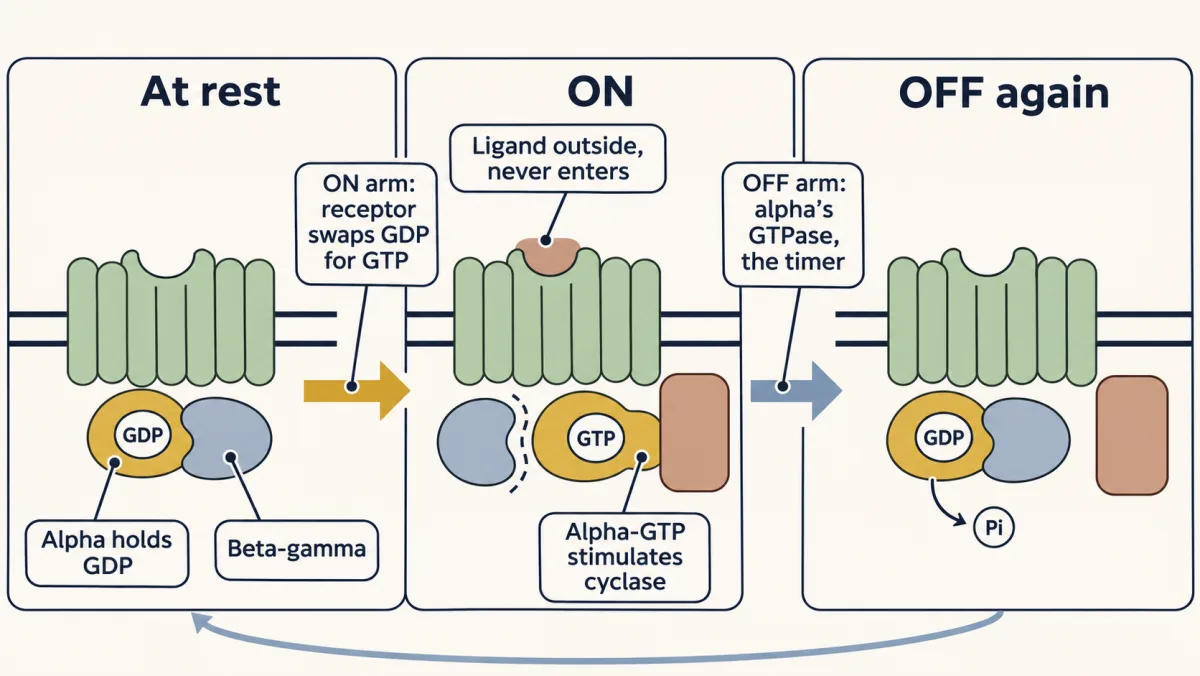
There are three logics, one per type of alpha. Gs stimulates adenylate cyclase and raises cAMP. Gi inhibits it and lowers cAMP. Gq activates phospholipase C and launches the calcium branch. So the same epinephrine can speed your heart, mobilize glucose and constrict a vessel, because the response is written into the receptor, its G protein and the machinery of that cell, not into the ligand.
The cAMP relay: a chain of switches
Earl Sutherland found the next switch through a contradiction. Adrenaline needed the membrane fraction of broken liver cells to work, yet the factor that switched on phosphorylase appeared in the soluble fraction. The membrane, it turned out, converts the hormone’s arrival into a small, water-soluble messenger that carries the command inward: cyclic AMP, the first second messenger ever identified.
Adenylate cyclase makes cAMP from ATP by clipping off two phosphates and cyclizing the rest. Think of cAMP as used ATP with a circular handle. The cyclic phosphate is the message; cAMP is not the same molecule as AMP.
The whole relay, epinephrine to glucose, is a row of switches. Each one has its own ON arm and OFF arm:
| Switch | Pushed ON by | Pulled OFF by | Acts on |
|---|---|---|---|
| Receptor | epinephrine binding | epinephrine leaving; desensitization and internalization under sustained stimulation | Gs (as its GEF) |
| Gs-alpha | the receptor swapping GDP for GTP | its own GTPase | adenylate cyclase |
| cAMP | adenylate cyclase, from ATP | phosphodiesterase (PDE), to AMP | PKA’s regulatory subunits |
| PKA | cAMP freeing its catalytic subunits | regulatory subunits clamping it again as cAMP falls | phosphorylase kinase, glycogen synthase |
| Phosphorylase kinase, phosphorylase | phosphorylation | protein phosphatases | glycogen, cut into glucose 1-phosphate |
Protein kinase A (PKA) sits as an R2C2 tetramer: two regulatory subunits clamping two catalytic subunits silent. cAMP binds the regulatory subunits, they let go, and the catalytic subunits come free and active.
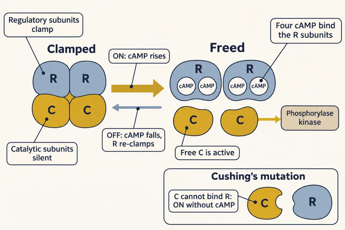
Free PKA then uses the cell’s universal toggle: reversible phosphorylation. Kinases add a phosphate using ATP; phosphatases take it off. The phosphate brings two negative charges and some bulk, which can change the protein’s shape or create a docking site for a partner, activating or inhibiting depending on where it lands. PKA uses both signs at once. It switches on breakdown (phosphorylase kinase, then phosphorylase) and switches off synthesis (glycogen synthase), so stored glucose is released and not rebuilt. Glucagon uses the same Gs → cAMP → PKA route in liver. The film also names lipolysis, the breakdown of stored fat, as a cAMP output; this lesson follows the glycogen branch.
The one idea to carry through this lesson. Every switch in a pathway is pushed ON by one reaction and pulled OFF by another, and how much of it is ON at any moment is set by the contest between the two. An active enzyme’s gain is how fast it works times how long it stays ON, and its OFF reaction sets that lifetime (binding steps just pass the signal on at a fixed ratio). So the pathway’s output is the product of its gains; once the signal is removed, the response dies away at about the pace of its slowest OFF step (the faster ones add a little); and a switch sticks ON when it loses its OFF reaction or stops needing its ON signal.
The rule covers membrane-receptor pathways: G-protein-coupled receptors (GPCRs), receptors that recruit a kinase, and receptors that are kinases. It does not cover nuclear receptors or ligand-gated channels. The film gives it in four beats: a switch turns on, the signal amplifies, the switch turns off, a stuck switch is disease.
How big: speed times lifetime, multiplied down the chain
An active enzyme does not make one product and stop; it keeps working until its own OFF reaction catches it. So the gain of an enzyme step is two numbers multiplied:
gain = how fast the active form works × how long it stays active
Take the cyclase. While Gs-alpha holds GTP, the cyclase it touches keeps turning ATP into cAMP. When alpha’s GTPase hydrolyzes that GTP, the cyclase stops. In round numbers, a cyclase making about 10 cAMP a second, switched on by a Gs-alpha that stays ON for about 10 seconds, makes about 100 cAMP. Slow the GTPase and the same cyclase makes more. The OFF arm sets the gain.
Now run the whole relay, in round teaching values; real gains differ between cell types.
| Step | What one active molecule does | Gain |
|---|---|---|
| Receptor → Gs | activates G proteins one after another, until the hormone leaves | ×10 |
| Gs → cyclase | one Gs-alpha switches on one cyclase | ×1 |
| Cyclase → cAMP | makes cAMP until its Gs-alpha switches off | ×100 |
| cAMP → PKA | four cAMP free the two catalytic subunits of one PKA | ×½ |
| PKA → phosphorylase kinase | one catalytic subunit phosphorylates many | ×10 |
| Phosphorylase kinase → phosphorylase | one kinase phosphorylates many | ×10 |
| Phosphorylase → glucose 1-phosphate | clips glucose units off glycogen, one after another | ×1,000 |
Multiply down the column: 10 × 1 × 100 × ½ × 10 × 10 × 1,000 = 50,000,000. One hormone molecule, tens of millions of glucose units. One row does not amplify (×1) and one loses half (×½). Both are binding steps, not enzymes: one Gs-alpha binds one cyclase, and four cAMP free two catalytic subunits. (The ×½ is a teaching conversion; in the cell, protein kinase A (PKA) follows the cAMP level, which phosphodiesterase (PDE) keeps trimming.) The total is still enormous, because the other steps multiply.
Predict. Make one change at a time. (a) Double the cyclase’s gain, from 100 to 200. (b) Cut it to 1, so each cyclase makes a single cAMP. What happens to the 50 million?
Check your prediction
(a) It doubles, to 100 million: doubling any one factor doubles a product. (b) It falls a hundredfold, to 500,000. If the gains added instead, the total would be about 1,100, and changing one step would hardly move it. Multiplying is why each step matters, and why a single step is where toxins, mutations and drugs act.
The product rule has a limit. It holds while each step has plenty of the next enzyme waiting to be switched on. Push in a big enough signal and the downstream enzyme is all ON already, so the output stops rising. The ceiling is set by whichever step runs out of enzyme first (Going deeper, “When the product rule stops working”).
The widget below starts from the table above; its caffeine preset is ×2 on cAMP, as halving PDE would give. Set the gains, then knock out one step (it becomes ×1) and check the total moves by exactly that step’s factor. The cyclase row is split into its two factors; drag the GTPase-set ON time and watch the gain, and everything below it, follow.
One molecule in, millions out: output is the product of the gains.
The same final number can come from a real hormone (Normal), from a switch that has lost its OFF arm (Cholera), or from one that no longer needs its ON signal (Cushing's). That is the molecular logic of disease.
How long: the slowest OFF step
A signal that cannot be turned off is not a signal; it is a stuck cell. Every switch in the relay table has its own OFF arm. Weaken it and that switch’s steady level rises, just as if you had strengthened the ON arm, though it now also takes longer to switch off.
Predict. At high concentrations, caffeine blocks phosphodiesterase. Add it while the hormone is present. What happens to cAMP, and will it last much longer after the hormone leaves?
Check your prediction
cAMP rises, as the OFF-arm rule predicts. In a simple model of this relay, halving PDE activity doubles the cAMP level. But cAMP lasts only a little longer after the hormone leaves, because cyclase keeps making cAMP until Gs-alpha switches off, and in that model the G protein is the slowest OFF step on the way to cAMP. So blocking PDE mostly makes cAMP bigger, not longer. The glycogen response can last longer still, because phosphatases must also undo the phosphorylations (Going deeper, “Size and duration in a model relay”; at coffee doses caffeine acts mainly elsewhere, see “cAMP and caffeine outside this relay”).
OFF arms are also placed in space. AKAP scaffold proteins tether protein kinase A (PKA) next to the specific targets it should phosphorylate, and they also recruit phosphatases, building the OFF switch right beside the ON switch. The scaffold is a surgical tray with every instrument pre-arranged.
Broken switches: four ways to break the contest
If each switch’s level is a contest, disease can come from either side of it. Cholera, some forms of Cushing’s syndrome and many cancers involve, at least in part, a switch that will not turn off, but not every break leaves a switch stuck ON:
| What broke | Example | What happens |
|---|---|---|
| Lost its OFF arm | cholera (Gs GTPase); oncogenic Ras (GAP-assisted GTPase); PTEN loss | the switch can turn ON but not OFF |
| Freed from its ON signal | Cushing’s (PKA catalytic subunit cannot bind its regulatory subunit); Bcr-Abl (an always-ON fusion kinase) | the switch is ON with no signal at all |
| A brake cut | pertussis (Gi cannot be switched ON) | the brake’s target runs high |
| A weakened ON arm | insulin resistance | too little response, not too much |
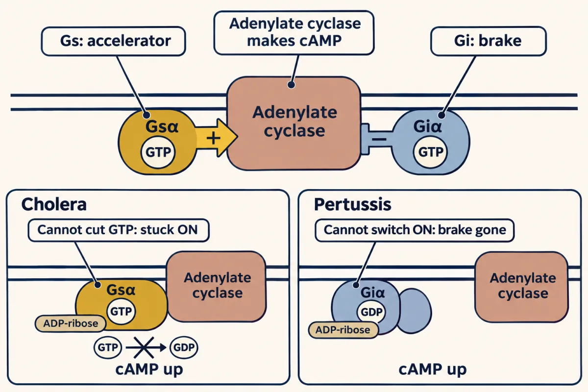
In cholera, choleragen chemically modifies Gs-alpha by ADP-ribosylation (attaching an ADP-ribose group) so it can no longer hydrolyze its GTP. Gs is locked ON, cAMP never stops, and chloride and water pour into the intestine. Severe dehydration can kill within hours.
In some cases of Cushing’s syndrome, a mutation leaves PKA’s catalytic subunit unable to bind its regulatory subunit. Protein kinase A (PKA) escapes its clamp, and cortisol production runs without a hormone telling it to. Nothing is wrong with phosphodiesterase (PDE) or the phosphatases here; the switch simply no longer needs cAMP. The clinical picture (muscle weakness, thin and easily bruised skin, osteoporosis) follows downstream.
Predict. Pertussis (whooping cough) toxin uses the same chemistry, ADP-ribosylation, on Gi. It locks Gi in its inactive form. Does cAMP go up or down?
Check your prediction
Up. Ask three questions for each toxin:
| Toxin | What it does to its G protein | What that G protein normally does to the cyclase | Net cAMP |
|---|---|---|---|
| Cholera | Gs stuck ON | pushes it ON | up |
| Pertussis | Gi stuck OFF | holds it back | up |
“Stuck OFF” sounds like less signal, but Gi is a brake that can no longer lower cAMP.
Same template, new parts
The rest of the chapter is the same rule with new parts: for each pathway, find the switch, its ON arm and its OFF arm. Learn the names in bold; the rest you only need to recognize.
The Gq branch: one cut, two messengers
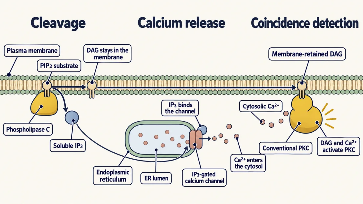
When Gq fires, it activates phospholipase C (PLC), which cleaves the membrane lipid phosphatidylinositol 4,5-bisphosphate (PIP2) into two messengers at once. Inositol trisphosphate (IP3) is soluble; it floats to the endoplasmic reticulum and opens an IP3-gated calcium channel, releasing a flood of Ca²⁺. Diacylglycerol (DAG) stays in the membrane, and together with that Ca²⁺ it activates protein kinase C (PKC). Conventional PKC needs both signals, which makes it a coincidence detector: it fires best when the two signals arrive together.
Calcium is a ubiquitous second messenger, and its common reader is calmodulin, which carries four Ca²⁺-binding EF-hand motifs, each a helix-loop-helix fold. Ca²⁺-loaded calmodulin activates targets including CaM kinase and the plasma-membrane Ca²⁺ ATPase (PMCA). The OFF arms are pumps: the PMCA pushes calcium out of the cell, and the SERCA pump returns it to the endoplasmic reticulum. DAG has its own OFF arm, DAG kinase. Calcium also ties straight back to the glycogen relay: in muscle, calmodulin is built into phosphorylase kinase itself.
Receptors that recruit a kinase: JAK-STAT
Not every receptor talks to a G protein. Growth hormone binds both chains of a receptor pair that is already assembled, with a JAK2 kinase attached to each tail. Binding moves the two JAK2s into position, and they phosphorylate each other (cross-phosphorylation), then the receptor tails. STAT5 docks there and is phosphorylated, and STAT5 dimers enter the nucleus and change gene expression. Too much growth hormone, usually from a pituitary tumor, overdrives this pathway and causes gigantism in children or acromegaly in adults.
Receptors that are kinases: EGFR and Ras
Receptor tyrosine kinases (RTKs) are kinases. EGFR dimerizes on ligand binding, the two halves cross-phosphorylate, and the phospho-tyrosines become docking sites for the adaptor Grb2, which brings in SOS. SOS is Ras’s guanine-nucleotide exchange factor (GEF): it pushes Ras ON. Ras is a monomeric small GTPase, a tiny version of G-alpha: GDP-off, GTP-on. Its OFF arm is its own GTPase, sped up by GAPs (GTPase-activating proteins). Active Ras drives the Raf-MEK-ERK cascade toward proliferation.
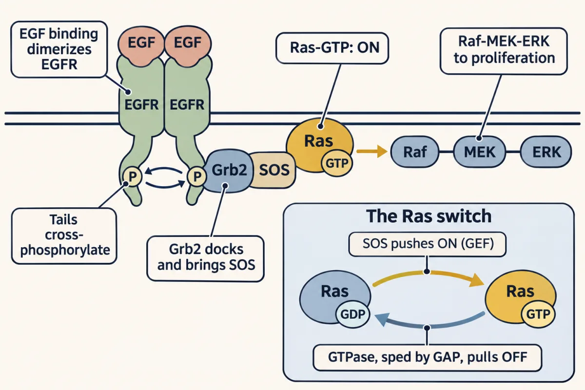
Predict. An oncogenic Ras mutation blocks the GAP-assisted GTPase but leaves SOS free to load GTP. Which switch earlier in this lesson does this mutant Ras resemble?
Check your prediction
Gs-alpha in cholera. Both can still be pushed ON, but neither can be pulled OFF efficiently, so each molecule that picks up GTP stays ON far longer than normal. About one in five people with cancer (roughly 19% in an incidence-weighted estimate) has a tumor with a Ras mutation of this kind. It is the lost-OFF-arm story again, one tier down.
Insulin: the lipid-kinase deep dive
Insulin’s receptor is also an RTK, but it routes mainly through a lipid switch, in five steps:
- Receptor. Unlike EGFR, the insulin receptor is a pre-formed dimer. Insulin binding triggers a quaternary rearrangement, and the halves cross-phosphorylate.
- IRS. The receptor phosphorylates adaptor proteins called IRS, which recruit phosphoinositide 3-kinase (PI3K). Both EGFR and the insulin receptor can activate Ras and PI3K; the IRS adaptors steer insulin strongly toward PI3K.
- Phosphatidylinositol 3,4,5-trisphosphate (PIP3). PI3K is a lipid kinase: it phosphorylates PIP2 to make PIP3, a membrane second messenger. Its OFF arm is the lipid phosphatase phosphatase and tensin homolog (PTEN), which converts PIP3 back to PIP2.
- Akt. PIP3 recruits the kinase Akt to the membrane, along with PDK1, which phosphorylates it; a second kinase, mTORC2, completes its activation. Protein phosphatases strip the activating phosphates.
- Output. Akt drives the two outcomes that define the fed state: GLUT4 translocation to the membrane for glucose uptake in muscle and fat, and glycogen synthesis in muscle and liver.
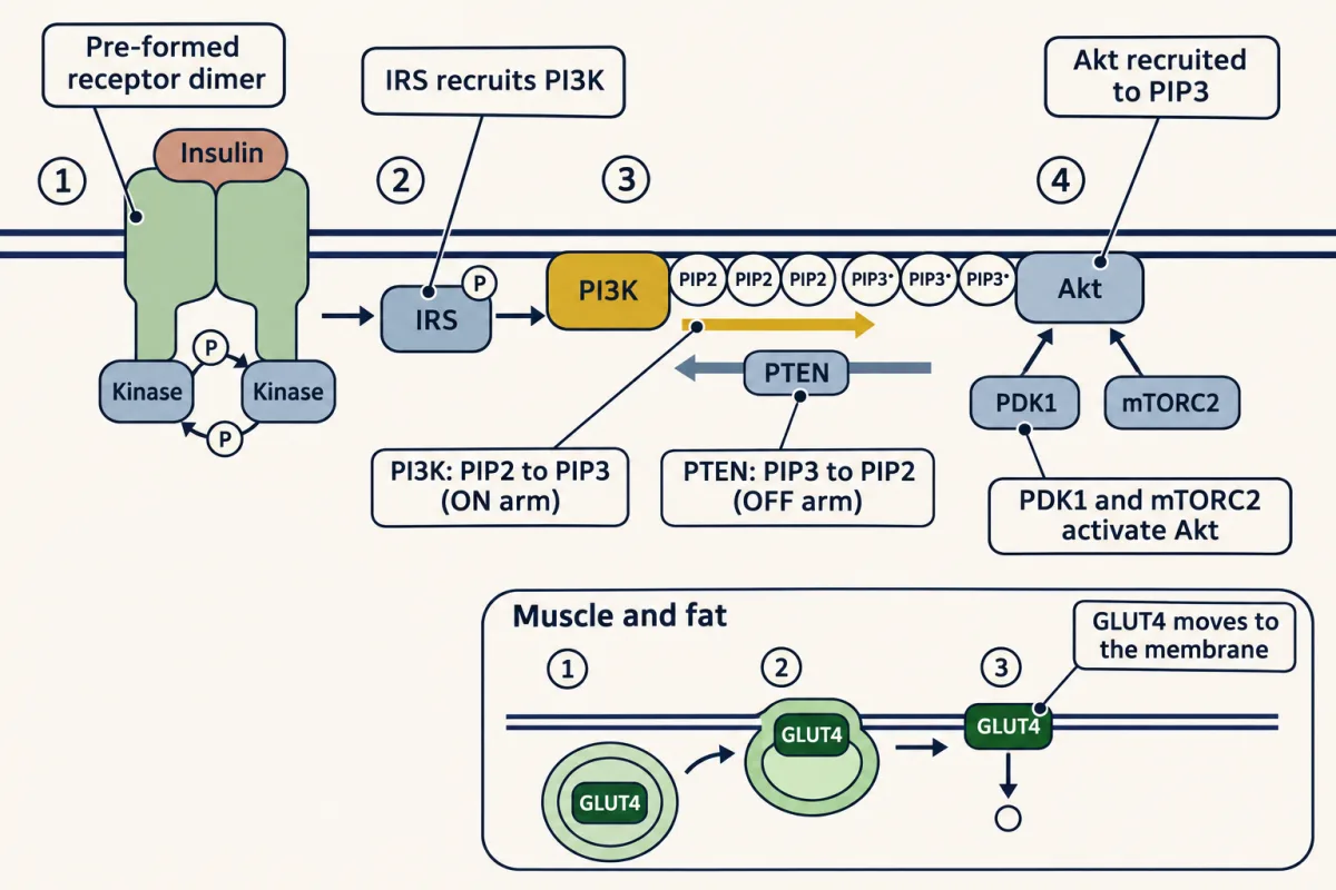
Hold the two PIP2 stories side by side. In the Gq branch, PLC cleaves PIP2 into IP3 and DAG. In insulin signaling, PI3K phosphorylates PIP2 into PIP3. Same substrate, different chemistry, different messengers.
Both sides of this contest cause disease. PTEN is a tumor suppressor: lose it, and PIP3 builds up and Akt stays active, which helps drive many cancers. On the ON side, a block at any node along insulin, receptor, IRS, PI3K, PIP3, Akt, GLUT4 reduces glucose uptake in muscle and fat. Weakened signaling along this chain (insulin resistance) is one half of type 2 diabetes; the other half is beta cells that can no longer make enough insulin to overcome it.
Drugs on the wiring
Much of cancer is signaling gone wrong. Proto-oncogenes promote growth and become oncogenes when stuck ON; tumor-suppressor genes brake growth or guard the genome, and help cause cancer when lost.
Two drug strategies attack the wiring from opposite sides. Gleevec (imatinib) treats chronic myelogenous leukemia, where the Philadelphia chromosome fuses two genes into a constitutively active Bcr-Abl tyrosine kinase. Gleevec wedges into the kinase’s ATP-binding pocket and shuts it off, turning a leukemia that usually killed within a few years into a manageable chronic condition. It acts from the inside, on the stuck switch itself. Cetuximab is an antibody that blocks epidermal growth factor (EGF) from binding EGFR, preventing dimerization from the outside. If Gleevec is like putting gum in a lock, cetuximab is like covering the doorbell so no one can ring it. But cetuximab only helps if the cancer is actually EGFR-driven.
Predict. A tumor carries a KRAS mutation that locks Ras ON. Will cetuximab, which blocks EGFR above Ras, help much?
Check your prediction
Usually not. The KRAS mutation leaves the pathway stuck ON below the receptor. Weakening an ON arm upstream does little to a switch that has lost its OFF arm, because loaded Ras is not switched back off (mutant KRAS is largely GTP-loaded without help from EGFR). That is why precision medicine requires knowing the tumor’s genetics, and why single-pathway drugs so often fail: pathways cross-talk, and the cell reroutes around a block.
Cells compute with molecular switches. Broken switches cause disease. Knowing the wiring is what makes a therapy targetable. That is signal transduction.
Going deeper
When the product rule stops working
In a simple model of the relay, the active G protein matches the linear prediction at small inputs, but at an input ten times the level that half-saturates it, it reaches about 0.9 of the total where the linear rule predicts ten times the total.
Saturation can also make a single switch sharp. When the kinase and phosphatase of one switch are both working near their maximum rate, the contest still decides the outcome, but abruptly: in one model where each enzyme’s Km is about 1% of the substrate it acts on, an ON/OFF rate ratio of 0.9 leaves the switch 8% ON and a ratio of 1.1 leaves it 91% ON, where unsaturated enzymes would give 47% and 52%. This is zero-order ultrasensitivity. It does not break the rule; ON still wins when it outpaces OFF.
Size and duration in a model relay
In a three-switch model (G-alpha, cAMP, a phosphorylated target) where the GTPase gives active G-alpha a half-life of about 7 seconds and phosphodiesterase (PDE) gives cAMP one of about 1.4 seconds, halving PDE lengthened cAMP’s half-time after the hormone left only from about 9 to about 11 seconds. The faster PDE step still added a little: about 9 seconds, not 7. Slowing only the phosphatase (here giving the phosphate a half-life of about 80 seconds) stretched the target’s half-time to about 90 seconds: the response can outlast its second messenger. With the GTPase blocked, as in cholera, cAMP stayed up after the hormone left, and even with no hormone, active G protein slowly built up from spontaneous GDP-to-GTP exchange.
Where the glucose goes
The opening figure multiplies round gains and counts glucose 1-phosphate. In muscle, that glucose 1-phosphate is burned in the cell, not released as blood glucose. In human liver, adrenaline acts partly through alpha-1 receptors (Gq and Ca²⁺) as well as beta-2 receptors.
The GPCR family
G-protein-coupled receptors (GPCRs) are the largest receptor family in humans (roughly 800 of them) and the target of about 35% of approved drugs. Once alpha-GTP leaves or rearranges, beta-gamma can signal to its own targets too. Light, which the film lists among first messengers, reads through one of them: in vision, light switches on rhodopsin, a GPCR, directly.
cAMP and caffeine outside this relay
In the glycogen cascade, cAMP’s job is to free protein kinase A (PKA); in other settings it also acts on Epac and some ion channels. Caffeine’s PDE block needs high concentrations; at the doses in a cup of coffee its main action is blocking adenosine receptors.
Kinases and phosphatases
The human genome encodes more kinases than phosphatases (roughly 500 versus roughly 200). That does not mean OFF control matters less. Phosphatase catalytic subunits such as PP1 and PP2A get their specificity from many regulatory subunits, so fewer catalytic genes still serve many targets.
GLP-1 drugs
Glucagon-like peptide 1 (GLP-1) receptor agonists such as semaglutide (Ozempic, Wegovy), and tirzepatide (Mounjaro), which also activates the glucose-dependent insulinotropic polypeptide (GIP) receptor, act on a Gs-coupled GPCR. GLP-1 is released from gut L-cells after a meal; its receptor shares the seven-transmembrane core of the beta-adrenergic receptor but belongs to a different family (class B), with a large outer domain that grips the peptide. Its safety feature is built into the wiring: GLP-1 boosts insulin release when blood glucose is high, and the boost fades as glucose falls. That glucose-dependence means these drugs rarely cause hypoglycemia, unlike the older sulfonylureas, while still acting on pancreas, brain and stomach.
Potency and efficacy
Every agonist, GLP-1 agonists included, is described in a given assay by two numbers. Potency is how much drug it takes to get half the maximal response (the EC₅₀); efficacy is how large that maximal response can get (Emax). In the explorer below, the potency slider moves the curve left or right and the efficacy slider sets its height. Then add a competitive antagonist and watch the curve slide right while its ceiling stays put: enough extra agonist always wins the competition back.
- Apparent EC₅₀ (relative units)
- 1.0
- Ceiling response
- 100%
The dose that gives half-maximal response is the EC₅₀ — the lower it is, the more potent the drug. Efficacy is how high the curve can reach.
Potency (EC₅₀, the curve's horizontal position) and efficacy (Emax, its height) are independent. A competitive antagonist slides the curve right but can be overcome with more agonist; it never lowers the ceiling.
The twist and the name
Growth hormone has two receptor-binding sites on one molecule. Because the two sites are not alike, binding twists the receptor chains relative to each other, and that twist is what brings the two JAK2s into position. Janus kinase (JAK) is named for Janus, the two-faced Roman god, because it carries two kinase-like domains though only one is a fully active kinase.
EF hands
The calmodulin EF-hand motif is named for helices E and F of parvalbumin, where it was first seen. Your own hand is the mnemonic for the fold: stretch out your index finger and thumb as the two helices and curl your middle finger as the calcium-binding loop between them.
Your notes
Select any text in the lesson to highlight it or add a note to it. Your highlights and notes are saved in this browser and collected in your notebook.
This browser is not letting the site save anything (a private window or strict privacy settings), so highlights and notes will be lost when you leave the page.
How we measure it
The cell-fractionation paradox (Sutherland's method)
Grind tissue, spin it, and split it into a membrane pellet and a soluble supernatant. Sutherland found adrenaline needed the membrane fraction to act but the response (a phosphorylase-activating factor) showed up in the supernatant. The hormone could not be in two places at once. The resolution was a diffusible second messenger made at the membrane.
Reconstitution with a broken cell (Gilman's cyc⁻ assay)
Start with membranes from a mutant cell that cannot raise cAMP, add back a protein extract from normal cells, and watch signaling return. Gilman's cyc⁻ S49 lymphoma restored cAMP only when GTP was present, which is how the G-protein transducer was first caught; Gilman purified Gs from rabbit liver some years later.
Radioligand receptor binding
Tag a hormone with a radioactive label and count how much sticks to membranes at equilibrium. Lefkowitz tagged the hormone ACTH with radioactive iodine, an early handle on a hormone receptor as a countable molecule. Receptor pharmacology itself was decades older.
Western blot readout of kinase inhibition
Run cell lysate on a gel, transfer it, and probe with a phospho-specific antibody to see whether a kinase is firing. Druker used this to show STI-571 (Gleevec) shut down Bcr-Abl in CML cells.
Practice questions on this lesson (8) → MCAT-style, original, each with an explanation
Where this lesson fits
Before thisReview first
- Enzyme Kinetics: The Michaelis–Menten ModelThis course · Lesson 8
You need saturation of enzymes by substrate from there to follow why the product rule stops working at large signals.
- Protein Binding and Allostery: How Hemoglobin Loads and Delivers OxygenThis course · Lesson 9
Ligand binding that changes a protein's shape is the idea behind a receptor switching on a G protein here.
- Membranes: The Impermeable Sheet That Builds ItselfThis course · Lesson 12
Why large polar signals cannot cross the membrane and need a spanning receptor comes from there.
After thisStudy next
- Integration of Metabolism: How the Body Switches FuelsThis course · Lesson 30
Insulin, glucagon and epinephrine signals turn pathways up or down here, using the switches built in this lesson.
- Gluconeogenesis: Pushing Glucose Back UphillThis course · Lesson 17
Glucagon's cAMP and PKA signal is what switches liver glucose building on during fasting.
Elsewhere on the siteFilms, simulations and method films in other courses
- GTPase timerSimulation · Cell Signaling
This simulation's cousin shows the G-protein timer that turns the signal off.
- The Messenger Level: Made and ErasedFilm · Cell Signaling
It follows the cAMP second messenger and PKA step by step, the core relay used here.
- A Membrane Address for AktFilm · Cell Signaling
It covers the PI3K, PIP3 and Akt steps that carry insulin to GLUT4 in this lesson.
- One Cut, Two DestinationsFilm · Cell Signaling
It shows PLC cutting PIP2 into IP3 and DAG, the Gq branch described here.
Suggested by an AI model from the lessons themselves, then checked. How we use AI
Finish line
Test yourself
8 questions
Practice and MCAT-style, with explained answers.
Next · Lesson 14
Digestion and Absorption: Turning a Meal into Molecules Your Cells Can Use
Digestion has two halves that only work together: enzymes cut a meal into small pieces, and the gut wall carries those pieces into the blood or lymph.
Continue →← Lesson 12 · Membranes: The Impermeable Sheet That Builds Itself