Sensory Systems · Unit 4: Proprioception and interoception
Sensing the Gut: Enteroendocrine Cells and Nutrient Signals
Under review Parts of this lesson are still being checked.
- This lesson has not yet been through the review-literature fact-check that the rest of the course had.
The text stays up while the check is done. If you spot a problem, report it. All lessons under review.
A mouse with no sweet taste still learns to prefer sugar over a sweetener. The sensor is in its gut, and it is not a neuron. How would you show that?
Pause when the reading reaches a film, and offer to play it. Saved on this device.
Watch the film now, or keep listening
Builds on:Vagal Sensory Neurons: The Body Reports to the BrainFive Ways to Turn a Stimulus into a CurrentSensory Neurons by Their Genes: the Atlas EraWhy these?
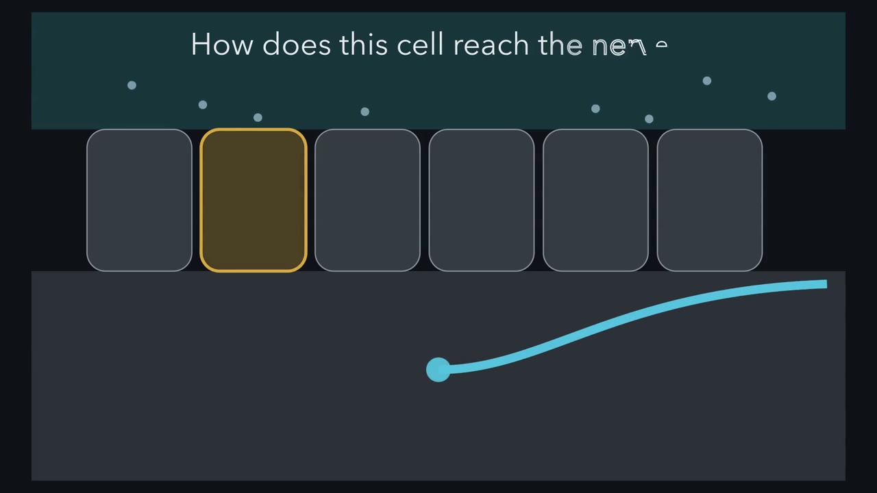 ▶ Watch How to Prove a Gut Synapse · 3:29
Read the transcript of “How to Prove a Gut Synapse”
Film transcript: How to Prove a Gut Synapse
A cell in the gut lining is not a neuron. How could it send a rapid message to a vagal sensory neuron? One possibility is a hormone released into nearby tissue. Another is a point-to-point synapse. Speed alone will not settle the choice. Start with anatomy.
Modified rabies virus begins in selected enteroendocrine cells and labels connected nerves. In some mice, labelling reaches vagal nodose neurons. Tracing can map a connection across the epithelial boundary. It cannot show rapid transmission, identify the messenger, or reveal whether the connection changes nerve firing. The picture must become physiology. That establishes connectivity, not the transmitter. The cells also carry transcripts for VGLUT1. Those transcripts fit glutamate release, but still do not show a current. Now move the cell and a nodose neuron into a dish. Light activates the gut cell. Within 60 to 800 milliseconds, an excitatory current appears in the neuron.
That current measures the neuron's response directly. Its sub-second latency supports a fast messenger in this dish. But one current still cannot establish which receptors carry it. Kynurenic acid blocks ionotropic glutamate receptors, and the current disappears. If receptors are blocked rather than cells damaged, what should happen after washout?
Washout removes the blocker, and the current returns. Recovery supports receptor block rather than cell damage. Together, latency and reversible pharmacology identify fast glutamate action on the neuron. Here is the rule. No single result proves the route. A fast synapse is established when anatomy, timing, pharmacology and causal perturbation converge. Causality comes from the living mouse. Intestinal sucrose raises cervical-vagus firing. Blocking SGLT1 removes that response, placing the sugar sensor upstream. Light that silences CCK-labelled gut cells abolishes the sucrose response.
Light that activates the same labelled population raises vagal firing. These tests ask necessity and sufficiency. The chain has boundaries. Cocultures are dishes. Only a minority of CCK cells contacted sensory fibres in fixed tissue. A synapse does not erase hormone signalling. Anatomy found the contact. Physiology identified rapid glutamate transmission. Acute manipulations connected the sensor to vagal firing. The next question is what this signal changes.
From the film
- Anatomy can show a gut cell contacts a nerve, but it cannot identify the transmitter.
- A fast current in a cocultured vagal neuron tests transmission directly.
- A glutamate-receptor blocker that silences the current, followed by washout recovery, supports glutamate transmission.
- In mice, silencing and activating CCK-labelled gut cells test necessity and sufficiency for a vagal sugar response.
The first film followed a cell in the intestinal lining to a vagal sensory neuron. The next task is to keep the claims at the level each experiment earns. Everything is mouse work unless a sentence says otherwise; smell and taste owns the tongue, so taste is a control here.
The one idea. In mice, a non-neuronal gut epithelial cell can act as a first-order sensory cell, converting a luminal nutrient signal into rapid excitation of a vagal afferent. The route is established only by linking evidence across cell, synapse, nerve and behaviour; no single experiment proves the whole path.
Scope: the mouse routes tested here, not every enteroendocrine cell, every gut stimulus or a demonstrated human behavioural pathway. A synaptic route can exist beside hormonal signalling.
First establish a sensory epithelial cell
Enterochromaffin (EC) cells make more than 90 percent of the body’s serotonin yet are under 1 percent of intestinal epithelial cells. Both are background statements in Bellono and colleagues (Cell, 2017), co-led by the laboratories of David Julius and Holly Ingraham. Their rarity had blocked single-cell work, so the authors genetically tagged EC cells in mouse intestinal organoids, cultured mini-intestines, and checked the findings in native tissue.
The tagged cells expressed chemosensory receptors: Transient receptor potential ankyrin 1 (TRPA1) acted as an irritant receptor, and the odorant receptor Olfr558 as a sensor of a microbial metabolite. Olfr558 was also tested in HEK293 cells. EC cells were electrically excitable. Fibres carrying 5-HT3 receptors made what the authors called synaptic-like contacts with them. In an ex vivo colon preparation, epithelial norepinephrine or isovalerate evoked firing in single mucosal afferent fibres, and the 5-HT3 antagonist alosetron blocked it.
Predict. Does this result show that EC cells synapse onto the vagus?
Check your prediction
No. The recorded fibres were pelvic, hence spinal, afferents; none was shown to be vagal. The experiment supports a sensory epithelial cell with an output onto a nerve, as taste cells have, but “onto the vagus” goes beyond it. Whether that output is read as a rate, pattern or labelled line is a question of sensory coding.
This case establishes the receptor-cell logic. The next experiments concern partly different enteroendocrine populations and a vagal connection.
What each synapse experiment earns
Before 2018 the prevailing view, as Kaelberer and colleagues put it, was that the brain senses gut stimuli only through passive hormone release. Bohorquez and colleagues (J. Clin. Invest., 2015) showed that a cytoplasmic process on enteroendocrine cells, earlier called a neuropod, contacts mucosal nerves. Kaelberer and colleagues (Science, 2018) asked whether the contact was a synapse and called synaptically connected cells neuropod cells.
The film built the argument; the exact observations below show where each inference stops.
| Test | Observation | Claim earned—and limit |
|---|---|---|
| Connectivity | Modified rabies virus given by enema to PYY-Cre pups, or by oral gavage to CCK-Cre pups, infected enteroendocrine cells and labelled nerves. In the colon experiment, labelling reached vagal nodose neurons in 2 of 5 mice with infected colon nerves using the pseudotyped virus. | A connection exists in some animals; tracing does not identify the transmitter or its behavioural role. |
| Transmitter machinery | Enteroendocrine cells expressed VGLUT1 transcripts. | This fits glutamate release but does not show a postsynaptic effect. |
| Speed | In 9 cocultured enteroendocrine-cell–nodose-neuron pairs, light activation evoked excitatory postsynaptic currents within 60 to 800 ms. | The gut cell can rapidly excite the neuron in a dish. |
| Pharmacology | Kynurenic acid at 3 mM, an ionotropic glutamate-receptor blocker, abolished the currents; washout restored them in 4 neurons. | Glutamate receptors carry the recorded current; recovery argues against damaged cells. |
| Acute causality | In anaesthetised mice, intestinal sucrose raised cervical-vagus firing within seconds and peaked about a minute or more later. Phloridzin, an SGLT1 blocker, abolished the response. Halorhodopsin silencing of CCK-labelled cells abolished it; channelrhodopsin activation raised firing. | The authors concluded that these cells were necessary and sufficient to transduce a glucose stimulus in this preparation. Firing within seconds alone could still fit a hormone or a synapse. |
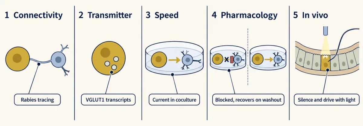
Use the rule once: name the link each experiment tests.
Predict. The contact result was 18.9 percent, measured from more than 100 cholecystokinin (CCK) cells per mouse in 3 mice. What is the biological replicate: each cell or each mouse?
Check your prediction
The mouse. Cells sampled from the same mouse are nested observations, not independent animals. The result establishes a minority contact fraction in 3 mice; because Pgp9.5 labels sensory fibres, it does not make every contact vagal or justify generalising to all enteroendocrine cells.
Two more limits matter. Coculture latency in milliseconds, in-vivo firing within seconds and learned preference over 24 to 48 hours measure different processes. Optogenetic silencing is acute, but a Cre line is not pure by mature cell type because it marks cells that expressed the driver at some time.
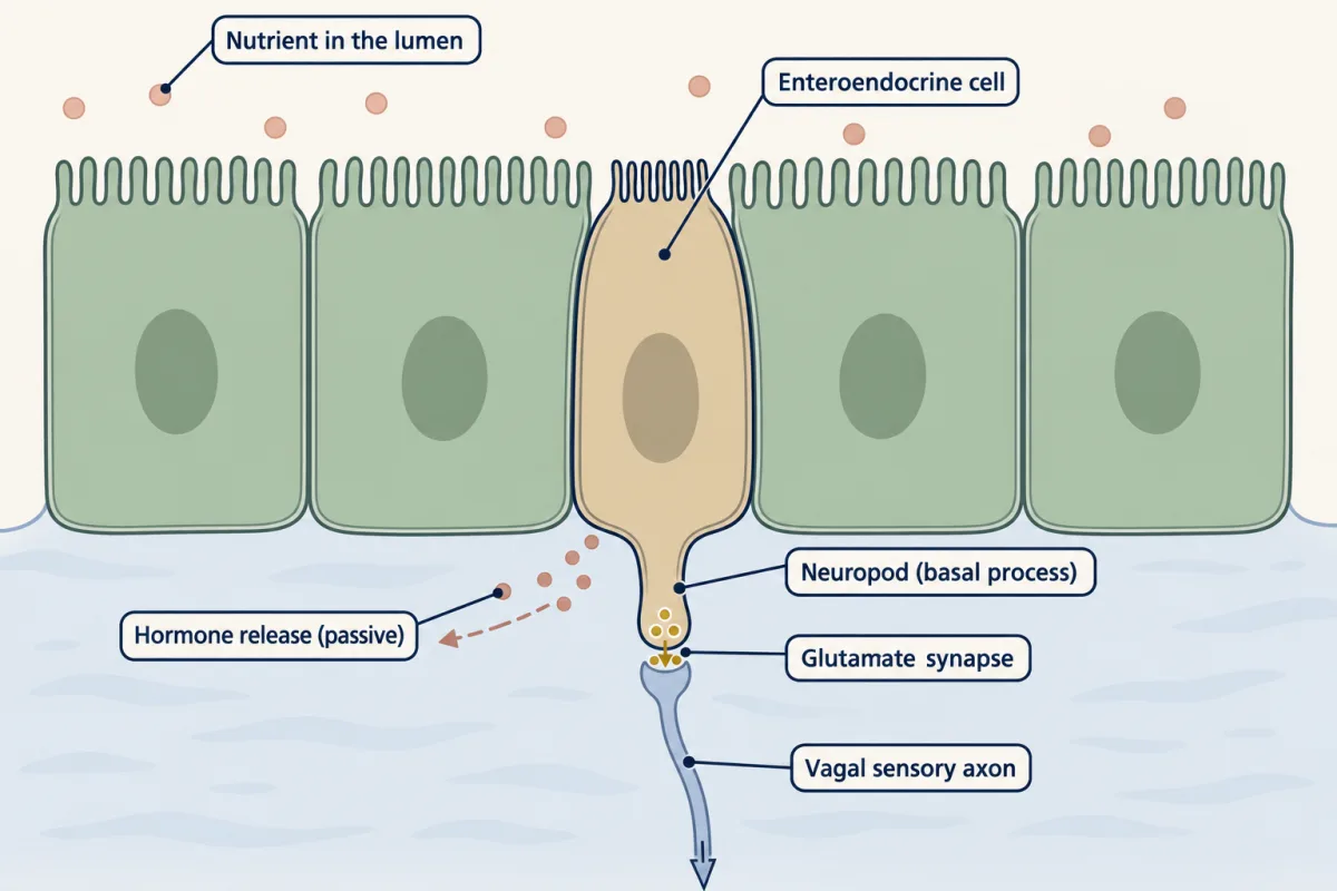
That fast link reaches the nerve; the second film asks what changes when the signal is used to choose between nutrients.
Read the transcript of “How the Gut Teaches Preference”
Film transcript: How the Gut Teaches Preference
Chapter one established a rapid route from a gut sensor cell to the vagus. How can that route teach sugar preference when sweet taste is absent? First separate two measurements. Initial attraction is what the mouse chooses at the first sip. Learned preference is how that choice shifts after signals arrive from the gut. Researchers matched sugar and a calorie-free sweetener for early attraction.
Naive mice initially drank at similar rates. Then the sugar bar rose over the choice period. Suppose sweet taste alone creates that later preference. What should happen in mice that cannot signal sweetness during the choice period?
It still develops. Mice lacking the sweet receptor or a taste-transduction channel still preferred sugar later. In mice, learned sugar preference can develop without sweet-taste signalling. In Tan and colleagues' assay, gut-delivered sugar activates vagal neurons that acesulfame K does not. Blocking SGLT1 sharply reduces glucose responses. Intestinal SGLT1 function is needed for this gut sugar signal. Silencing sugar-activated brainstem neurons prevents the learned preference but leaves initial attraction. Activating another population can create preference for a previously less-preferred stimulus. A later experiment targeted CCK-labelled duodenal sensor cells.
Silencing them strongly reduced the preference advantage above chance. Initial attraction and learned preference are different measurements. Sugar is not the only nutrient lesson. Fat preference survives loss of fat taste.
Imaging then reveals broad nutrient responders and a distinct fat-only vagal population. The broad population also answers sugar and amino acids, so fat is not carried only by fat-only cells. Remove SGLT1, and gut sugar responses disappear while fat responses remain. Remove combinations of fat receptors, and fat responses fall. The inputs can separate. Here is the guiding idea in action. Match each claim to its link: epithelial sensor, vagal response, brainstem learning, or behaviour. Do not merge their timescales. These are mouse circuits.
Germline deletions do not identify the required cell type. Silencing labelled populations is acute but not perfectly pure. Human behaviour remains untested here. The moving picture leaves exact genotypes, sample sizes and the preference calculation to the text. It also leaves a harder naming problem: three different GLP-1 routes.
From the film
- In mice, learned sugar preference can develop without sweet-taste signalling.
- Initial attraction and learned preference are different measurements.
- Intestinal SGLT1 function is needed for the gut sugar signal in these mouse studies.
- Sugar and fat have separable receptor requirements and partly distinct vagal populations.
Sugar without taste: locate each link
The chapter left out the exact genotypes and interventions. Tan and colleagues (Nature, 2020), from Charles Zuker’s laboratory, offered sugar against acesulfame K at a concentration equally attractive at first. Preference shifted within 24 hours, and by 48 hours mice drank almost exclusively sugar. Mice lacking T1R2/T1R3 (n = 5) or TRPM5 (n = 7), which cannot taste sweetness, still developed the preference. Learned preference in this assay therefore does not require sweet-taste signalling.
Vagal-ganglion imaging found neurons that responded to gut-delivered sugar but not sweeteners; intestinal phlorizin caused a dramatic loss of glucose responses. Silencing transmitter release in sugar-activated neurons of the caudal nucleus of the solitary tract, labelled by the activity-dependent driver TRAP2 (n = 6), abolished development of sugar preference without removing innate attraction to sugar or sweeteners. Chemogenetic activation of Penk-Cre neurons in the same region created preference for a previously less-preferred stimulus. Tan and colleagues thus separated first-sip attraction from learning but left the intestinal cell type open.
Buchanan and colleagues (Nature Neuroscience, 2022) tied CCK-labelled duodenal neuropod cells to the choice. They reported glutamatergic transmission to the vagus for sugar and purinergic transmission for sweetener. Of 132 CCK-GFP small-intestinal cells from 3 mice, 60.1 percent expressed only Slc5a1, which encodes sodium-glucose cotransporter 1 (SGLT1); 19.6 percent expressed SGLT1 and Tas1r3, which encodes T1R3; 1.2 percent only T1R3; and 19.1 percent neither. In these mouse studies the sugar transducer is epithelial SGLT1, not a sweet receptor. The only human result is glutamate release from organoids of one donor. The authors say it suggests conservation; it is not human behaviour.
Predict. Light activation of gut-innervating vagal neurons sustains reward behaviour. Does that show a nutrient normally activates the same route?
Check your prediction
No. Han and colleagues (Cell, 2018) used light in adult male mice, not a nutrient. Right-side, but not left-side, activation sustained self-stimulation, flavour and place preference and dopamine release. The route ran through glutamatergic neurons of the dorsolateral parabrachial region to dopamine cells in the substantia nigra. The result shows that this route can sustain reward, not which nutrient normally recruits it. See motivation and reward.
Sugar and fat can separate
Li and colleagues (Nature, 2022) tracked preference for 1.5 percent Intralipid against 3 mM acesulfame K with lick counters over 48 hours. TRPM5-mutant mice, blind to fat taste, still developed a strong preference. Vagal imaging revealed a general nutrient population responding to sugar, fat and amino acids, plus a fat-only, TRPA1-expressing cluster. Silencing VIP vagal neurons markedly impaired nutrient preference while leaving immediate attraction to sugar and fat intact.
The receptor knockouts separate the inputs. In SGLT1-knockout mice no vagal neurons responded to intestinal sugar, while fat responses remained. Fat responses fell substantially only in GPR40/GPR120 double and CD36/GPR40/GPR120 triple knockouts. A germline deletion is neither acute nor restricted to one cell type: it shows that a receptor is needed in the animal, not which cell needs it.
Worked problem: preference above chance
Buchanan and colleagues used halorhodopsin in CCK-labelled neuropod cells, an intraluminal fibre and a one-hour choice between 300 mM sucrose and 15 mM sucralose. Preference was the share of total intake. Under the same light, controls showed 90.8 percent sucrose preference (N = 5), silenced mice 58.9 percent (N = 8), and mice with local duodenal glutamate-receptor block 44.0 percent (N = 4).
Predict. The glutamate-block group chose sucrose 44.0 percent of the time. To estimate the effect of the block, should you compare it with 50 percent chance, the 90.8 percent control from the silencing cohort, or its own 82.4 percent vehicle control?
Check your prediction
Use its own 82.4 percent vehicle control. The 50 percent benchmark shows that 44.0 percent is 6.0 points below chance, but it is not the treatment control. The 90.8 percent value came from a separate cohort.
For the silencing comparison, use the advantage that could be lost: 90.8 − 50 = 40.8 percentage points. The silenced advantage is 58.9 − 50 = 8.9 points. The loss is 90.8 − 58.9 = 31.9 points, so
fraction lost = (control − silenced) / (control − 50) = 31.9 / 40.8 = 0.78, about 78 percent
Dividing by 90.8 would mix the chance half of the choice with the above-chance preference.
Do not round the 44.0 percent value to indifference. Locomotion and 24-hour food and water intake were unchanged, arguing against malaise or reduced general drinking. With N of 4 to 8, the percentages carry wide uncertainty that the arithmetic hides.
- Control advantage, C − 50
- Manipulated advantage, M − 50
- Points lost, C − M
- Fraction lost, (C − M) / (C − 50)
In this sketch: mice, a one-hour choice between 300 mM sucrose and 15 mM sucralose, preference scored as sucrose’s share of total intake, with 50 percent as chance, and the lesson’s rule applied to whatever values are set. Any value other than the lesson’s is labelled as set in this sketch.
Rule to keep: measure preference as the advantage over 50 percent chance, then divide the points lost by the control advantage, not by the raw preference; and compare each group only with its own matching control. Small groups (N of 4 to 8) leave wide uncertainty.
Li and colleagues provide a second reading. Across 9 ganglia, 60 of 203 VIP neurons responded to nutrients: 60 / 203 = 29.6 percent, matching “approximately 30 percent.” About 4 percent were fat-only responders. If that means 4 percent of all 203, then 0.04 × 203 is about 8 neurons. About 8 / 60, or 13 percent, of the nutrient-responsive group would be fat-only; the broad responders that make up the rest also answer sugar and amino acids.
GLP-1: three routes, not one relay
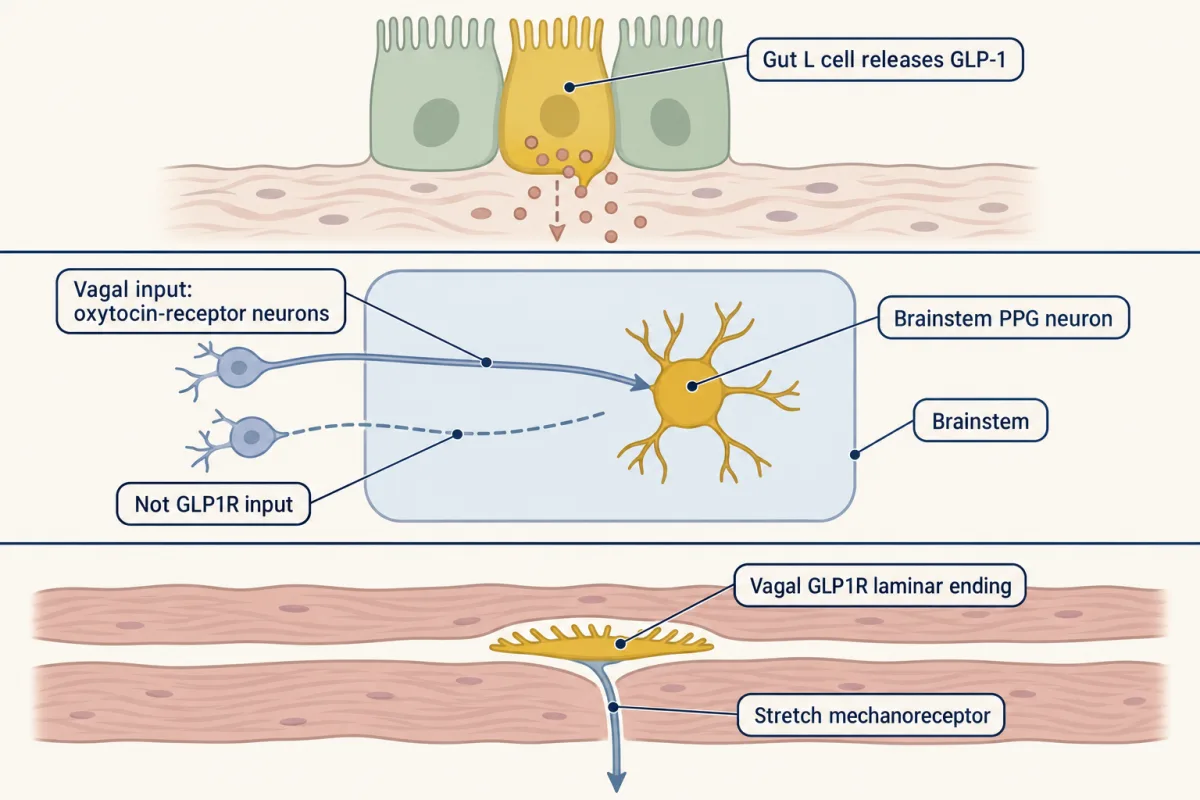
The name glucagon-like peptide 1 (GLP-1) appears in three different places. Keep them in separate lanes before drawing arrows.
| Lane | Cells and location | Supported role in the rodent evidence here |
|---|---|---|
| Peripheral peptide | Enteroendocrine L cells in the gut | Nutrients trigger GLP-1 release, background in a rat study by Krieger and colleagues (Diabetes, 2016). |
| Central peptide | Preproglucagon (PPG) neurons in the brainstem | Brierley and colleagues (Nature Metabolism, 2021) concluded that central and peripheral GLP-1 systems suppress eating through independent gut-brain circuits. |
| Receptor-defined afferent | Vagal neurons expressing GLP1R | Williams and colleagues (Cell, 2016) reported intraganglionic laminar endings and a role as gastrointestinal mechanoreceptors. |
Predict. A proposed chain is “gut L-cell GLP-1 → vagal GLP1R neuron → brainstem PPG neuron → eating falls.” Which observations would break it?
Check your prediction
Brainstem PPG neurons receive vagal input predominantly from oxytocin-receptor neurons rather than GLP1R neurons, and they are not necessary for eating suppression by GLP-1 receptor agonists in mice. Those observations break the proposed last two links. The evidence supports several GLP-1-related routes, not one serial relay.
The third lane needs its own controls. GPR65 neurons supply most villi but were insensitive to GLP-1 or cholecystokinin. Over 95 percent of GLP1R neurons and over 97 percent of GPR65 neurons were slow C fibres. A 2025 erratum corrects only an EKG trace in Fig. 3A; those percentages are unaffected. The vagal sensory neurons lesson develops their endings.
Design problem
A reader says that a gut hormone “tells the brain through the vagus.” Design experiments that decide, for one nutrient, whether the message is hormonal or synaptic and where the receptor that matters sits.
One way to do it
Separate transmitter from hormone first. Look for a vesicular-transporter transcript in the cell. Record a fast current with measured latency in a coculture, block it with an ionotropic glutamate-receptor antagonist and show recovery on washout. Test whether silencing the cells removes the vagal response, then drive them for sufficiency.
State the limits: optogenetic silencing of a Cre-labelled population is acute but not pure by mature type; a germline receptor deletion is neither acute nor cell-specific. Next compare a receptor deletion restricted to one neuronal class with interruption of afferent supply to the gut, measuring the same behaviour. Include a malaise control and separate innate attraction from learned preference. All route evidence here is rodent. The human data are one organoid donor and one pharmacokinetic measurement, so repeat the route tests before making a statement about people.
Going deeper
Enterochromaffin cells in an intact mouse
Bayrer and colleagues (Nature, 2023) reported that enhancing EC-cell function was sufficient to cause hypersensitivity to gut distension in mice. EC-cell activity was necessary for the sensitising action of isovalerate, a bacterial short-chain fatty acid, and the EC-to-mucosal-afferent circuit appeared tonically engaged in females.
GLP-1 drugs do not identify one physiological route
In rats, vagal deafferentation did not block the weight effect of a long-acting glucagon-like peptide 1 (GLP-1) receptor agonist given for 14 days: weight gain was about 10 percent lower than controls in both groups (Secher and colleagues, J. Clin. Invest., 2014). In mice, fluorescently labelled liraglutide bound arcuate neurons after peripheral injection (Secher and colleagues, 2014). Deleting GLP1R from glutamatergic, vGlut2, neurons eliminated liraglutide’s weight effect, while deletion from GABAergic, vGAT, neurons did not (Adams and colleagues, Diabetes, 2018). None of this is a statement about people.
The one human measurement here is pharmacokinetic. After intravenous GLP-1(7-36) amide, intact peptide was 19.9 percent of the rise in immunoreactivity measured with a COOH-terminal assay (Deacon and colleagues, Diabetes, 1995; n = 8 normal subjects). Vagal GLP-1 receptors still matter physiologically: knockdown in rat nodose neurons enlarged meals, sped gastric emptying and raised postmeal glucose, with normal long-term energy balance (Krieger and colleagues, 2016).
Your notes
Select any text in the lesson to highlight it or add a note to it. Your highlights and notes are saved in this browser and collected in your notebook.
This browser is not letting the site save anything (a private window or strict privacy settings), so highlights and notes will be lost when you leave the page.
How we measure it
Intestinal organoids with tagged cells, patch clamp and calcium imaging
Rare cells are tagged genetically in cultured mini-intestines so that single cells can be recorded and imaged (and, in the Kaelberer work, cocultured with nodose neurons). It tests what a cell detects and whether it is excitable, in a dish rather than in the intact animal.
Monosynaptic rabies tracing
A modified rabies virus infects chosen starter cells and crosses synapses to the cells connected to them, which are then labelled. It shows connectivity, not the transmitter and not whether the connection matters for behaviour.
Optogenetics with an intraluminal optical fibre
A light-gated channel or pump is placed in a cell type, and a fibre delivers light to it, here inside the intestine. Driving the cells tests sufficiency; silencing them tests necessity, for those cells, that light and that behaviour only.
Two-bottle choice with lick counters and activity-dependent genetic access
Mice choose between two drinks while licks are counted electronically, separating a first-sip attraction from a preference that builds over hours or days. Neurons active during a stimulus can be given a genetic silencing or activating tool and the choice tested again.
Practice questions on this lesson (3) → MCAT-style, original, each with an explanation
Where this lesson fits
Before thisReview first
- Vagal Sensory Neurons: The Body Reports to the BrainThis course · Lesson 16
This lesson takes the vagal sensory neurons it names and asks which gut cells excite them.
- Five Ways to Turn a Stimulus into a CurrentThis course · Lesson 1
You need the ways a sensory cell turns a stimulus into a current to see how a gut cell does it.
- Sensory Neurons by Their Genes: the Atlas EraThis course · Lesson 3
Reading vagal neuron populations by gene markers, such as VIP or TRPA1 clusters, builds on this atlas approach.
After thisStudy next
- Combining Senses and Predicting ThemThis course · Lesson 29
Combining gut signals with other senses by reliability takes the idea of weighing evidence further.
- Restoring Senses: Implants, Prostheses and Optogenetic VisionThis course · Lesson 30
Restoring or driving sensory pathways by optogenetic activation builds on the stimulation logic used here.
Suggested by an AI model from the lessons themselves, then checked. How we use AI
Finish line
Test yourself
8 questions
Practice and MCAT-style, with explained answers.
Next · Lesson 18
Parallel Channels Out of the Eye
A hawk's shadow slides across a meadow. What does a mouse's optic nerve tell its brain about it? Not a picture: more than 40 kinds of ganglion cell each report the scene through their own filter.
Continue →← Lesson 16 · Vagal Sensory Neurons: The Body Reports to the Brain