Cell Biology · Unit 2: Membranes and compartments
New to this? Review it at high school level: Organelles as a System
Organelles as Distinct Environments
Rewritten on : new text, flashcards and film. Your notes and highlights stay, but the text around them may have moved.
A lysosome near pH 4.7 and a mitochondrial matrix near pH 8 share one cytosol near 7.2. What holds each at its own number?
Pause when the reading reaches a film, and offer to play it. Saved on this device.
Watch the film now, or keep listening
Builds on:Transport Across MembranesExperiments That Distinguish MechanismsMembrane Organization and TopologyWhy these?
 ▶ Watch A Tap, a Hole, a Level · 3:56
Read the transcript of “A Tap, a Hole, a Level”
Film transcript: A Tap, a Hole, a Level
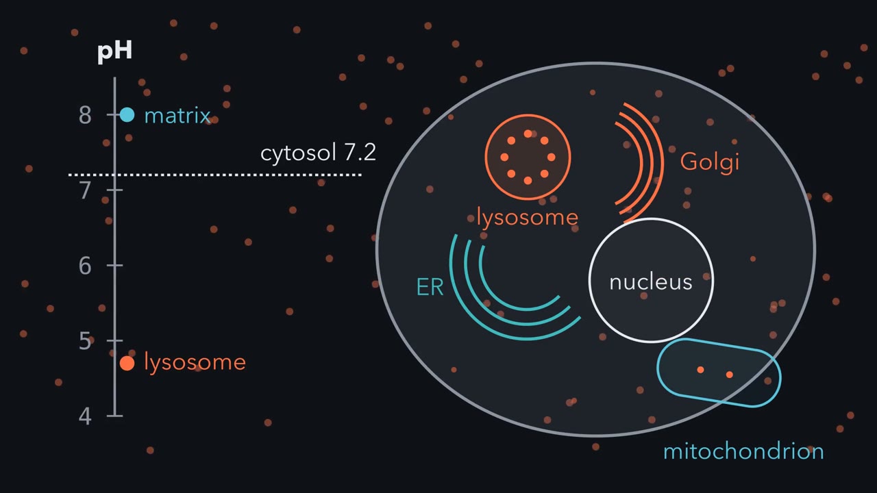
How can one cell hold a lysosome at pH 4.7 and a mitochondrion near 8, in one cytosol? Why does each settle at its own number? This is the cell's pH ladder, with typical values from different cells. Each marker is one compartment. The white line is the cytosol. Acid compartments sit below. The matrix, the mitochondrion's inside, sits above. Start with the wall. A membrane lets ions through slowly, but not never. Protons trickle back toward the cytosol. Picture the lysosome as a container with a tap and a hole. The tap is a proton pump, the V-ATPase, spending ATP to push protons in.
As protons pile up, more leak back out through the hole. At some level, out matches in, and the level stops rising. This picture needs no sensor to set that level. That is the idea, for this acid lumen. The level sits where pumping in meets leaking out. Other things equal, a stronger pump makes it more acid. A leakier wall makes it less acid. Now a drug, bafilomycin, clamps the V-ATPase tap shut. Watch the ladder. Which way will the lysosome's marker move, up or down?
Up, toward the line: less acid. With the tap off, the hole keeps draining. In HeLa cells, bafilomycin made the Golgi less acid. So that acid is actively kept. The Golgi uses the same pump family, yet it settles higher, near six point six. Does the lysosome have more pumps, a tighter wall, or something else? That is still being worked out. The nucleus is different. Its envelope is full of pores, and small ions, protons included, pass through them quickly. So its pH stays close to the cytosol's. The ER is also a nonequilibrium store. A calcium pump called SERCA fills it to thousands of times the cytosol's level, while leaks and release channels move calcium out.
Thapsigargin, a plant compound, blocks SERCA. Calcium keeps leaking out, and nothing pumps it back. The store runs down, and cytosol calcium rises for a while. The mitochondrion's pumps face the other way. They are the respiratory chain, driven by electron transport. While it runs, they push protons out of the matrix, so the matrix sits above the line. Why the other way? The matrix was once a bacterium's cytoplasm, and during respiration its electron-transport chain pumped protons outward. Chloroplasts did the same. Their stroma, the fluid inside, was once a cyanobacterium's cytoplasm, and in light it turns alkaline. Every proton pump here pushes out of a cytoplasm.
The acid lumens, the matrix and the stroma need ongoing energy-dependent transport. Acid lumens need their V-ATPase. The matrix and stroma need respiration or light. Stop that input, and those gradients run down.
From the film
- A membrane leaks, so each gradient here lasts only while a pump keeps paying to balance the leak.
- Block the pump and the gradient runs down: bafilomycin made the HeLa Golgi less acid, and thapsigargin, by blocking SERCA, lets the ER's calcium store drain.
- Small ions pass quickly through nuclear pores, so the nucleus stays close to the cytosol's pH.
- While respiration runs, and in chloroplasts in the light, pumps push protons out of the matrix and stroma, once a bacterium's cytoplasm, so those spaces are alkaline.
A lysosome near pH 4.7 and a mitochondrial matrix near pH 8 share one cytosol near pH 7.2, yet differ about 2,000-fold in free protons. What holds each at its own number?
The first film built the tap-and-hole picture for that puzzle; this text puts numbers on it, tests it with drugs and asks where the outward-facing pumps came from.
The one idea to carry through this lesson. A membrane leaks, so each gradient here (an acid lumen, the ER’s calcium, a respiring matrix, a stroma in the light) lasts only while a pump keeps paying to balance the leak. Stop the pump and the gradient runs down.
A toy model with numbers
The lumen is the space enclosed by an organelle’s membrane; the cytosol is the fluid part of the cell outside all organelles. As the first film showed, a lipid bilayer lets ions through only slowly (membrane transport).
In words In this toy model, the lumen's free protons equal the cytosol's, plus the pump's push divided by how leaky the wall is.
- [H+]lumen“H plus in the lumen”
- free-proton activity, approximated here by an effective molar concentration; pH = −log10 of the activityeffective M
- [H+]cytosol“H plus in the cytosol”
- free proton concentration of the cytosol, about 6.3 × 10⁻⁸ M at pH 7.2, held fixedM
- push / leak“push over leak”
- the extra free protons the pump holds above the cytosol's; a rate divided by a rate per unit of concentration, so a concentrationM
Sanity check No push, or a huge leak: the lumen matches the cytosol. Double the push or halve the leak: the extra free protons double. The model ignores voltage, counterions, other transporters and a pump that slows as its gradient grows: it shows directions, not real pump strengths.
Try one number
Golgi at pH 6.58: [H+] = 10−6.58 = 2.6 × 10⁻⁷ M. Subtract the cytosol’s 0.6 × 10⁻⁷ M: in the model, push/leak ≈ 2.0 × 10⁻⁷ M.
Where it comes from optional
Step 1 (the assumptions): protons pumped in per second = push, a constant. Protons leaking out per second = leak × ([H+]lumen − [H+]cytosol).
Step 2: at steady state, in = out, so push = leak × ([H+]lumen − [H+]cytosol).
Step 3: divide by leak and add [H+]cytosol to both sides.
Need a refresher? rates and “per” · log scales
- A level costs energy for as long as it lasts. It is a steady state: flow in matches flow out, and the pump pays in ATP or electron transport the whole time. An equilibrium has no net flow and costs nothing; things drift toward it once the paying stops. No sensor or set point is needed, though cells do regulate pumps, for example by assembling and disassembling the V-ATPase.
- Different jobs need different levels. Lysosomal hydrolases such as the protease cathepsin D work best in acid. A calcium store sends a signal only if its calcium is far above the cytosol’s, so that opening a channel releases it (calcium signalling). ATP synthesis needs a proton gradient across the inner mitochondrial membrane (oxidative phosphorylation).
This is a first, qualitative model of ion gradients; real organelles add membrane voltage, counterions and other transporters. Redox, a level of a different kind, is in Going deeper.
Predict. To calibrate a pH reporter (a fluorescent protein whose signal changes with pH) inside cells, you bathe them in buffers of known pH and add ionophores: here, carriers that swap protons for potassium or sodium ions across membranes, which opens a wide path for protons in and out when the bath’s ions match the cell’s inside. Where does the reporter’s compartment settle, and why does that give you a calibration?
Check your prediction
Near the bath’s pH. With a huge leak, push/leak shrinks toward zero, so no pump can hold a gap. Read the reporter at several bath pH values and you have a curve that turns its signal into pH. The clamp needs matched conditions: these ionophores trade protons for potassium or sodium (nigericin plus monensin is one pair used), so the bath is high in potassium like the cell’s inside, and the cells need time to equilibrate.
The pH ladder
| Compartment | pH | Main proton pump, direction | System |
|---|---|---|---|
| Mitochondrial matrix | about 7.98 (estimated) | respiratory chain, out of the matrix | HeLa cells, targeted reporter |
| Chloroplast stroma, in light | about 7.8 to 8.0 | photosynthetic chain, out of the stroma | Older plant measurements, reviewed |
| Cytosol (the line) | about 7.2 | Reference value | |
| Nucleus | near 7.2 | none needed: pores pass small ions quickly | Mammalian cells, review |
| ER lumen | near neutral | none named here | HeLa cells, targeted reporter |
| Medial/trans Golgi | 6.58 | V-ATPase, into the lumen | HeLa cells, targeted reporter |
| Early and recycling endosomes | about 6.3 to 6.5 | V-ATPase, into the lumen | Mammalian cells, review |
| Late endosomes | about 5.5 | V-ATPase, into the lumen | Mammalian cells, review |
| Lysosomes | 4.5 to 5.0 | V-ATPase, into the lumen | Mammalian cells, review |
This is the first film’s ladder with the source system for each value; its 4.7 is the middle of the lysosomal range. For calcium, free Ca2+ is about 100 nM in the resting cytosol and about 0.5 to 1 mM in the ER lumen, a 5,000- to 10,000-fold difference; lysosomes hold Ca2+ about 0.5 mM and Cl− 80 to 100 mM (mammalian reviews).
Two traps: not every organelle is acidic, and a small gap in pH hides a large gap in protons.
Worked problem: reading the ladder in free protons
How many more free protons are in the Golgi than in the matrix? A gap of 1.4 pH units looks small; the ratio shows how large it is. Use the two HeLa means, 6.58 and 7.98. They come from separate measurements, not one cell read twice.
pH = −log10(aH+), where aH+ is the activity of free protons (their effective concentration), so aH+(Golgi) / aH+(matrix) = 10pH matrix − pH Golgi
- Subtract: 7.98 − 6.58 = 1.40.
- Raise ten to that power: 101.40 = 25.1.
- Check, treating activity as concentration: 2.63 × 10⁻⁷ M / 1.05 × 10⁻⁸ M ≈ 25.
The Golgi lumen has about 25 times the free proton activity of the matrix. This is free activity only; it says nothing about protons held by buffers or how fast protons move. The same arithmetic puts a lysosome at 4.7 about 300 times above the cytosol.
Each pH unit is a tenfold change in free protons, so the ratio is 10 raised to the pH difference. It compares free proton activity only; it says nothing about the total protons held by buffers or how fast they move. This is a sketch of the logic, and the values come from different systems.
Turning the knobs
The first film predicted what bafilomycin A1 does to the V-ATPase; here is what it leaves out. The V-ATPase is a rotary pump that spends ATP to move protons from the cytosol into a lumen, and a mammalian review names it the main mechanism keeping endosomes and lysosomes acid. How far a lumen drifts depends on its other fluxes: predict “less acid”, not “exactly 7.2”.
Charge is part of the push. Each proton pumped in makes the lumen more positive, and a positive lumen pushes back on the pump. So acidification is thought to need a counterion, an ion whose movement cancels that charge: chloride following the protons in, or another cation moving out.
Predict. A lysosome loses most of its counterion flow, no other path makes up for it, and its V-ATPase is untouched. Where does its pH settle?
Check your prediction
Less acid, closer to the cytosol line: the charge the pump builds holds it back, so its effective push falls against the same leak. Real lysosomes have several counterion paths, some of them exchangers rather than simple channels, so blocking one need not do this; an experiment must check whether another path took over.
SERCA, the sarco/endoplasmic reticulum Ca2+-ATPase, pumps calcium from the cytosol into the ER and builds that 5,000- to 10,000-fold gradient.
Predict. After thapsigargin has emptied the ER, a hormone opens the ER’s calcium-release channels. What signal does the cytosol see?
Check your prediction
Little or none from the ER: an empty store has nothing to release. Emptying the store also switches on store-operated calcium entry across the plasma membrane (calcium signalling).
Same pump, different rungs
The first film left open why one pump family puts the Golgi, endosomes and lysosome on different rungs. That is still studied: the candidates include how many pumps a membrane carries and how they are regulated, how leaky the membrane is to protons, how easily counterions move, and the lumen’s own ionic properties.
Predict. In the toy model, could pH readings alone tell you whether the lysosome has more pumps or a tighter wall than the Golgi?
Check your prediction
No. In the model only push/leak sets the level, so a strong pump with a leaky wall and a weak pump with a tight wall give the same pH. You need a second measurement: count the pumps, say, or block them with bafilomycin and time the drift back, which in the model depends on the leak and the lumen’s buffering. Real pumps also slow as their gradient grows (Going deeper).
Pumps facing out, because they were bacteria
The first film used the simplest picture of the matrix; here is the fuller one. The pH difference across the inner mitochondrial membrane is under one unit, and most of the mitochondrial proton-motive force, the pH difference plus the voltage across the membrane, is voltage (the proton-motive force). In chloroplasts, light drives protons from the stroma into the thylakoid lumen, and there too the proton-motive force includes both a pH difference and a voltage (the light reactions).
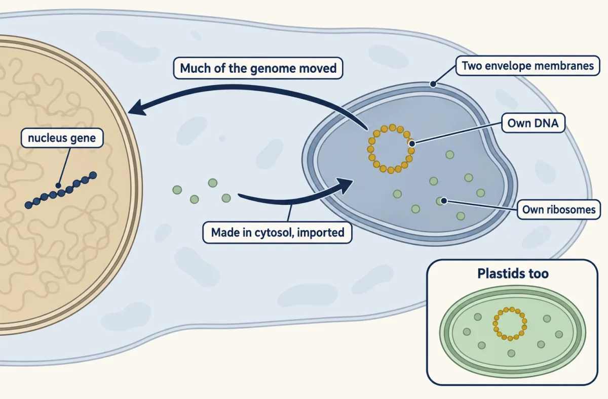
The outward direction is a bacterial inheritance: the inner mitochondrial membrane was the bacterium’s plasma membrane. The evidence for that ancestry is strong. Unlike most organelles, mitochondria and plastids have their own DNA and ribosomes, inside two envelope membranes (three or four around some algal plastids).
Molecular sequences tested the idea. Trees of mitochondrial ribosomal RNA placed mitochondrial sequences within the Bacteria rather than with archaea or the eukaryotic cytosol, and later protein data pointed to the Alphaproteobacteria; the exact branch is still debated. Trees built from many proteins supported a cyanobacterial origin for plastids. Who proposed the idea, the closest living relatives and the branch debate are in Going deeper.
Many ancestral mitochondrial genes were lost, and many others moved to the nucleus by endosymbiotic gene transfer. Most mitochondrial proteins are now encoded in the nucleus, made in the cytosol and imported (protein sorting); human mitochondrial DNA encodes 13 proteins, of more than a thousand that work in mitochondria. Even the respiratory chain’s outward-facing pumps are built mostly from imported parts. The proteome is a mosaic, with many proteins of origins outside the Alphaproteobacteria.
Every argument above assumed you knew which compartment a protein sits in. The second film picks up here: how a lab finds out.
Read the transcript of “Follow the Markers”
Film transcript: Follow the Markers
How do you find which compartment holds a protein? Chapter 1 showed how pumps and leaks help compartments keep distinct chemistries. To relate that chemistry to a protein, first find where the protein sits. A fluorescence image can show a protein at the lysosome. But at is not inside. That image alone cannot tell which side of the membrane it is on. So labs also take cells apart. They break cells and separate the pieces by a series of centrifuge spins.
Organelles overlap across these crude fractions; no fraction is assumed pure. To read the fractions, measure markers, proteins whose home is already known. Histone H3 marks the nucleus. Cathepsin D marks the lysosome. Pyruvate kinase marks the cytosol. Each marker spreads across the fractions in its own shape. A protein that stays associated tends to travel with its compartment. So an unknown is assigned provisionally to the marker group whose full profile it most closely shares. In 2016, Georg Borner's group did this with mass spectrometry. They reported localization information for more than 8,700 HeLa proteins.
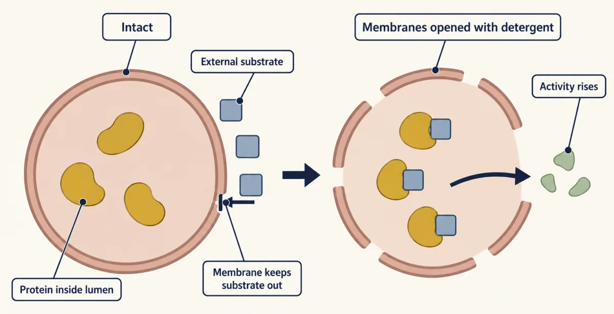
Curated markers guided organellar assignments. Proteins split between compartments can yield mixed profiles and uncertain assignments. So a match is provisional. You would want a second marker that agrees, repeated fractionations, and an imaging check in intact cells. One more test asks whether an enzyme is enclosed by a membrane. In a 1951 report, de Duve's group found acid phosphatase acting less than expected in fresh fractions. More activity appeared once the particles were damaged. Picture the enzyme sealed in the lumen, its substrate outside and unable to cross.
Now add detergent to open the membranes. What happens to the activity? It rises, as long as detergent does not change the enzyme itself. The substrate can now reach it. Activity hidden behind a membrane is called latency.
Following these hidden enzymes across fractions led his group to propose the lysosome in 1955. De Duve later shared the 1974 Nobel Prize in Physiology or Medicine. The wall that holds a lumen's chemistry also hides what sits inside it. So place the protein first, by its whole profile and independent checks. Then ask what its compartment's chemistry does for it.
From the film
- Cell fractionation separates broken cells by repeated centrifuge spins; crude fractions overlap and are not assumed pure.
- Marker proteins of known home, such as histone H3, cathepsin D and pyruvate kinase, each spread across the fractions in their own shape.
- An unknown protein is assigned provisionally by comparing its whole profile with marker groups; split locations can yield mixed, uncertain profiles.
- Latency supports membrane enclosure: if detergent does not alter the enzyme itself, opening the membrane lets an outside substrate reach it.
Which compartment holds a protein?
Now try the second film’s method with numbers: four fractions, F1 to F4, and each protein’s share in each. The numbers are made up for practice; the three markers are ones used in a real HeLa map.
| Protein | F1 | F2 | F3 | F4 |
|---|---|---|---|---|
| Histone H3 (nucleus) | 0.80 | 0.10 | 0.05 | 0.05 |
| Cathepsin D (lysosome) | 0.05 | 0.20 | 0.60 | 0.15 |
| Pyruvate kinase (cytosol) | 0.05 | 0.05 | 0.10 | 0.80 |
| Unknown X | 0.06 | 0.18 | 0.61 | 0.15 |
Predict. Which marker does X travel with, and why is its F3 peak alone not enough to say?
Check your prediction
Compare whole shapes, not one peak. Summed fraction-by-fraction differences: 0.04 against cathepsin D (0.01, 0.02, 0.01, 0.00), 1.48 against histone H3 and 1.30 against pyruvate kinase. X is provisionally lysosomal. Before concluding, run the second film’s confirmation checks (a second matching lysosomal marker, replicate fractionations, imaging in intact cells). Large studies leave some proteins unclassified because they live in more than one compartment (experiments that distinguish).
Design problem
You suspect that a protein lives in the lysosome lumen and needs low pH to work. Design experiments to test both parts.
Saved in this browser only.
One way to do it
Confirm the environment with a lysosome-targeted pH reporter, calibrated in the cells with ionophores.
Place the protein: compare its fractionation profile with at least two lysosomal markers, such as cathepsin D, and with markers of nearby compartments. Test latency, the second film’s detergent test (figure below), using an added substrate that cannot cross the membrane, and check separately that detergent does not change the protein’s activity by itself. Imaging shows the protein is at lysosomes, not inside them; on isolated lysosomes, a protease added outside reaches a lumenal protein only once detergent opens the membrane (protease protection).
Test the pH requirement: add bafilomycin A1 and follow lysosomal pH and the protein’s activity, checking that the protein still reaches lysosomes (its lysosomal-fraction amount is unchanged). In fly cells bafilomycin also inhibits SERCA, so repeat with a second way of raising lysosomal pH, such as depleting a V-ATPase subunit. Activity that falls as pH rises is a correlation; the purified protein’s activity across a pH range in vitro, and a rescue (restoring activity by restoring acidity), argue for cause.
Going deeper
Redox: a level of a different kind
The ER lumen is oxidizing, which lets disulfide bonds form, yet near neutral. Redox describes electrons, not protons, and is kept by ongoing reactions rather than a pump against a leak. In cultured CHO cells, a redox reporter called CY-RL7 was about 88% oxidized in the ER lumen and fully reduced in the cytosol. Redox also depends on which chemical couple you measure. In budding yeast, a glutathione-coupled reporter estimated the cytosol at about −306 mV and the matrix at about −301 mV, so the matrix pool was strongly reducing even though mitochondria make reactive oxygen species; the intermembrane-space pool exchanged with the cytosol through porins, but no appreciable exchange with the matrix was detected. In mammalian cultured cells, the redox state of the peroxisome depended strongly on growth conditions. No reliable peroxisomal pH is given here, so none is stated.
When the pump slows as the gradient grows
The toy model holds the push fixed and ignores membrane voltage. A real V-ATPase moves fewer protons as the electrochemical gradient it works against grows, so a tenfold change in the pump’s capacity, or in the leak, need not shift the pH by a full unit. The direction of each change still follows the balance; the card’s exact numbers do not.
Calcium that rides on another pump’s gradient
A pump that sets one gradient need not set another. In one HEK293 assay, the lysosome’s calcium store refilled after release even with the V-ATPase blocked, but failed to refill when ER calcium was depleted or chelated. That holds for that cell line and assay, and the transporter that takes calcium up was not identified. The lysosome is also a platform for nutrient signals (mTOR and AMPK).
Who proposed it, and where on the bacterial tree
Konstantin Mereschkowski proposed a bacterial origin for plastids early in the twentieth century; in 1967 Lynn Margulis, writing as Lynn Sagan, made the case for both mitochondria and plastids (her spirochete origin for flagella has not been supported).
A 2022 analysis of 108 mitochondrial proteins, using a model that allows for changes in amino-acid composition across sites and branches, favoured mitochondria as sister to the Alphaproteobacteria, excluding two related lineages (MarineProteo1 and Magnetococcia), rather than inside any living group. Other analyses have placed mitochondria inside the Alphaproteobacteria, near the Rickettsiales, or deeper still.
For plastids, the closest cultured relative in 2017 was Gloeomargarita lithophora, a cousin of the plastid line, not its ancestor. Peroxisomes, by contrast with mitochondria and plastids, have no genome; they share fatty acid breakdown with mitochondria (fatty acid degradation).
Your notes
Select any text in the lesson to highlight it or add a note to it. Your highlights and notes are saved in this browser and collected in your notebook.
This browser is not letting the site save anything (a private window or strict privacy settings), so highlights and notes will be lost when you leave the page.
How we measure it
Targeted, calibrated fluorescent pH reporters
A pH-sensitive fluorescent protein is fused to a sequence that sends it to one compartment, its location is checked, and its signal is calibrated inside the cell with ionophores that set the compartment to known pH values. This gives the pH of one compartment in a living cell, as for the HeLa Golgi and matrix.
Compartment-targeted redox reporters
Reporters such as CY-RL7 or Grx1-roGFP2 change their fluorescence with their own oxidation state. Aimed at the ER, the cytosol or the matrix, they show which compartments are oxidizing and which are reducing, and how quickly each recovers from an oxidant. Each reporter reads only a limited range of one redox couple, so the probe must suit the compartment; CY-RL7 was built for the oxidizing ER.
Fractionation profiling against organelle markers
Broken cells are separated into a series of fractions, and the amount of each protein in each fraction is measured, often by quantitative mass spectrometry. A protein is assigned to the compartment whose marker proteins share its whole profile; proteins in more than one compartment may stay unclassified.
Molecular phylogenetics
Ribosomal RNA or protein sequences from organelles, bacteria and the nucleus are aligned and built into trees. Where organelle sequences fall shows their ancestry; composition-aware models test how sensitive that placement is to assumptions.
Practice questions on this lesson (3) → MCAT-style, original, each with an explanation
Where this lesson fits
Before thisReview first
- Transport Across MembranesThis course · Lesson 5
You need pumps, leak fluxes and gradients across a membrane from there to model each organelle's level here.
- Experiments That Distinguish MechanismsThis course · Lesson 3
The fractionation profiles and latency test here apply its logic of choosing experiments that separate mechanisms.
- Membrane Organization and TopologyThis course · Lesson 4
This lesson uses the idea that a bilayer keeps its two faces and lumen separate.
After thisStudy next
- Protein Sorting into CompartmentsThis course · Lesson 7
It explains how proteins made in the cytosol reach the compartment where you have just assigned them.
- Protein Quality Control and Organelle TurnoverThis course · Lesson 9
It builds on the acid lysosome whose pump-maintained pH lets hydrolases degrade material.
- Endocytosis, Recycling and DegradationThis course · Lesson 13
It follows cargo through endosomes whose falling pH, set by the V-ATPase, releases ligands and receptors.
Elsewhere on the siteFilms, simulations and method films in other courses
- The proton-gradient battery — a cross-section of the inner membrane: run electrons through Complexes I→III→IV to charge the gradient, mint ATP, then add an uncoupler and watch ATP collapse while electrons keep flowing as heatSimulation · Biochemistry
It shows the respiratory chain pumping protons out of the matrix, which sets the alkaline matrix pH here.
- Breaking the proton gradientFilm · Biochemistry
It shows how a proton gradient across the inner membrane is built and broken, as with ionophores here.
- A Path Is Not a PumpFilm · Biochemistry
It shows how a leaky bilayer needs pumps to keep a gradient, the core idea of this lesson.
Suggested by an AI model from the lessons themselves, then checked. How we use AI
Finish line
Test yourself
8 questions
Practice and MCAT-style, with explained answers.
Next · Lesson 7
Protein Sorting into Compartments
Made in a test tube with no membranes, a secretory protein comes out longer than the one the cell secretes. Why?
Continue →