Physiology · Skim version
Calcium, Phosphate and Bone
Read the full lesson → Skim the whole course →
The guiding idea
Your kidneys filter about ten grams of calcium a day and you lose about a fifth of a gram in urine. What sets that last fraction?
Almost all the body's calcium sits in bone, yet the parathyroid glands regulate only the small free fraction in plasma.
-
What is regulated: ionized calcium
-
One sensor, one hormone, three doors
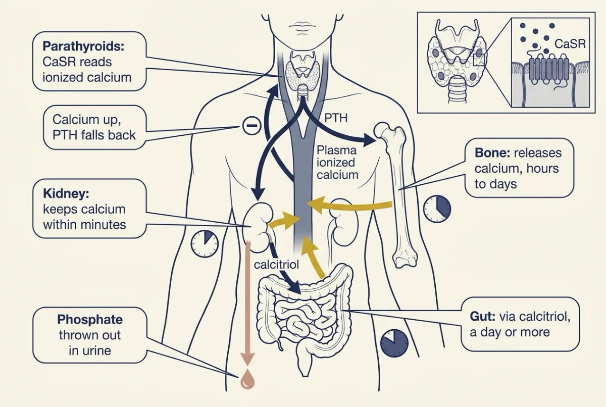
One sensor, one hormone, three doors: when ionized calcium dips, the parathyroid's calcium-sensing receptor lets PTH rise, and PTH brings calcium back through the kidney within minutes, bone over hours to days and the gut, through calcitriol, over a day or more, while the kidney throws phosphate out. AI-generated illustration -
The kidney door, in numbers
-
The bone door: RANKL and OPG
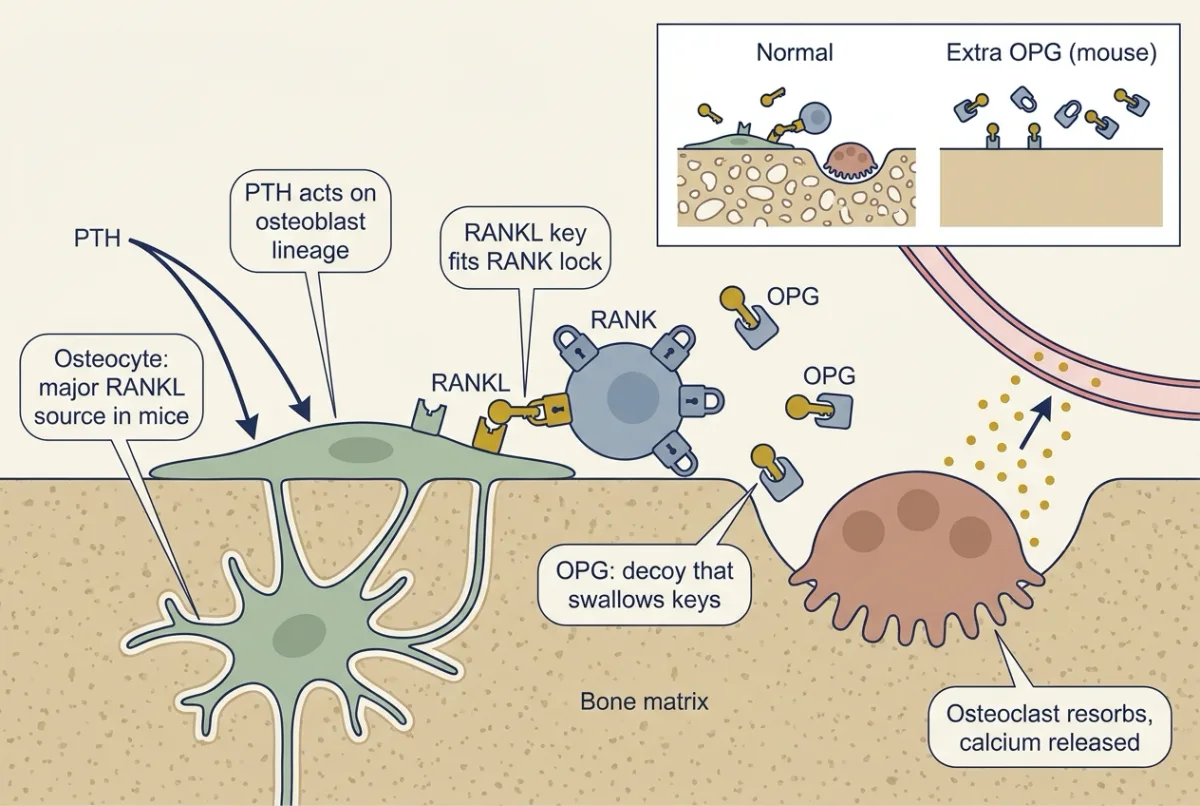
PTH reaches osteoclasts mainly through osteoblast-lineage cells and osteocytes, which display RANKL (the key) for RANK on osteoclast precursors (the lock). OPG is a decoy that swallows the key, so mice making extra OPG resorb too little bone and develop osteopetrosis, an excess of dense bone. AI-generated illustration -
Phosphate’s own brake: FGF23
-
Predicting the disorders from the loop
-
Going deeper
Where this lesson fits
Before thisReview first
- Glomerular Filtration and Renal Blood FlowThis course · Lesson 15
You need GFR and filtered load from there to calculate how much calcium and phosphate the kidney filters.
- Tubular Transport, Segment by SegmentThis course · Lesson 16
The distal and proximal tubule steps for calcium and phosphate here build on how each nephron segment handles ions.
- Endocrine Axes and How to Test ThemThis course · Lesson 2
Reading PTH as high or inappropriately normal uses how feedback axes are tested.
After thisStudy next
- Exercise: The Whole Body at WorkThis course · Lesson 30
Whole-body exercise work draws on the muscle and bone calcium handling set up here, though only loosely.
Elsewhere on the siteFilms, simulations and method films in other courses
- Tap, Drain and a SensorFilm · Organismal Biology
Hormone released into blood, carried to cells with a receptor: the basic pattern PTH and calcitriol follow.
- Cells Compute With SwitchesFilm · Biochemistry
PTH acts through a cell-surface receptor and cAMP signalling, so this shows how that kind of relay works.
- Calcium gradientSimulation · Cell Signaling
Calcium kept low inside cells explains why the free calcium fraction is a regulated signal.
Suggested by an AI model from the lessons themselves, then checked. How we use AI