Developmental Neuroscience · Unit 6: Axons find their way
Choosing a Partner: Layers, Self-Avoidance and Smell
Rewritten on : new text, figures, flashcards and film. Your notes and highlights stay, but the text around them may have moved.
How does a neuron stop in the right layer, find the right partner and keep its own branches from tangling? One fly gene that can make tens of thousands of different surface proteins is part of the answer.
Pause when the reading reaches a film, and offer to play it. Saved on this device.
Watch the film now, or keep listening
Builds on:Pioneers, Guideposts and the Logic of Guidance CuesWhy these?
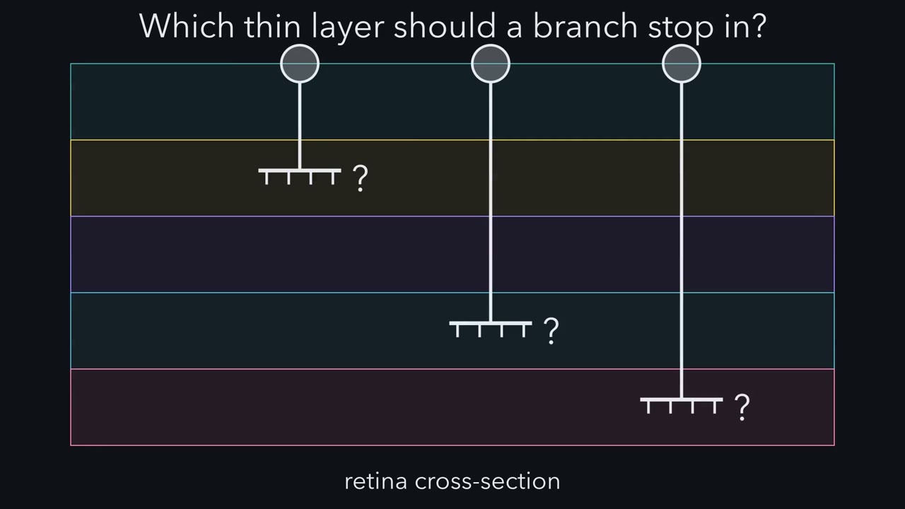 ▶ Watch Same Means Stick · 2:09
Read the transcript of “Same Means Stick”
Film transcript: Same Means Stick
How does a growing branch know which thin layer to stop in? A gradient can bring an axon near its target. But a thin layer is either the right one or not. Choices like that often call for discrete labels. In the retina, the inner plexiform layer is a thin band where three kinds of cell make synapses. It is divided into thinner sublaminae. Each cell type sends its processes into one or a few. In chick retina, two related proteins are Sidekick-1 and Sidekick-2. Largely separate groups of neurons make them. Each group's processes end in different sublaminae. Each protein binds its own kind, which is called homophilic binding. Sidekick-1 sticks to Sidekick-1.
Does the label only mark the band, or help choose it? Predict. Make neurons produce a Sidekick they normally lack. Where should their processes end? Their processes shifted toward the sublamina where that Sidekick was already concentrated. Adding the label shifted the arbor.
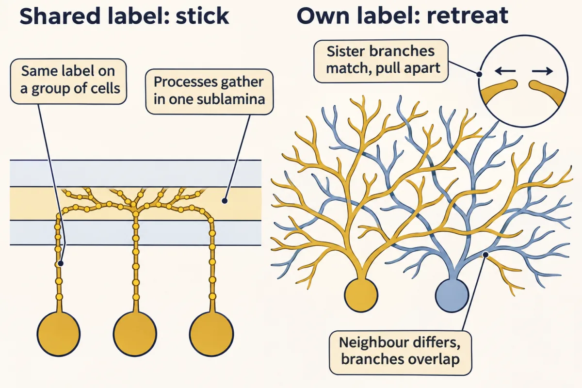
Later, Yamagata and Sanes added Dscam and DscamL, making four labels. These molecules bias a choice. They are one part of how layers form. In mouse retina, cells lacking DSCAM or DSCAML1 still reach the right sublaminae. Here is the idea to carry through this lesson. A match between identical proteins says only, we are the same. The cell decides what same means. When a group of cells shares a sticky label, their processes are drawn together. Here, same means stick. Takeaway: in chick retina, shared sticky labels help like processes settle in matching sublaminae, alongside other cues.
From the film
- A gradient suits continuous order but is poor at discrete choices such as which layer, so discrete labels are often needed.
- In chick retina, making neurons produce a Sidekick they normally lack shifted their processes toward the sublamina rich in that Sidekick.
- A match between identical proteins only says the cells are the same, and the cell decides what same means.
- When a group of cells shares a sticky label, their processes are drawn together; such labels help layers form alongside other cues.
An axon that has reached the right neighbourhood still has to stop in the right layer, find the right partner and keep its own branches apart. Cells do this with molecular labels. One fly gene, Dscam1, can be spliced into 38,016 different proteins, and a neuron uses them to recognise its own branches.
The previous lesson ended with axons arriving at roughly the right place in a target, sorted by gradients. Arriving is not the end of the trip, for a retinal axon reaching the tectum or a bipolar cell process reaching the inner retina. This lesson is about the labels that finish the job.
Two words first. A neuron’s processes are its axons and dendrites together. Its arbor is its whole branched tree.
Labels, not gradients
A neuron can learn where it belongs from a gradient or from a label. The topographic maps lesson was about gradients. A gradient is good at continuous order: neighbouring points in the eye map to neighbouring points in the tectum. It is poor at discrete choices. A thin layer is either the right one or not, and a glomerulus either belongs to one receptor channel or does not. Discrete choices often call for discrete labels.
Think of a conference with name tags. A gradient is like telling people to sit in rows by age. A label is like seating people at the table whose sign matches their tag.
Most of the labels in this lesson are proteins that bind identical copies of themselves, and the same kind of match turns up in two jobs that look like opposites.
The one idea to carry through this lesson. A match between identical surface proteins only tells two membranes “we are the same”. The cell decides what “same” means. If a group of cells shares the label and the response is to stick, their processes are drawn together, which helps them settle in the same sublamina. If each cell carries its own combination and the response is to retreat, that cell’s own branches spread apart while it still overlaps its neighbours.
The rule comes from a few tested systems: Sidekick-1, Sidekick-2, Dscam and DscamL in chick retina (stick), and fly Dscam1 and mouse gamma protocadherins in self-avoidance (retreat). These labels are a bias, not an address book: they shift where processes end, and they are one part of how layers form, not the whole of it. Two cases stretch the rule, mouse DSCAM and smell, and each is flagged where it comes up.
Same means stick: layers in the retina
The inner plexiform layer of the retina is a thin band where bipolar cells, amacrine cells and retinal ganglion cells make synapses. It is divided into thinner sublaminae, and each cell type sends its processes into one or a few of them. A cell that ends in the wrong sublamina meets the wrong partners.
In chick retina, Masahito Yamagata, Joshua Weiner and Joshua Sanes found in 2002 that two related proteins, Sidekick-1 and Sidekick-2, are made by largely separate groups of retinal neurons, and that the processes of each group end in different sublaminae. Both are members of the immunoglobulin (Ig) superfamily, cell-surface proteins built from repeated Ig-like domains. Each binds its own kind, which is called homophilic binding. Cells carrying Sidekick-1 stick to other cells carrying Sidekick-1.
A protein found in a layer might only mark it, or it might help choose it. The key test was misexpression: making a protein in a cell that normally lacks it.
Predict. Make a group of chick retinal neurons produce a Sidekick they do not normally make. If the label only marks the layer, what happens to their processes? If it helps choose the layer, where should they end?
Check your prediction
The processes did not stay where they were. They shifted toward the sublamina where that Sidekick was already concentrated: adding the label moved the arbor.
This is a test worth recognising: move the signal, and see whether the outcome follows. A signal that passes it is called instructive, a term the lesson on the neurotrophic hypothesis uses later.
In 2008 Yamagata and Sanes extended the set to four proteins, adding Dscam and DscamL, each marking a different group of cells and sublaminae. Reducing their levels disturbed the order.
Keep the scope in view. The four-protein code was described in chick. Later work in mouse retina from the same laboratory tied Sidekick-2 to one particular circuit between an amacrine cell type and a ganglion cell type, but mouse cells lacking DSCAM or DSCAML1 still reach the right sublaminae, so mouse sublaminae are not simply assigned by these four proteins.
A badge only you wear: self-avoidance
Read the transcript of “A Badge Only You Wear”
Film transcript: A Badge Only You Wear
How does a neuron keep its own branches from tangling, while still overlapping its neighbours? Take one neuron's arbor. Its branches often spread evenly, without crossing or bundling. This is self-avoidance. Sister branches from the same cell touch, then move apart.
Yet fly sensory neurons share the body wall with other classes, and their dendrites overlap. At every contact, a branch must ask: is this me, or someone else? Predict. A protein binds only identical copies, and binding makes branches pull apart. What must it be like?
Every branch of one neuron must carry the same version, so sister branches always match. Neighbouring neurons must carry different versions, so their branches seldom match. The fly has such a protein, from one gene, Dscam1. It comes in many isoforms.
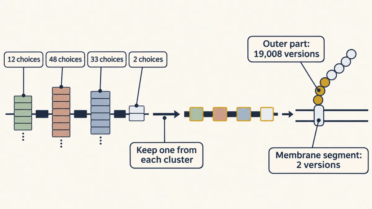
Exons are the stretches of a gene kept in its messenger RNA. Four of Dscam1's exons come as clusters of alternatives. Each messenger RNA keeps one exon from each cluster. That gives 19,008 outer surfaces, and 38,016 isoforms in all. The fourth cluster builds the stretch that crosses the membrane, so it does not change the outer surface. Each isoform binds mainly to identical isoforms. But matched branches pull apart. Same means retreat. Remove Dscam1, and a neuron's own branches cross and bundle. Take a neuron that lacks Dscam1 and give it a single isoform, and its own branches spread apart again. Predict. Now give every neuron the same single isoform.
What happens between neighbours? Neighbours that carry the same single isoform cannot tell themselves from each other. The logic predicts they avoid each other. Flies whose Dscam1 could make only one outer surface had severely disorganised circuits. Which isoform a neuron carries matters little. Differing from its neighbours is what matters.
Why thousands? Picture each neuron drawing about twenty badges from a bowl. From a small bowl, a neighbour carries many of your badges. From a huge bowl, a neighbour carries almost none. In the huge bowl, sister branches match completely and a neighbour only slightly. Flies need thousands of isoforms to wire normally. Mouse DSCAM is not spliced this way. Instead, starburst amacrine cells use gamma protocadherins, adhesion proteins of the cadherin family. Without them, each cell's own dendrites cross and clump. In starburst cells lacking the protocadherins, give every cell the same single isoform. Each arbor separates, but neighbours avoid each other too.
With only about 58 genes, recognition is thought to compare the whole combination, not single isoforms. Takeaway: a label unique to one cell, read as retreat, keeps that cell's own branches apart.
From the film
- Self-avoidance means a neuron's own sister branches move apart while its branches still overlap those of other neurons.
- Fly Dscam1 can be spliced into 38,016 isoforms that bind mainly to identical isoforms, and matched branches pull apart.
- Which isoform a neuron carries matters little; what matters is that its own branches match and its neighbours carry different ones.
- In the mouse starburst amacrine cells tested, gamma protocadherins are needed for a cell's own dendrites to avoid each other.
A layer needs a label that many cells share. The next problem needs the reverse: a label that belongs to one cell alone.
Take a single neuron’s arbor of dendrites. In many neurons the branches spread evenly over the cell’s territory, without crossing or bundling with one another. This is self-avoidance. It requires each branch to recognise sister branches from the same cell and move away from them. Yet some neurons must do this while their branches lie across the branches of other neurons. Fly sensory neurons share the body wall with sensory neurons of other classes, and their dendrites overlap. In mouse retina, each starburst amacrine cell, a retinal interneuron named for its radiating dendrites, overlaps many neighbours of its own type.
So at every contact a branch faces one question: is this me, or someone else?
Predict. Suppose every branch carries a surface protein that binds only identical copies of itself, and that binding makes the touching branches pull apart. What must be true of the protein if a neuron is to avoid itself but still overlap its neighbours?
Check your prediction
Every branch of one neuron must carry the same version, so that sister branches always match. Neighbouring neurons must carry different versions, so that their branches seldom match. The protein has to come in many versions, and each neuron needs a surface identity that differs from its neighbours’.
The fly has such a protein. Forms of one protein made from a single gene are called isoforms. In 2000 Dietmar Schmucker, James Clemens, Huidy Shu, Lawrence Zipursky and colleagues described Dscam in Drosophila, now called Dscam1, and found that it makes isoforms on a vast scale. Exons are the stretches of a gene that are kept in its messenger RNA. Four of Dscam1’s exons come as clusters of alternatives, with 12, 48, 33 and 2 members, 95 alternative exons in all. Each messenger RNA keeps exactly one exon from each cluster, a choice called alternative splicing:
12 × 48 × 33 = 19,008 outer domains; × 2 membrane-spanning choices = 38,016 isoforms
The first three clusters build parts of the ectodomain, the outer part of the protein, the part that meets other cells. The fourth cluster builds the stretch that crosses the membrane, so it does not change the outer surface. In 2004 Woj Wojtowicz, Clemens, Zipursky and colleagues showed that Dscam1 isoforms bind mainly to identical isoforms. Binding is homophilic, as with Sidekick, but here the outcome is repulsion: when two branches carrying the same isoforms touch, they pull apart.
The two requirements, recognition (sister branches match) and diversity (neighbours differ), can be tested separately.
| What the fly neurons carry | A neuron’s own branches | Branches of neurons that normally overlap it |
|---|---|---|
| Many isoforms, a different mix in each neuron (normal) | spread apart | overlap |
| No Dscam1 | cross and bundle | overlap |
| One neuron lacking Dscam1, given back a single isoform; its neighbours normal | spread apart | overlap |
| The same single isoform in every neuron | spread apart | ? |
The second row is the 2007 loss-of-function result, reported by three groups (Matthews, Grueber and colleagues; Hughes, Schmucker and colleagues; Soba, the Jans and colleagues). The third row shows that one isoform is enough for self-recognition: what matters is that every branch of one cell carries the same one.
Predict. Fill in the last row.
Check your prediction
Neighbours that carry the same single isoform cannot tell themselves from each other, so their branches meet as if they were sister branches, and the logic predicts they avoid each other. In 2007 Daisuke Hattori, Ebru Demir and colleagues made flies whose Dscam1 could produce only one ectodomain isoform. Circuits in these flies were severely disorganised. Which isoform any one neuron carries matters little. What matters is that its neighbours carry different ones.
Why thousands? The size of the menu matters only for telling yourself from others. Each neuron appears to express a small, partly random subset. Suppose it uses about 20 isoforms chosen at random from the menu. Then a randomly chosen neighbour carries only a small fraction of them:
| Menu size | Expected shared isoforms (about 20 × 20 ÷ menu) | Fraction of a neuron’s own isoforms that a neighbour also carries |
|---|---|---|
| 48 | about 8 | about 40% |
| 1,152 (an intermediate menu, for comparison) | about 0.35 | under 2% |
| 19,008 | about 0.02 | about 0.1% |
Sister branches match on every isoform, so a large menu keeps accidental matches with neighbours rare. In 2009 Hattori and colleagues found that thousands of isoforms are needed for normal wiring.
Read the table as a scale argument, not a model of the real threshold. Estimates of how many isoforms one neuron makes range from a handful to tens, and some exons are used far more often than others, which makes the effective menu smaller. Because a neuron touches many neighbours, it will often share an isoform with one of them by chance, which suggests that normal recognition tolerates some sharing (Going deeper works the numbers). One reading is that one shared isoform is a weak signal and a mostly matching set a strong one. So “unique” means a distinguishable combination, not zero overlap: sister branches carry matching sets, while a neighbour can share some isoforms and still differ.
Same problem, different molecules: the mouse
Watch a trap here. Vertebrate DSCAM and DSCAML1 are not spliced this way and cannot supply this diversity. They still matter for spacing: in mouse retina, loss of DSCAM or DSCAML1 makes the dendrites of particular cell types bundle and their cell bodies clump. There DSCAM acts as a label shared by a whole cell type, not as a per-cell identity, and its job seems to be to stop like cells sticking to each other too much rather than to push them apart, unlike the family’s sticky role in the chick layer code. Whether a DSCAM match is what triggers that effect is not established, so this case only stretches the rule.
For individual identity in the cases tested, such as starburst amacrine cells, mouse relies on the clustered protocadherins, adhesion proteins of the cadherin family, about 58 genes in three clusters (alpha, beta and gamma), expressed in different combinations in different neurons. In 2012 Julie Lefebvre, Sanes and colleagues showed that starburst cells need the gamma protocadherins for their dendrites to avoid each other. Without them, each cell’s own dendrites cross and clump.
Predict. Now, in starburst cells lacking the gamma protocadherins, give every cell back the same single isoform. What happens to each cell’s own branches, and to the overlap between neighbouring cells?
Check your prediction
Each cell’s branches separated, but neighbouring cells also began to avoid each other. It is the fly’s last row again, with different molecules: matching isoforms make neighbours look like self.
A sharp student will ask how 58 genes can do the job of 19,008 ectodomains. If one shared isoform were enough to count as “same”, a family this small would make most pairs of neurons match. Comparing whole combinations, rather than single isoforms, would help, and it is a leading model: recognition between cells is thought to require a match across the whole assembly of protocadherins on each membrane. In one proposed picture, protocadherins from the two membranes link into a zipper-like chain that keeps growing only while the isoforms match, and a mismatched isoform can stop it (Going deeper has the arithmetic).
A receptor that is also an address: smell
Read the transcript of “A Receptor That Is Also an Address”
Film transcript: A Receptor That Is Also an Address
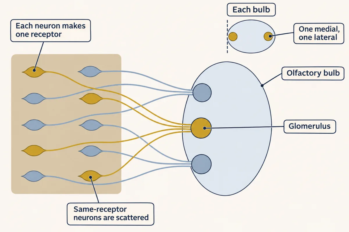
Can the protein that detects a smell also tell its axon where to go? In 1991, Linda Buck and Richard Axel found the odorant receptor genes. In the nose, the rule is one neuron, one receptor, with rare exceptions. Neurons with the same receptor are scattered within a zone of the nose.
Yet their axons gather in a few glomeruli in the olfactory bulb. A glomerulus is a ball of neuropil, where sensory axons meet bulb neurons. In 1996, Peter Mombaerts and colleagues marked axons from neurons using the P2 promoter.
They converged on two glomeruli in each bulb, one medial, one lateral. One receptor, about two glomeruli. The receptor protein also reaches the axon terminals. Could it be part of the address? Predict. Delete the P2 receptor, but keep the marker. Where do the axons go?
They wandered, and failed to form a P2 glomerulus. So the receptor is needed. A caveat: neurons without a working receptor often switch on a second receptor gene. Predict. Put the M12 receptor in place of P2. Where do the axons go now?
They converged near the P2 glomerulus, and far from M12's own. So the receptor helps choose, and other features of the neuron count too. Here the shared label is the receptor itself. Unlike Sidekick, it is not known to bind matching receptors on other axons.
In layers and in self-avoidance, a match says same, and the cell decides what same means. Smell shares the logic of a shared label, but no receptor-to-receptor match is known; how the receptor sorts axons is still being worked out.
Takeaway: in smell, receptor identity helps axons of one channel converge. Other features of the neuron count too.
From the film
- In mouse, olfactory sensory neurons that express the same odorant receptor are scattered in the nose, yet their axons gather in about two glomeruli per bulb.
- Deleting the P2 receptor left its axons wandering without forming a P2 glomerulus, so the receptor is needed.
- Putting M12 in place of P2 sent axons near the P2 glomerulus, not M12's own, so the receptor helps choose the target and other features of the neuron also count.
The retina-to-tectum map preserves where things are. The olfactory map sorts what things are: it is mainly a map of qualities, with only coarse spatial order.
In 1991 Linda Buck and Richard Axel identified a large family of genes in rat that encode odorant receptors. They are G protein-coupled receptors, and in rodents the family runs to about a thousand genes. How binding an odour leads to an electrical signal is covered in the senses lesson. Here the question is wiring.
Two rules describe the wiring, one in the nose and one in the bulb.
- One neuron, one receptor. Each olfactory sensory neuron in the nose expresses one odorant receptor gene, with rare exceptions.
- One receptor, a few glomeruli. Neurons expressing a given receptor are scattered within a zone of the epithelium, yet their axons gather in a few glomeruli in the olfactory bulb.
A glomerulus is a ball of neuropil, a tangle of axons, dendrites and synapses with no cell bodies, where many sensory axons meet the dendrites of bulb neurons. The second rule came first from in situ hybridisation by Vassar, Axel and colleagues and by Ressler, Sullivan and Buck in 1994. In 1996 Peter Mombaerts and colleagues in Axel’s laboratory engineered mice in which neurons using the P2 receptor promoter, the stretch of DNA that switches the gene on, also made a marker that fills the axon. The P2 axons converged on a pair of glomeruli in each bulb, one on its medial side and one on its lateral side. In the mature bulb each glomerulus receives axons that mostly express one receptor, and each receptor’s axons reach about two glomeruli per bulb.
So the receptor is the shared identity of a smell channel. It fits this lesson’s rule only by analogy: unlike Sidekick, it is not known to bind matching receptors on other axons. It is thought to act through other surface molecules it controls, which sort the axons; one version of that idea is disputed (Going deeper). And the receptor protein is found on the axon terminals as well as the dendrites. Could the smell detector also be part of the address?
Predict. You replace the coding sequence of the P2 odorant receptor in mouse with a marker, so neurons that use the P2 promoter make the marker but no P2 receptor. Predict where their axons go. Then predict what happens if, instead, you put a different receptor’s coding sequence in place of P2’s.
Check your prediction
Start from what each experiment keeps and what it removes. Both keep the P2 promoter, so the labelled neurons are those that chose the P2 gene, in the same zone of the nose. The intended change is the receptor protein.
If the promoter or the neuron’s position decided the target, the receptor-less axons would still converge on the P2 glomerulus. They do not. In the deletion experiments reported by Fan Wang, Axel and colleagues in 1998, the labelled axons failed to form a P2 glomerulus and wandered instead of converging. So the receptor is needed. One complication: neurons left without a working receptor often switch on a second receptor gene, which makes the deletion harder to read than the swap.
If the receptor were the whole address, a swapped receptor would send the axons to the donor receptor’s own glomerulus. That is not quite what happened. In the 1996 swap, with M12 in place of P2, the axons converged near the P2 glomerulus and far from M12’s own. In a 1998 swap, with P3 in place of P2, they converged on a glomerulus touching the P3 glomerulus. So the receptor moved the axons, but not to a fixed donor address: it is part of the label, and other features of the neuron also count.
Putting it together
Gradients give continuous position across a sheet; labels give discrete membership: a layer, a glomerulus, or self versus other. The same homophilic chemistry draws Sidekick processes together and drives Dscam1 branches apart, because the cell decides what a match means.
| System | Label | Shared by | Response to a match | Key experiment | What it does not establish |
|---|---|---|---|---|---|
| Chick inner plexiform layer | Sidekick-1, Sidekick-2, Dscam, DscamL | a group of cell types | stick | misexpression shifts the arbor toward the matching sublamina (2002); reducing the proteins disturbs the order (2008) | that these proteins assign layers on their own; mouse cells lacking DSCAM or DSCAML1 still reach the right sublaminae |
| Fly sensory neurons | Dscam1 isoforms, thousands of them | one neuron (its own mix) | retreat | no Dscam1: own branches cross and bundle; one ectodomain in every neuron: circuits disorganised (2007); thousands needed (2009) | how much matching triggers retreat |
| Mouse retinal spacing | DSCAM, DSCAML1 | a whole cell type | like cells stick less (whether a match triggers this is not known) | loss: dendrites of particular types bundle and cell bodies clump | a per-cell identity, active retreat, or that a DSCAM match triggers the effect |
| Mouse starburst amacrine cells | gamma protocadherins | one neuron (its combination) | retreat | loss: own dendrites cross and clump; one isoform in every cell: neighbours avoid each other (2012) | that every mouse neuron works this way |
| Mouse olfactory sensory neurons | odorant receptor | all neurons that chose one receptor gene | no receptor-to-receptor match known; axons converge on about two glomeruli per bulb | deletion: axons wander (1998); M12 in place of P2: a new glomerulus near P2 (1996); P3 in place of P2: one touching P3 (1998) | that the receptor binds other receptors, or that it is the whole address |
None of this is final. Maps and connections set up by these molecules are refined later in life by activity, a subject for the lessons on spontaneous activity and synapses.
Design problem
In the retina of a new model animal you find a cell-surface protein, Z, that binds only identical copies of itself. Design experiments that tell whether Z is a layer label (shared, stick), a spacing label shared by one cell type (shared, stops like cells over-sticking) or a per-cell identity (unique to each cell, retreat). Say what each experiment can and cannot show.
Saved in this browser only.
One way to do it
Walk the table above, with Z in each row.
- Who carries it, in what forms? Map which cells make Z, and whether it comes in many forms used in different mixes by different neurons. One form shared by a group of cells points to a layer or spacing label. Many forms in different mixes point to a per-cell identity. Expression alone shows neither a role nor a response.
- Test the binding. Use homophilic binding assays: cells or outer domains carrying one form, tested against the same and different forms. This shows how strictly each form binds its own kind, not what the cell does after binding.
- Label single neurons. With single-neuron labelling, score three things separately: where the arbor ends in the layer, how often the cell’s own branches cross or bundle, and how much it overlaps neighbours of its type.
- Remove Z. Arbors in the wrong sublaminae suggest a layer label. Own branches that cross and bundle while still overlapping neighbours suggest self-avoidance. Dendrites of one type bundling together, with cell bodies clumping, suggest a shared spacing label that normally stops excess adhesion; bundling alone does not show active retreat. A normal-looking result does not rule out a role. Confirm that Z was removed at the right stage, then consider redundant cues, compensation and how sensitive the assay is.
- Move Z. Make Z in a cell type that lacks it. A layer label should shift the arbor toward the sublamina rich in Z: the misexpression test from the Sidekick section.
- Remove the diversity. If Z comes in many forms, make every neuron carry the same one. A per-cell identity predicts that each cell still avoids itself, but neighbours now avoid each other too.
Going deeper
Stages of targeting
Targeting can be broken into four stages: reach the target region, slow down and branch, find the right place, and choose the right partner. In cortical axons, branches often form at sites where the growth cone paused.
Position from neighbours
Besides labels and gradients, a neuron’s position can be set by its neighbours. Competition between neighbouring axons is taken up in the lesson on competition for targets.
Why some isoform sharing may be tolerated
Take the full fly menu of 19,008 ectodomains and suppose each neuron uses 20 of them at random. The chance that one neighbour shares at least one isoform is about 2%. But a neuron touches many neighbours. With 20 touching neighbours, the chance that at least one shares at least one isoform is about a third. With 50 isoforms per neuron it is above 90%. If a single shared isoform were enough to cause retreat, many neurons would be pushed away from some neighbour. These calculations motivate the idea that recognition tolerates some sharing; they do not measure the biological trigger.
How 58 protocadherin genes can be enough
The roughly 58 clustered protocadherin genes include about 53 alternate ones, generally chosen at random in each neuron, and five C-type ones whose expression is more fixed. Suppose, as a model, that each neuron uses 10 to 15 of the 53 alternate ones at random. Two neurons would then share at least one isoform 90% to 99.8% of the time, while the chance that they carry exactly the same set is between about 1 in 20 billion and 1 in 6 trillion. The arithmetic shows why comparing whole combinations helps; it does not prove that an exact match is the only rule that could work.
A disputed model of receptor-driven targeting
One model holds that cAMP made by the receptor’s odour-independent activity, not by odour binding, sets how much of some guidance receptors the axon makes (see the cAMP lesson). That model rests on a few receptor lines, and an independent test of its neuropilin-1 step did not support it.
Your notes
Select any text in the lesson to highlight it or add a note to it. Your highlights and notes are saved in this browser and collected in your notebook.
This browser is not letting the site save anything (a private window or strict privacy settings), so highlights and notes will be lost when you leave the page.
How we measure it
Gene-targeted knock-in reporters
A marker such as tau-lacZ is placed into a gene in mouse so that every neuron using that gene's promoter also makes the marker, which fills the axon. Staining then shows where those axons end. Replacing the gene's coding sequence at the same time shows what the protein itself contributes.
Misexpression and knockdown in developing retina
An adhesion protein is made in cells that normally lack it, or reduced in cells that normally have it, while the cells are still growing. Comparing where their processes end in the inner plexiform layer shows whether the protein biases sublaminar choice.
Single-neuron labelling
Only one or a few neurons in a field are made to express a fluorescent protein, so that one cell's whole arbor can be traced. This is how self-avoidance is scored: you count how often a cell's own branches cross or bundle, separately from its overlap with neighbours.
Homophilic binding assays
Purified outer domains or whole cells carrying one isoform are tested for binding to the same or a different isoform, by aggregation or by binding of soluble protein. A grid of such tests shows how strictly each form binds only its own kind.
Practice questions on this lesson (3) → MCAT-style, original, each with an explanation
Where this lesson fits
Before thisReview first
- Pioneers, Guideposts and the Logic of Guidance CuesThis course · Lesson 22
It sets out how axons read cues along their path, which this lesson extends to the final choice of layer and partner.
After thisStudy next
- Synapse Elimination: One Muscle Fiber, One AxonThis course · Lesson 37
It takes the matching of axon and target further, as competing axons on one muscle fiber lose or keep their synaptic sites.
- Synapses in the Brain: Adhesion, Scaffolds and AstrocytesThis course · Lesson 36
It builds on partner choice by showing how adhesion proteins and scaffolds then turn a contact into a synapse.
- Activity Before Experience: Spontaneous Retinal WavesThis course · Lesson 38
It covers the activity-driven refinement of maps that this lesson says follows label-based targeting.
Elsewhere on the siteFilms, simulations and method films in other courses
- One in Two ThousandFilm · Sensory Systems
It shows how each olfactory neuron picks one receptor gene, the choice that later sets its glomerulus address.
- What the Bulb SendsFilm · Sensory Systems
It follows the glomerular map of receptor identity that these axons build in the bulb.
- A Cell Cannot Name a MoleculeFilm · Neuroscience
It shows one receptor type per olfactory neuron, with separate lines for each receptor, the basis of glomerular sorting.
Suggested by an AI model from the lessons themselves, then checked. How we use AI
Finish line
Test yourself
8 questions
Practice and MCAT-style, with explained answers.
Next · Lesson 28
Too Many Neurons: Naturally Occurring Cell Death
In Hamburger's raw counts, a chick embryo has just over 20,000 motor neurons in the column that serves its leg at the peak, and about 40% of them die over the next three days. Nothing is wrong with the embryo. How could anyone tell those neurons died, and what decides how many stay?
Continue →← Lesson 26 · Topographic Maps: Sperry's Eye and Eph Gradients