Developmental Neuroscience · Unit 8: Building synapses
Synapses in the Brain: Adhesion, Scaffolds and Astrocytes
Rewritten on : new text, flashcards and film. Your notes and highlights stay, but the text around them may have moved.
A kidney cell that makes one neuronal protein, neuroligin, can persuade an axon to build a nerve terminal on it. So what happens to the synapses of a mouse that lacks neuroligins 1, 2 and 3?
Pause when the reading reaches a film, and offer to play it. Saved on this device.
Watch the film now, or keep listening
Builds on:The Neurotrophic Hypothesis TestedThe Growth Cone: The Tip That Steers the AxonWhy these?
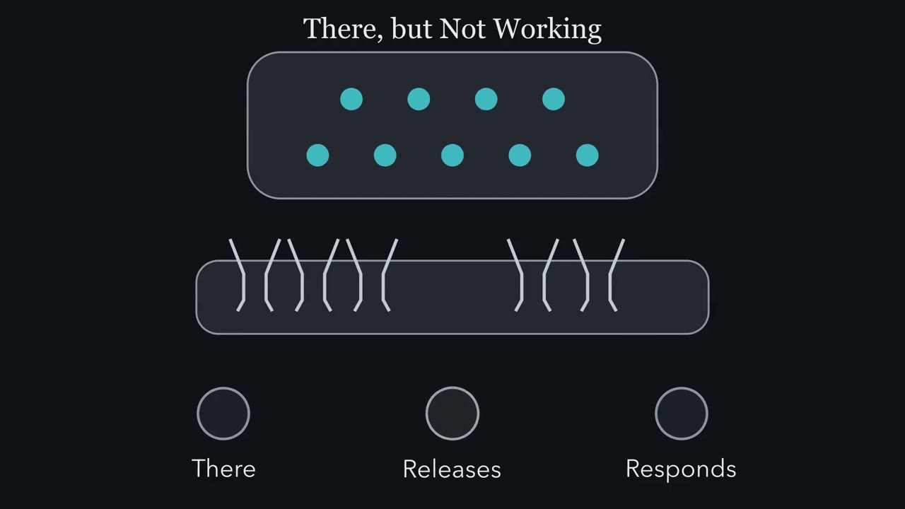 ▶ Watch There, but Not Working · 2:34
Read the transcript of “There, but Not Working”
Film transcript: There, but Not Working
When is a synapse finished? When it is there, when it releases, or when the other side responds? A mature muscle fiber is usually contacted by one motor axon, at one site, using one transmitter. A neuron in the brain receives thousands of synapses. Excitatory and inhibitory synapses share one dendrite, so each contact must specify its kind. A finished synapse does three things. It is there. It releases transmitter. And the other side responds. They can come apart, and each needs its own measurement. A microscope shows it is there. A dye taken up by recycling vesicles shows release. A recording shows the response.
Take stargazer, a mouse that tosses its head upward and has seizures. In its cerebellum, granule cells receive synapses from mossy fibers. AMPA receptors carry most fast excitatory current. NMDA receptors open when glutamate and a co-agonist are bound. Near rest, magnesium from outside blocks most of their current.
In stargazer, these synapses give NMDA responses but no AMPA responses. Predict: which property failed? Not being there. The synapses are present. Not release either. An NMDA response needs glutamate, so transmitter is being released. What failed is one kind of response. The same synapse responds through one receptor and not the other.
The gene encodes stargazin, a small membrane protein. It brings AMPA receptors to the cell surface. Its tail binds PSD-95, a scaffold under the synapse, and that holds the receptors there. A count would miss this defect. A recording shows it, and so does labeling the receptors. So carry one idea through this lesson. A synapse that is there is not yet a synapse that works. Takeaway: each experiment sees only the properties it measures.
From the film
- A synapse that is there is not yet a synapse that works.
- A finished synapse is there, releases transmitter and responds, and each property needs its own measurement.
- In stargazer cerebellar granule cells the synapses are present and release but give NMDA and no AMPA responses, so a count alone would miss the defect.
A kidney cell does not make synapses. Give it one neuronal protein, neuroligin, and the axons that touch it in a dish build nerve terminals on it.
The result came from Peter Scheiffele and Tito Serafini’s laboratory in 2000, and it sets the questions for this lesson. What tells a growth cone to become a nerve terminal? What builds the receiving side? What do astrocytes add? And one question to hold: if neuroligin can start a terminal on a kidney cell, what do you expect in mice that lack neuroligins 1, 2 and 3? Fewer synapses, normal synapses, or synapses that are there but do not work? Keep your answer for the section on dish and mouse.
Three things a working synapse does
A finished synapse has three properties, and each needs its own measurement.
- It is there. A nerve terminal sits against a receiving site. You see it by electron microscopy, or by staining for presynaptic and postsynaptic proteins that sit side by side.
- It releases. The terminal releases transmitter from vesicles and retrieves them. You see it with a dye or antibody label that recycling vesicles take up, or by a response on the other side.
- It responds. Receptors on the receiving side open when transmitter arrives. You see it by recording currents from the receiving cell. A synapse can respond through one kind of receptor and not another.
Two more words come from the lesson on the neurotrophic hypothesis. A factor is sufficient if adding it is enough to produce an effect in a system that is otherwise ready, under the conditions tested. It is necessary if the effect does not happen without it. A dish with one protein on a bare cell tests sufficiency. A knockout mouse, with a gene disrupted, tests necessity.
The one idea to carry through this lesson. A synapse that is there is not yet a synapse that works. A signal can change more than one of the three properties, and each experiment sees only the ones it measures. The rule is about central excitatory and inhibitory synapses as they form and first mature, in the systems this lesson uses: brainstem and cortex in the knockouts, the cerebellar mossy fiber synapse, and purified retinal ganglion cells in culture.
Why central synapses are harder than the neuromuscular junction
Synapse formation has three jobs: (1) making selective connections, (2) differentiating a nerve terminal, and (3) building a postsynaptic apparatus. Partner choice, the first, is the subject of the lesson on partners, layers and self-avoidance. This lesson takes the second and third in the brain and spinal cord, the central nervous system.
At a mature mammalian neuromuscular junction (see the lesson on the neuromuscular junction), a muscle fiber is usually contacted by one motor axon at one site, and the synapse uses one transmitter, acetylcholine, with one main receptor.
Predict. A cortical pyramidal neuron receives thousands of synapses from many neurons. Excitatory synapses that use glutamate and inhibitory synapses that use GABA sit on the same dendrite. Why can a single signal that says “cluster receptors here” not be enough?
Check your prediction
A neuron in the brain differs from the muscle fiber on every count. Each kind of synapse on that dendrite has its own receptors, so “cluster receptors here” does not say which receptors. Each contact has to specify what kind of synapse it will be.
Both partners also arrive partly ready. Neurons manufacture many synaptic building blocks before contact. Presynaptic proteins travel down the axon in preassembled transport packets. In culture, a growth cone that never meets a partner can still cluster vesicles and release transmitter, and a dendrite with no axon nearby can carry small clusters of receptors, sometimes with scaffold proteins. Each side can form its specialization alone, but stable synapses require both: contact enlarges and stabilizes what each side began. Think of two halves of a bridge built out from opposite banks: each can stand for a while, but only joined do they carry traffic and last.
Signals that turn a growth cone into a nerve terminal
Many adhesion proteins span the synaptic cleft, among them cadherins, nectins and SynCAM. The question is which of them only hold membranes together and which also instruct, acting as organizers: signals that tell a partner to build its side of a synapse.
A well-studied case is the mossy fiber synapse of the cerebellum. Neurons in the pons, part of the brainstem, send axons called mossy fibers to cerebellar granule cells, and the granule cells make three signals that act on them:
- Neurexin and neuroligin. Neurexins are axon-side membrane proteins; neuroligins, made by granule cells, are their dendrite-side partners. In Scheiffele’s experiment, kidney cells made neuroligin and were grown under axons from explants of the pons. Where an axon crossed such a cell, vesicle proteins clustered. When the axons were depolarized, the clusters took up antibodies against the inside face of the vesicle protein synaptotagmin, which they can reach only when a vesicle opens to the outside. So vesicles were being released and retrieved there. Control cells, which the axons also touched, did not do this. Soluble beta-neurexin, which binds neuroligin and keeps the axon’s own neurexin from reaching it, blocked the effect. The signal also runs the other way: Ann Marie Craig’s laboratory reported in 2004 that neurexin on a non-neuronal cell clusters postsynaptic proteins in dendrites that touch it.
- Wnt7a and its receptor. In 2000 Anna Hall, Frances Lucas and Patricia Salinas reported that Wnt7a from granule cells makes mossy fiber ends spread and branch and gather the vesicle protein synapsin I. In mice lacking Wnt7a, maturation of the mossy fiber terminals was delayed. Wnt7a signals through Frizzled receptors in other neurons (see the lesson on Wnt signaling), but which receptor the mossy fibers use has not been identified.
- FGF22 and FGFR2. In 2004 Hisashi Umemori, Joshua Sanes and colleagues reported that FGF22, made by granule cells, promotes vesicle clustering in mossy fiber axons through FGFR2, a receptor tyrosine kinase (see the lesson on receptor tyrosine kinases), and that in mice, neutralizing FGF22 and its relatives FGF7 and FGF10, or inactivating FGFR2, reduced presynaptic differentiation of mossy fibers.
Whether the brain needs neuroligin waits until you know how the receiving side is built and how its responses are read.
The receiving side: a scaffold that holds receptors
Two kinds of glutamate receptor matter here. AMPA receptors are the glutamate-gated channels that carry most fast excitatory current (see the lesson on ion channels as receptors). N-methyl-D-aspartate (NMDA) receptors open when glutamate and a co-agonist are bound, but in physiological extracellular magnesium most of their current is blocked near the resting potential; depolarization relieves the block.
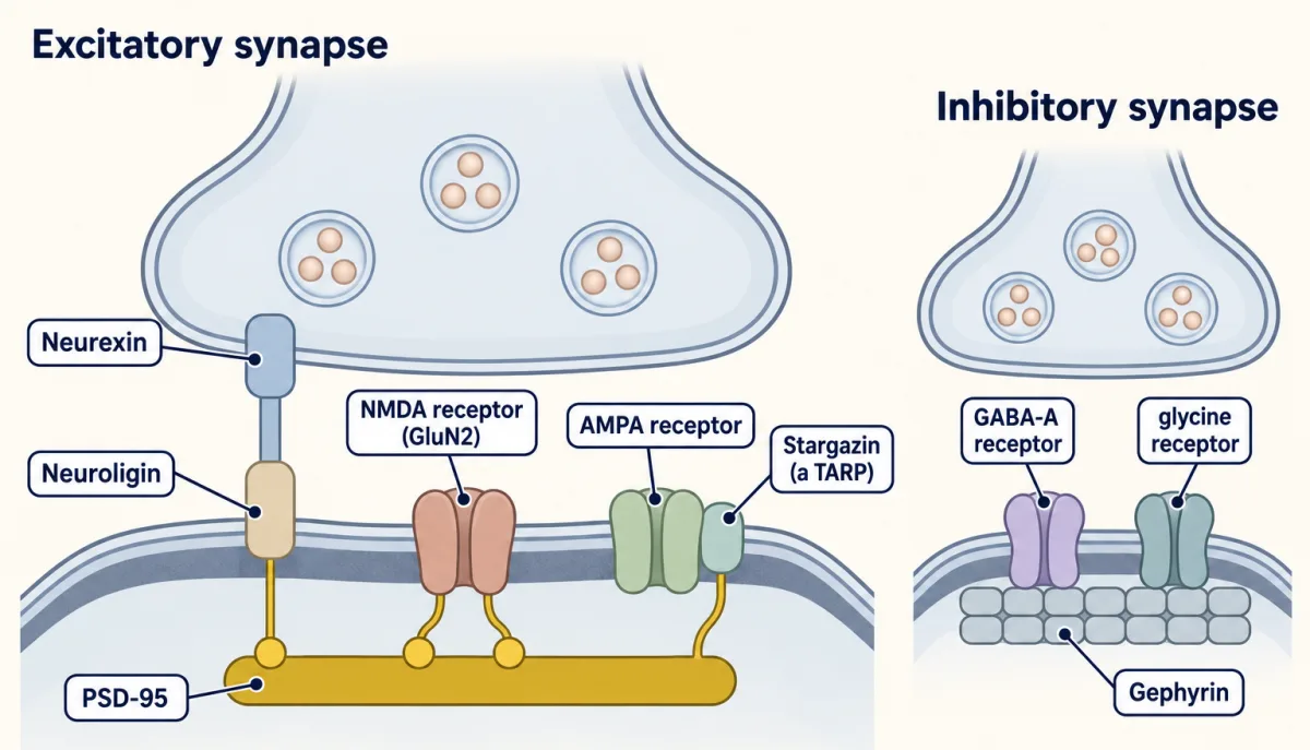
Under an excitatory terminal sits the postsynaptic density, a dense layer of protein under the membrane that shows as a dark thickening in electron micrographs. One of its most abundant proteins is PSD-95, a membrane-associated scaffold (a MAGUK) with three PDZ domains, short modules that grip the last few amino acids of a partner protein (see the lesson on protein interaction modules). The first two PDZ domains bind the tail of NMDA receptor GluN2 subunits. The third binds the tail of neuroligin. A scaffold works like a pegboard: whatever has the right hook hangs in the right place (see the lesson on adaptors and scaffolds).
AMPA receptors do not bind PSD-95 directly. The clue to how they get there came from a mouse. Stargazer is a spontaneous mutant that tosses its head upward and has seizures. Its gene encodes stargazin, a small membrane protein.
Predict. In stargazer cerebellar granule cells, mossy fiber synapses give NMDA receptor responses but no AMPA receptor responses. Which of the three properties has failed: being there, releasing, or responding?
Check your prediction
The synapses are there, and they release: an NMDA response needs glutamate, so transmitter is being released. What fails is one kind of response: the same synapse responds through NMDA receptors but not through AMPA receptors. A count of synapses would miss this defect. A recording shows it, and so does labeling the AMPA receptors.
In 2000 the laboratories of David Bredt and Roger Nicoll reported that stargazin binds AMPA receptors and brings them to the cell surface, and that its tail, which binds PSD-95’s PDZ domains, holds them at synapses. Stargazin became the first of the transmembrane AMPA receptor regulatory proteins, or TARPs. Other TARPs serve other neurons, which is one reason the stargazer defect shows most clearly in cerebellar granule cells.
Inhibitory synapses use a different scaffold. Gephyrin forms dense clusters under the terminal that hold glycine receptors and many GABA-A receptors.
A dish shows what a protein can do; a mouse shows what the brain needs
Read the transcript of “What a Dish Can Show”
Film transcript: What a Dish Can Show
A kidney cell does not make synapses. Can one neuronal protein persuade an axon to build a terminal on it? Neurexin sits on the axon. Neuroligin is its partner on the dendrite. In 2000, Peter Scheiffele and Tito Serafini's laboratory made kidney cells produce neuroligin. They grew these cells under axons from the pons. Where an axon crossed a neuroligin cell, synaptic vesicle proteins gathered into clusters. When the axons were depolarized, the clusters took up antibodies to the inside face of a vesicle protein. So vesicles were being released and retrieved. Control cells did not do this. Soluble beta-neurexin, which binds neuroligin, blocked the effect.
The signal also runs the other way. Neurexin on a bare cell clusters postsynaptic proteins in dendrites. So neuroligin is sufficient, under these conditions, to start a terminal on a bare cell. But is it necessary? Predict. Mice lack the alpha-neurexins, or neuroligins 1, 2 and 3. Fewer synapses, normal ones, or synapses that are there but fail?
You need two measurements. Count the synapses, and record what they do. In 2003, Markus Missler, Thomas Südhof and colleagues reported mice lacking all three alpha-neurexins. Excitatory synapses were near normal in density. Inhibitory synapses were moderately fewer. And the synapses released transmitter poorly.
In 2006, mice lacking neuroligins 1, 2 and 3 were reported. Brainstem synapses were normal in number. But transmission was impaired, more so at inhibitory synapses. Add stargazer, from the first film. Counting found most synapses in place in every mouse. Recording found something wrong in every one. This does not mean deletions never change number. Deleting every neurexin from some interneurons does lower synapse number. Why would the brain manage without it? One likely reason is overlap. Several organizers can start contacts, so removing one family leaves others. Another is that some organizers specify or check contacts, rather than start them.
Takeaway: a dish tests what one protein can start. A mouse tests what the brain cannot do without. Measure function, as well as count.
From the film
- In a dish, neuroligin on a kidney cell is sufficient, under those conditions, to start a presynaptic terminal on it.
- In mice lacking alpha-neurexins or neuroligins 1 to 3, most synapses were present but transmission was impaired.
- A dish tests what one protein can start, and a mouse tests what the brain cannot do without, so measure function as well as number.
The kidney-cell result makes neuroligin sufficient, under these conditions, to start presynaptic differentiation. It does not say whether neuroligin is necessary.
Predict. Mice are made that lack the alpha-neurexins, or that lack neuroligins 1, 2 and 3. What two measurements would tell “fewer synapses” from “synapses that are there but do not work”? Then check the answer you gave at the start of the lesson.
Check your prediction
Count the synapses, and record the currents they produce in the receiving cell.
In 2003 Markus Missler, Thomas Südhof and colleagues reported mice lacking all three alpha-neurexins. They died soon after birth. Their excitatory synapses were present at close to normal density, inhibitory synapses were moderately fewer, and the synapses released transmitter poorly. In 2006 mice lacking neuroligins 1, 2 and 3 were reported to have synapses in normal number in the brainstem, with impaired transmission, more so at inhibitory synapses. They died because breathing failed.
Both results fit best with the answer that these proteins are needed for normal function and maturation more than for forming most synapses.
With stargazer added:
| Result | Synapses there? | Do they release? | Do they respond? |
|---|---|---|---|
| Neuroligin on a kidney cell (dish) | presynaptic vesicle clusters form on the non-neuronal cell | yes: recycling vesicles | postsynaptic response not tested by this assay |
| No alpha-neurexins (mouse) | excitatory near normal; inhibitory moderately fewer | poorly | — |
| No neuroligins 1 to 3 (mouse brainstem) | normal number | not separated: transmission impaired, inhibitory most | not separated |
| No stargazin (stargazer granule cells) | yes | yes: NMDA receptors respond | NMDA yes, AMPA no |
A dash means the result as given here does not test that property separately.
Read down the columns. In every mouse most synapses are present (in stargazer, shown by their N-methyl-D-aspartate (NMDA) responses), and recording found something wrong in every one. A synapse can be present without working, so normal counts never prove normal function. The reverse also matters: without the contact there is nothing to work, which is why deletions that lower number do cause deficits.
Why would the brain manage without a protein that works so well in a dish? One likely reason is that several organizers overlap, so removing one family leaves others to start contacts; those usually named are SynCAM, LRRTMs, cadherins and the secreted Wnts and fibroblast growth factors (FGFs). Another is that some organizers specify or check contacts rather than start them. Neither reading says these proteins never affect number. “No alpha-neurexins” is not “no neurexins”: beta-neurexins were still made in the alpha-neurexin triple knockout, as neuroligin 4 was in the neuroligin 1 to 3 knockout. Later deletions of all neurexins showed that the effect on number depends on the cell type, and in some kinds of interneuron synapse number falls.
Astrocytes: building and switching on
Read the transcript of “Astrocytes Build and Switch On”
Film transcript: Astrocytes Build and Switch On
Purified neurons fire, yet barely talk to each other. What are they missing? Ben Barres's laboratory purified rat retinal ganglion cells, leaving cultures with almost no glia. In 1997, Frank Pfrieger and Barres reported that these neurons fired action potentials. But they showed little synaptic activity. Astrocytes are not the only source. Neurons form some synapses without them. The readout is the mini: the small current from one vesicle released on its own.
Tetrodotoxin blocks action potentials. What remains depends on the synapses, not on how busy the neurons are. With firing blocked, mini frequency is roughly the number of synapses giving detectable minis, times their average release rate. Mini size reflects the receptors facing the vesicle, and the transmitter inside it. Now add a layer of glia that does not touch the neurons. Mini frequency rose about twelvefold. Survival did not change. Predict. More synapses, or more release from each one?
Mini frequency alone cannot say. More minis can mean more synapses, more release, or synapses that start responding. So Pfrieger and Barres also counted synapses by electron microscopy. Glia raised the count only about 2.3 times. If the same share of synapses gave detectable minis, each would release about five times as often. So the paper read most of the rise as more effective synapses. In 2001, Erik Ullian and colleagues reported a sevenfold rise in synapses, in their own cultures. That is far more than the 1997 count, so how much is number and how much is efficacy depends on the culture.
In 2005, Karen Christopherson and colleagues identified thrombospondins as astrocyte proteins that induce synapses. Those synapses looked normal by electron microscopy. They released transmitter. Yet they gave no AMPA receptor responses. They were silent near rest. They lack working AMPA receptors, and any NMDA receptors there would be mostly blocked by magnesium. They add to the count, but add no AMPA minis. Later work found glypicans 4 and 6, also from astrocytes. They recruit AMPA receptors, and the synapses respond.
So astrocyte signals share the work. Thrombospondin mainly builds. The glypicans mainly switch synapses on. Takeaway: a synapse that is there is not yet a synapse that works. Count it, and record from it.
From the film
- Astrocytes add to synapse formation but are not its sole cause, since neurons form some synapses without them.
- In purified retinal ganglion cell cultures, glia raised mini frequency about twelvefold but synapse count about 2.3 times, and the paper read most of the rise as more effective synapses.
- Thrombospondins build synapses that release but are silent near rest, and glypicans 4 and 6 recruit AMPA receptors and switch them on.
Astrocytes add to synapse formation but are not its sole cause: neurons form some synapses without them, and the size of the glial effect depends on the culture system.
Ben Barres’s laboratory could purify rat retinal ganglion cells by immunopanning, which left cultures with almost no glia. In 1997 Frank Pfrieger and Barres reported that these neurons survived, grew and fired action potentials, yet barely spoke to one another.
To read their result you need one tool. A miniature current, or mini, is the small current from one vesicle released on its own. Tetrodotoxin blocks action potentials, which removes release driven by firing, so what remains depends on the synapses and not on how busy the neurons are. Then:
detected mini frequency ≈ (synapses giving detectable minis) × (their average rate of releasing a vesicle on its own)
mini size: with recording conditions fixed, set mainly by the receptors facing the vesicle and the transmitter in it
Detection also depends on recording noise.
A layer of glia across the dish, not touching the neurons, or medium the glia had conditioned, changed the picture. With action potentials blocked, mini frequency rose about twelvefold. Evoked transmission, a separate measurement, failed much less often. Neuron survival did not change, so glia were not simply keeping more neurons alive.
Predict. Did the glia make more synapses, or make each synapse release more often? What would you measure to decide?
Check your prediction
Mini frequency alone cannot say: more minis can come from more synapses, from each synapse releasing more often, or from synapses that become detectable. So Pfrieger and Barres also counted synapses by electron microscopy, and found about 2.3 times as many with glia.
That is far short of twelvefold. If the same share of synapses gave detectable minis in both conditions, each would release about 5 times as often (12 ÷ 2.3 ≈ 5.2), and the paper read most of the rise as more effective synapses. The minis also got larger, so some extra events may simply have become big enough to detect. In 2001 Erik Ullian and colleagues in the same laboratory reported that astrocytes raised synapse number about sevenfold under their own culture conditions and were needed to keep them. That is much larger than the 2.3-fold count of 1997, so how much of the rise is number and how much is efficacy depends on the culture, and neither paper’s count settles it alone.
In 2005 Karen Christopherson and colleagues identified thrombospondins 1 and 2 as astrocyte-secreted proteins that induce synapses. Those synapses looked normal by electron microscopy and released transmitter but were postsynaptically silent: they give no AMPA response near rest. They lack working AMPA receptors, and any N-methyl-D-aspartate (NMDA) receptors present would be mostly blocked by magnesium at that voltage. Such a synapse adds to the anatomical count but not to the synapses giving detectable AMPA minis near rest. And thrombospondin is one of the cases where number does fall: mice lacking both thrombospondins had fewer excitatory synapses.
Later work identified other astrocyte factors, including glypicans 4 and 6, which recruit AMPA receptors and make such synapses respond, and cholesterol carried in lipoproteins. Thrombospondin mainly builds; the glypicans mainly switch synapses on, and these assays do not show a fixed order.
Maturation: GABA changes sign
Even after a synapse responds, what the response does to the cell can change. At the neuromuscular junction the acetylcholine receptor swaps its fetal gamma subunit for the adult epsilon subunit. In the brain, the best-known change is in GABA. In immature rodent hippocampal and cortical neurons, chloride inside the cell is high, because the transporter NKCC1 brings chloride in and the exporter potassium-chloride cotransporter 2 (KCC2) is scarce.
Predict. When GABA-A channels, which pass chloride, open in such a neuron, which way does chloride flow, and what does that do to the membrane voltage? What should change as KCC2 rises?
Check your prediction
Chloride flows out when E(Cl) is above the membrane potential, as it is when internal chloride is high, so GABA depolarizes the cell. E(Cl), the voltage that open chloride channels pull the membrane toward, comes from the Nernst equation for chloride at 37 °C:
E(Cl) = 61 mV × log10([Cl] inside / [Cl] outside)
With 30 mM chloride inside and 130 mM outside, E(Cl) is about −39 mV, above a resting potential near −60 mV, so opening the channel depolarizes. As KCC2 rises over the first postnatal weeks, internal chloride falls. With 7 mM inside, E(Cl) is about −77 mV, and the same channel now hyperpolarizes.
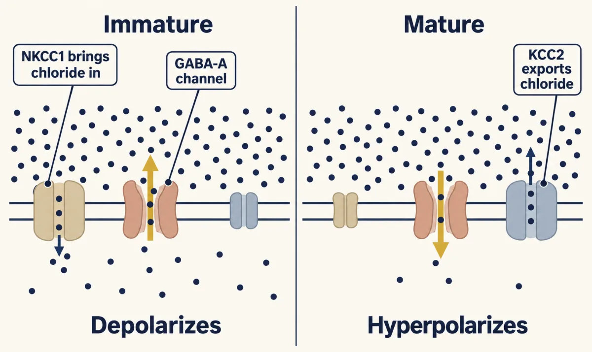
Depolarizing is not the same as exciting, though. An open GABA-A channel also damps firing by shunting current, and in the cortex of newborn mice GABA depolarizes most neurons yet still holds network activity down.
Putting it together
Each signal in the lesson, read against the three properties:
| Signal | Made by | What it changes | Evidence in this lesson |
|---|---|---|---|
| Neuroligin | granule cell (dendrite side) | starts a terminal in the dish; needed for normal transmission | kidney-cell coculture (2000); neuroligin 1 to 3 knockout (2006) |
| Neurexin | axon | clusters postsynaptic proteins in the dish; alpha-neurexins needed for normal calcium-triggered release | non-neuronal cell assay (2004); alpha-neurexin knockout (2003) |
| Wnt7a | granule cell | spreading, branching and synapsin I gathering in mossy fiber ends | terminal maturation delayed without it (2000) |
| FGF22, through FGFR2 | granule cell | vesicle clustering, presynaptic differentiation | reduced when FGF22, FGF7 and FGF10 are neutralized or FGFR2 is inactivated (2004) |
| PSD-95 with stargazin | receiving neuron | holds NMDA receptors and neuroligin; stargazin brings AMPA receptors | stargazer: NMDA responses, no AMPA responses |
| Gephyrin | receiving neuron | holds glycine and many GABA-A receptors | inhibitory synapses |
| Thrombospondins 1 and 2 | astrocyte | build synapses that release but are silent | identified in 2005; double knockout has fewer excitatory synapses |
| Glypicans 4 and 6 | astrocyte | recruit AMPA receptors, so silent synapses respond | later work |
| KCC2 | receiving neuron | lowers internal chloride and moves E(Cl) below rest, so GABA hyperpolarizes | rises over the first postnatal weeks in rodent cortex and hippocampus |
Design problem
You make a kidney cell line produce neuroligin and grow it under cultured pontine or hippocampal axons. What do you measure to show that an axon has built a real presynaptic terminal at the contact? Which two controls rule out mere adhesion? And once the dish works, what experiment in the animal comes next, and what two things must you measure in it?
Saved in this browser only.
One way to do it
Measure two things at each contact. First, stain for synaptic vesicle proteins such as synapsin and synaptophysin, and ask whether they form clusters where the axon crosses a neuroligin-expressing cell more often than where it crosses other cells. Second, test function. Stimulate the axons in the presence of an FM dye, wash, and look for bright spots at the contacts. Dye taken up during stimulation marks vesicles that were released and retrieved, so the cluster is a working terminal and not a pile of parts.
For controls, use cells that are untransfected or that make a different adhesion molecule, which the axons also touch and stick to. If clusters form only on neuroligin cells, stickiness alone does not explain them. Then add soluble neurexin to block the bond in parallel dishes. If clusters fail to form, the effect runs through the neurexin and neuroligin bond. Scheiffele and colleagues used tests of this kind in 2000.
To test necessity, make mice lacking the protein, or, since there are several neuroligins, lacking neuroligins 1, 2 and 3 together. Then both count synapses, by electron microscopy or staining, and record transmission, with evoked and miniature currents. Counting alone would have missed the 2006 result.
Going deeper
Adhesion proteins that span the cleft
Classical cadherins are membrane proteins with five extracellular repeats that bind the same cadherin on the facing cell (homophilic binding) only when calcium is present. Nectins are held inside the cell by afadin. SynCAM is a member of the immunoglobulin superfamily, and SynCAM made by a non-neuronal cell induced presynaptic terminals in contacting axons in a 2002 study, so it passes the same sufficiency test as neuroligin.
Early growth-cone experiments and the calcium signal
In 1983 two groups showed that growth cones of cultured frog and chick neurons release acetylcholine before reaching a muscle, detected with a patch of receptor-rich membrane held near the growth cone.
In cultured hippocampal neurons, Stephen Smith’s laboratory watched the packets carrying active-zone proteins arrive at new contacts.
Is the decision to form a synapse signalled by calcium? In cultured frog spinal neurons, contact with a muscle cell raised free calcium in the neurite, but contact with another neuron did not; in a cultured snail motor neuron, a cAMP analogue mimicked the boost in calcium entry that contact with its target produced. In these experiments the trigger came from the partner, from its surface or from the space between the cells.
Target-derived NGF and postsynaptic assembly in sympathetic ganglia
The signal for the receiving side can come from far away. In mouse sympathetic ganglia, Sharma, Deppmann, Harrington and colleagues reported in 2010 that nerve growth factor from the distant target tissue is needed for postsynaptic assembly in the ganglion neurons, and that postsynaptic assembly is in turn a prerequisite for presynaptic assembly, although preganglionic axons still grow into the ganglion without the growth factor. Signaling endosomes carrying its receptor TrkA travel back from the axon into the dendrites.
Three readouts in the glial add-back experiments
In the 1997 cultures, at one stimulus per second, failures of evoked transmission fell from about one stimulus in five to about one in fifty. Under a simple Poisson model, in which a stimulus fails only when no vesicle is released, those failure rates correspond to an average of about 1.6 rising to about 3.9 vesicles per stimulus, about 2.4-fold. That is close to the 2.3-fold rise in synapse number in the same cultures and far from the twelvefold rise in minis. At faster stimulation the paper’s binomial analysis, which assumed unchanged release probability, required about four to five times as many release sites (4.0 at 5 Hz, 4.8 at 10 Hz) against 2.3 times observed, which it read as more effective release.
The reversal potential of the GABA current
GABA-A channels also pass bicarbonate, so the reversal potential of the GABA current sits a little above E(Cl). If bicarbonate is about a fifth to a third as permeant as chloride, with about 15 mM inside and 25 mM outside, the reversal potential is about −65 to −69 mV in the mature example, still below a −60 mV rest, so the conclusion holds: the channel hyperpolarizes. The lesson uses E(Cl) to keep the arithmetic to one ion.
Your notes
Select any text in the lesson to highlight it or add a note to it. Your highlights and notes are saved in this browser and collected in your notebook.
This browser is not letting the site save anything (a private window or strict privacy settings), so highlights and notes will be lost when you leave the page.
How we measure it
Heterologous synapse-formation assay
A non-neuronal cell line is made to produce one candidate protein and grown with neurons. If axons or dendrites build synaptic specializations only on cells carrying the protein, the protein is sufficient to induce them under those conditions. It says nothing about whether the animal needs it.
Immunopanning
A cell suspension is passed over dishes coated with antibodies that bind unwanted cell types, then over a dish that binds the wanted type. The result is a culture of one kind of neuron with almost no glia, to which glia or single factors can be added back.
Whole-cell recording of miniature currents
A glass pipette sealed onto a neuron records the small currents caused by single vesicles released at its synapses, with action potentials blocked by tetrodotoxin. Their frequency reflects the number of active synapses and how often each releases; their size reflects the receptors on the receiving side and the amount of transmitter in each vesicle.
FM dye uptake
A fluorescent dye in the bath is taken into vesicles as they are retrieved after release. Bright spots after stimulation mark terminals that are cycling vesicles, a functional test that a cluster of vesicle proteins is a working terminal.
Method films · the methods library
Practice questions on this lesson (3) → MCAT-style, original, each with an explanation
Where this lesson fits
Before thisReview first
- The Neurotrophic Hypothesis TestedThis course · Lesson 29
The lesson uses the sufficient versus necessary distinction that was set up there.
- The Growth Cone: The Tip That Steers the AxonThis course · Lesson 3
You need to know how a growth cone behaves before asking what turns it into a nerve terminal.
After thisStudy next
- Synapse Elimination: One Muscle Fiber, One AxonThis course · Lesson 37
Once synapses are built and working, this lesson asks how inputs on one target compete and lose sites.
- Microglia and Complement Prune SynapsesThis course · Lesson 41
It takes up what happens after formation, when glia remove synapses that were built.
- Opening and Closing a Critical PeriodThis course · Lesson 40
It uses the maturing GABA synapse and its shift in sign to explain when plasticity opens.
Elsewhere on the siteFilms, simulations and method films in other courses
- Blips from a Silent NerveFilm · Neuroscience
It shows quantal release and response at a synapse, the release and response properties this lesson separates.
- Three Steps, One DrugFilm · Neuroscience
It covers GABA receptors and how the transmitter is read, linking to GABA changing sign as synapses mature.
Suggested by an AI model from the lessons themselves, then checked. How we use AI
Finish line
Test yourself
8 questions
Practice and MCAT-style, with explained answers.
Next · Lesson 37
Synapse Elimination: One Muscle Fiber, One Axon
A newborn rat muscle fiber has two or more motor axons on one synapse, and every one of them works. Within weeks all but one are gone. What decides which one stays?
Continue →← Lesson 35 · Building the Neuromuscular Junction: Agrin, Lrp4 and MuSK