Developmental Neuroscience · Unit 6: Axons find their way
Topographic Maps: Sperry's Eye and Eph Gradients
Rewritten on : new text, flashcards and film. Your notes and highlights stay, but the text around them may have moved.
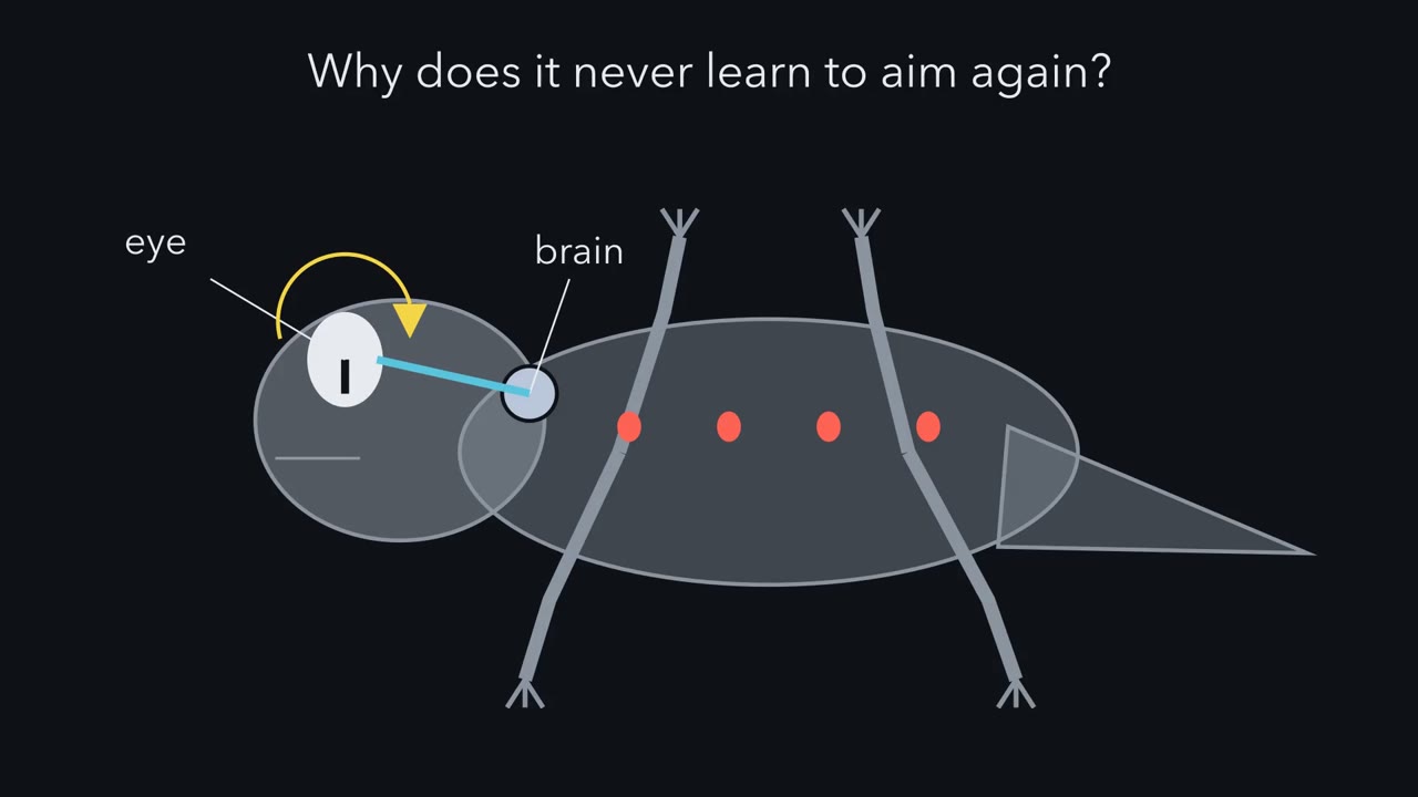
Turn a newt's eye upside down, let the nerve regrow, and it turns its head down toward a lure held above it. Why does it not learn?
Pause when the reading reaches a film, and offer to play it. Saved on this device.
Watch the film now, or keep listening
Builds on:The Growth Cone: The Tip That Steers the AxonPioneers, Guideposts and the Logic of Guidance CuesWhy these?
 ▶ Watch A Rank Against a Slope · 2:16
Read the transcript of “A Rank Against a Slope”
Film transcript: A Rank Against a Slope
Turn a newt's eye upside down and let its nerve regrow. Why does it never learn to aim again? In 1943, Roger Sperry rotated the eyes of adult newts. He cut the optic nerve and let it regrow. When sight returned, a lure held above its head made it turn its head downward. If the fibres regrew at random and use then matched them, what should weeks of missed prey teach?
To aim correctly. It never learned. It kept making the mistake for as long as anyone watched. Sperry favoured the simplest reading: each regrown fibre went back to its old place in the brain. That place is part of a map. Neighbouring cells in the retina connect to neighbouring places in the target. Temporal retina, the side nearer the temple, maps to the front. Nasal retina maps to the back. A growth cone, the moving tip of an axon, senses only what is close. Yet the map spans the target. So how does an axon know where to stop? Three ways are possible. Labels: one matching tag for each cell and each target site. Gradients: a smooth slope in the amount of a molecule gives each position a value.
Neighbours: each axon places itself relative to the axons around it. In chick and mouse, the retina uses gradients and neighbours together. Each axon carries an amount of a receptor called EphA. The target carries a slope of a repellent, ephrin-A. The repellent is low at the front and high at the back. The axon with the most receptor ends where it is lowest. More receptor, less ligand. But where each axon lands also depends on the other axons.
The takeaway: an axon's place is not an address in its own receptor level. It is a rank, set against a slope.
From the film
- An axon's place in the map is a rank set against a slope, not an address in its own receptor level alone.
- In chick and mouse, the retina uses gradients and neighbours together to place its axons.
- More receptor, less ligand: the axon with the most EphA ends where ephrin-A is lowest.
Turn an adult newt’s eye upside down in its socket, cut the optic nerve, and let it regrow. When the newt can see again and you hold a lure above its head, it turns its head downward. It keeps making that mistake for as long as anyone watched.
The newt is not blind and not confused. Its vision has come back in its original order, only rotated, as if the image had been flipped before it reached the brain. So the question for this lesson is: how does an axon know where to stop?
The map, and three ways to build it
The retina sends its axons to a target in the midbrain called the optic tectum in fish, amphibians and birds, and the superior colliculus in mammals. The projection is a topographic map: neighbouring ganglion cells in the retina connect to neighbouring places in the target, so the image on the retina is laid out in order on the brain.
The map has two axes. This lesson follows one of them and states it the same way throughout: temporal retina maps to the anterior target, nasal retina to the posterior target. (The temporal retina is the side nearer the temple.) The second axis runs at right angles: dorsal retina maps to the lateral tectum, ventral retina to the medial tectum.
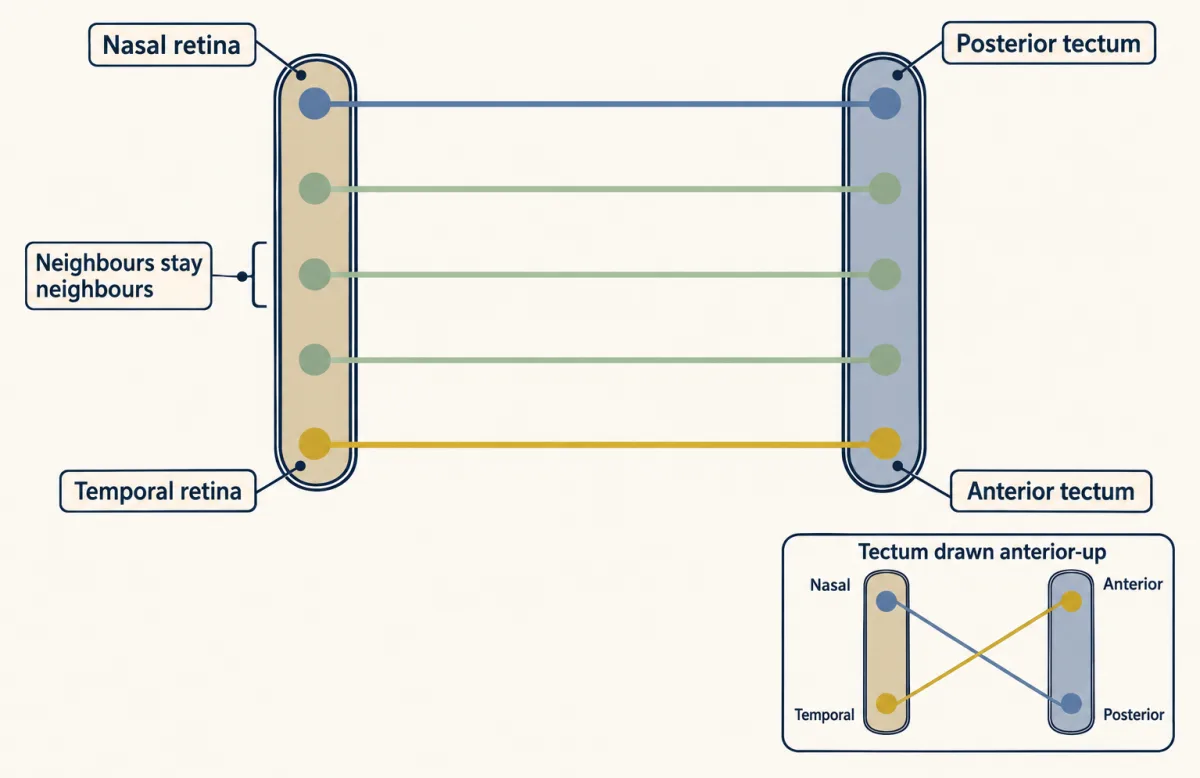
One word needs care. In chick and mouse the main axon does not simply halt at the right place. It first grows past it, then sprouts side branches there. So in this lesson, where an axon “stops” means where it keeps its branches, its termination zone.
A growth cone (the moving tip of a growing axon) can sense only what is close to it, yet the map spans the whole target. There are three ways an axon could know where to stop.
- Individual labels. One matching tag per cell and per target site.
- Gradients. Smooth slopes in the amount of a molecule, which give each position a value.
- Neighbours. Each axon places itself relative to the other retinal axons arriving in the target.
Picture seats in a theatre. Every ticket could name a seat. The lights could dim toward the back, and each person could sit where it is as dark as they like. Or people could line up by height, tallest at the front, and take the seats that order gives them. The rest of the lesson shows that the retina uses the second and third together.
The one idea to carry through this lesson. An axon’s place in the map is a rank set against a slope. Each retinal axon carries an amount of a receptor, EphA; the target carries a slope of its ligand, ephrin-A, which repels the axon and is low at the front and high at the back (both are introduced below). Rank the axons by how much receptor they carry: the axon with the most ends where the repellent is lowest. Where exactly each axon lands depends both on how much ligand is there and on how its signal compares with the other axons’. In short: more receptor, less ligand, and no axon’s place is an address in its own receptor level alone.
What the rule covers. The anterior-posterior axis of the coarse map in chick and mouse. It does not cover the other axis, which uses other cues (Going deeper), or the fine scale, where correlated activity sharpens termination zones (the last section, and the next unit). And how much weight falls on the rank differs between preparations: in zebrafish, a retinal axon with no neighbours can still find roughly the right place, so competition among axons is not essential for coarse targeting there.
Sperry’s newts: does the wiring follow the eye or the world?
Roger Sperry put this question to adult red-spotted newts, Triturus viridescens, in 1943. An adult newt’s optic nerve regenerates, so these were experiments on regrowth, which were then read as a window on how the map is built in the embryo. Many textbooks draw his later frog and goldfish versions, but the 1943 papers used adult newts.
In one paper he rotated the eye 180 degrees and left the optic nerve intact. The newts’ responses to moving objects were inverted and reversed, and stayed that way, which proves little, since the old connections were never broken. In the companion paper he also cut the optic nerve. In one group he cut through the nerve’s sheath as well and twisted the two stumps out of line, so that fibres could not simply follow old channels back. Vision returned after the nerve regrew.
Predict. Suppose regrowing fibres connect more or less at random, and useful function is then matched to them by experience. What should a newt with a rotated eye be doing after weeks of missed prey?
Check your prediction
It should have learned to aim correctly. It had not. Animals with a rotated eye aimed in the wrong direction, by an amount that matched the rotation (those with an unrotated eye aimed correctly), and experience did not correct them. The result argued against random wiring matched by use.
Sperry favoured the simplest reading: the regrown fibres had rebuilt the old map. The behaviour supported position-specific reconnection without proving a molecular mechanism (his adviser Paul Weiss’s resonance idea could also fit it; see Going deeper). Sperry proposed that retinal cells acquire chemical differences according to their position and that their targets carry matching differences. He developed this into the chemoaffinity hypothesis, set out in full in 1963, and suggested that two crossing gradients, one per axis, could provide the coordinates without a separate label for every cell. He did not have a molecule.
Is the cue in the target? The stripe assay
Read the transcript of “Reading the Slope”
Film transcript: Reading the Slope
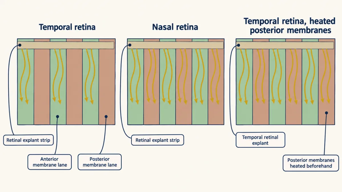
If Sperry was right, the target itself carries a cue. Can we ask an axon to read it? In 1987, Friedrich Bonhoeffer's group built a striped carpet. Membranes from the front and back of chick tectum lay in alternating narrow lanes. A strip of retina lay across them, so each growing axon met border after border. Where will axons from the temporal retina grow?
Temporal axons grew along the front lanes and stayed off the back ones. Nasal axons grew on both. On either membrane alone, both grew well. So the back membrane was not simply a poor surface. Now heat the back membranes first. What will the temporal axons do?
The avoidance vanished. An attractant on the unheated front lanes would have kept it. So the cue is a heat-sensitive repellent at the back of the tectum. In 1995, one paper found a repellent at the back of the tectum, now called ephrin-A5. A second paper mapped the gradient of ephrin-A2, which had been cloned the year before. Ephrin-A rises from front to back across the tectum. Its receptor, EphA, runs the opposite way, high in temporal retina. Why does more receptor mean ending at the front? Take toy numbers. Repulsion is the receptor level times the local ligand. Each axon enters at the front and grows back. It keeps its branches where the repulsion reaches the limit.
Three axons carry low, middle and high receptor. Which reaches the limit first? The axon with the most receptor hits the limit first, at the front edge. The axon with the least ends at the back. One correction. In chick and mouse, axons first overshoot their zone, then sprout side branches. Ephrin-A keeps branches from forming and surviving behind the right zone. The overshooting part is later removed. So where an axon stops means where it keeps its branches, its termination zone. The takeaway: more receptor, less ligand. In the toy, two opposite slopes put the axons in order.
From the film
- In the chick stripe assay, temporal retinal axons avoided posterior tectal membrane, while nasal axons grew on both kinds.
- Heating the posterior membrane removed the avoidance, which pointed to a heat-sensitive repellent at the back of the tectum.
- In the toy model, two opposite slopes, EphA in the retina and ephrin-A in the target, put axons in order.
- Where an axon stops means where it keeps its branches, its termination zone.
Chemoaffinity predicts that the tectum itself carries a position-dependent cue that retinal axons can read. Friedrich Bonhoeffer’s group in Germany built an assay to ask the axons directly. In 1987 Jochen Walter, Bonhoeffer and colleagues made a striped carpet: membranes from anterior and posterior chick tectum laid down in alternating narrow lanes. A strip of retina was placed across the lanes so that each growing axon met border after border.
| Axons from | Offered | Where they grew |
|---|---|---|
| Temporal retina | Alternating anterior and posterior lanes | Along the anterior lanes, off the posterior ones |
| Nasal retina | The same lanes | On both kinds |
| Either part of the retina | One kind of membrane alone | Well on either kind |
The last row matters: posterior membrane was not simply a poor surface. So either anterior membrane carries something temporal axons follow, or posterior membrane carries something they avoid.
Predict. A companion paper heated the posterior membranes before making the lanes. If the cue were an attractant on anterior membrane, what would happen to the temporal axons’ choice? If it were a repellent on posterior membrane?
Check your prediction
Heating the posterior membranes abolished the temporal axons’ avoidance. An attractant on the anterior lanes, which were not heated, would have kept the preference. So the result pointed to a heat-sensitive repellent on posterior membranes rather than an attractant on anterior ones.
The molecules: two opposite slopes
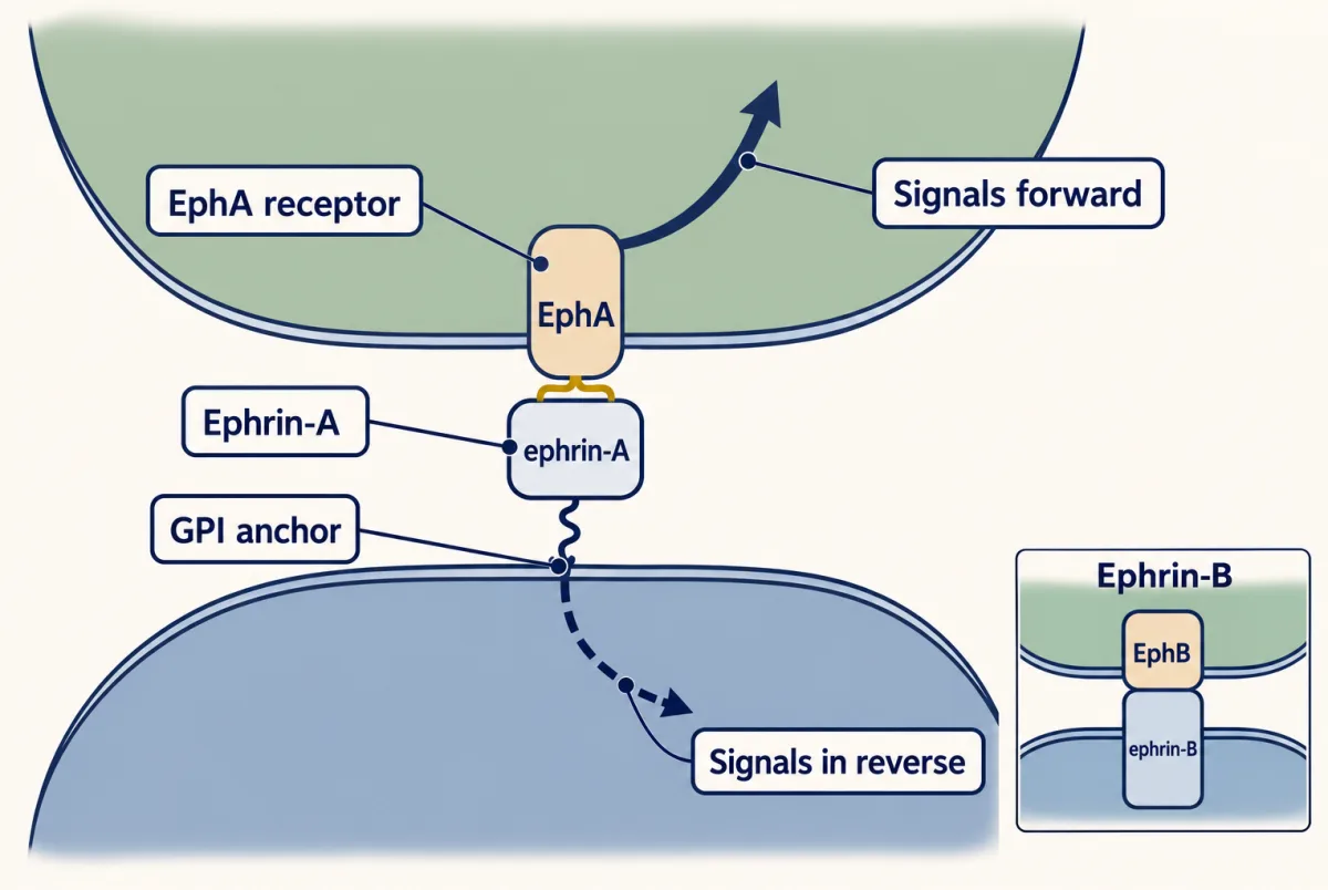
The stripe assay also gave biochemists a test to follow during purification. In 1995 two papers appeared back to back in Cell. Uwe Drescher, Bonhoeffer and colleagues purified a membrane-anchored protein from the posterior tectum that collapsed and repelled retinal growth cones in culture, now called ephrin-A5. Hwai-Jong Cheng, John Flanagan and colleagues reported that a ligand of the Eph receptor family, now called ephrin-A2, rose in a gradient from anterior to posterior across the chick tectum, while its receptor, now called EphA3, ran the opposite way in the retina, high in temporal ganglion cells and low in nasal ones.
Eph receptors are the largest family of receptor tyrosine kinases (that lesson covers how such receptors are switched on). Their ligands, the ephrins, are themselves membrane proteins. Ephrin-As are held to the outer surface by a GPI anchor (a lipid tail) and bind mainly EphA receptors; ephrin-Bs cross the membrane and bind mainly EphB receptors. Because both partners sit on cell surfaces, signalling needs contact, and it can run both ways: the receptor signals forward into its own cell, and the ephrin can signal in reverse into the cell that carries it (Egea and Klein reviewed this bidirectional signalling in 2007; that lesson explains how).
Why more receptor means less ligand
Build the pairing of opposite slopes with toy numbers, chosen to make the arithmetic easy, not measured.
- Let retinal position t run from 0 at the nasal pole to 1 at the temporal pole, with equal numbers of ganglion cells in equal intervals of t. EphA rises in a straight line, 1 + 2t: from 1 unit in the most nasal ganglion cells to 3 in the most temporal. These are toy assumptions, not measured receptor profiles or cell densities.
- Ephrin-A rises in a straight line from 1 at the front edge of the target to 3 at the back edge.
- An axon enters at the front and grows back. The repulsion it feels is its EphA level times the local ephrin-A (more receptor or more ligand, more push). It keeps its branches where that product reaches a limit of 3. An axon whose limit falls outside the target is drawn at the nearest edge.
Predict. Three axons carry 1, 2 and 3 units of EphA. At what ephrin-A level does each reach the limit, and which one ends furthest forward?
Check your prediction
| EphA on the axon | Ephrin-A where EphA × ephrin-A = 3 | Where it keeps its branches |
|---|---|---|
| 3 (temporal) | 1 | At the front edge |
| 2 (middle) | 1.5 | A quarter of the way back |
| 1 (nasal) | 3 | At the back edge |
The axon with the most receptor reaches the limit soonest, so a little ephrin-A repels it and it ends in front. The nasal axon carries little EphA, tolerates more ephrin and ends at the back. In the toy, two slopes running in opposite directions put the axons in order.
Two cautions about the toy. First, ephrin-A keeps branches from forming and surviving behind the right zone, and the overshooting part of the axon is later removed, so the “limit” is where branches can survive, not a wall the growth cone hits. Second, a repellent alone says only how far back an axon may branch, not why it does not branch further forward. The toy simply places each axon at its limit; what pushes axons back toward it, counter-gradients and comparison among axons, comes later. The gradients fit the map; proving them necessary took a mouse.
Is the slope needed? Ephrin knockouts
In 2000 David Feldheim, Jonas Frisén, John Flanagan and colleagues disrupted the genes for ephrin-A2 and ephrin-A5 in mice and traced axons from small patches of retina.
- With either gene gone, the map in the superior colliculus was moderately abnormal.
- With one working copy of each gene, mice still had a defect.
- With both gone, axons from one small patch of retina ended at several places scattered along the axis.
The map loses much of its anterior-posterior order, but not all of it. Termination zones are ectopic (in the wrong place) and multiple, and the projection still reaches the colliculus. The defect grows as ligand is lowered, which says that the amount of ephrin, not merely its presence, carries position. One caveat: the knockout removed the ephrins from the retina as well as the target.
Absolute or relative? Two maps from one retina
Read the transcript of “Two Maps from One Retina”
Film transcript: Two Maps from One Retina
Does an axon read an address in its own receptor level, or its rank among the others? In a normal animal, both readings put the axons in the same order. To tell them apart, change some axons but not their neighbours. In 2000, Arthur Brown, Greg Lemke and colleagues built such a mouse. Isl2 is a gene switched on in about half of the retinal ganglion cells, scattered across the retina. They inserted the receptor EphA3 into that gene. Mouse ganglion cells do not normally make it. Now neighbours at every point in the retina differed.
Use the toy numbers. The knock-in adds four units, so knock-in axons carry five to seven. Their unchanged neighbours carry one to three. First, the absolute reading, a fixed threshold. Where do the knock-in axons go, and their unchanged neighbours?
In the toy, the knock-in axons crowd toward the front. The unchanged axons keep exactly their normal places. Now the relative reading, in its simplest form: pure rank. Where does each group go? Every knock-in axon outranks every unchanged one. So knock-in axons take the front half, and unchanged axons the back. In mice with two copies of the knock-in, Brown and colleagues found two orderly maps. Look at the unchanged axons. Nothing about them was altered, yet they ended somewhere new. An absolute reading cannot produce that. Their place depended on the other axons.
So is the amount of ligand irrelevant? Try a thought test. Lower the ephrin-A everywhere, say by half. What does each reading predict now? The fixed threshold says the map shifts back. Pure rank says nothing moves, because no rank has changed.
Real mice with one working copy of each of two ephrin-A genes kept a normal main zone. But in some animals, temporal axons also formed an extra patch further back. Pure rank cannot give that. The amount of ligand affects how accurately axons map.
So neither toy is the whole story. In mouse and chick, the slope sets the scale. Comparison among axons sets who goes where. Slopes help lay out the coarse map. Correlated activity in the retina helps sharpen it, partly at the same time. That is the next unit.
The takeaway: an axon's place is a rank, set against a slope.
From the film
- In mice with extra EphA3 in a scattered half of ganglion cells, two orderly maps formed, so an axon's place depends on the other axons and not on its own receptor level alone.
- The amount of ephrin-A also matters: with less ephrin-A, mapping was less accurate.
- The ligand slope sets the scale, and comparison among axons decides who goes where on it.
- Slopes help lay out the coarse map, and correlated retinal activity helps sharpen it.
The rule so far can be read in two ways.
- Absolute reading (a fixed threshold). Each axon grows into the target from the front, meets more ephrin-A the further back it goes, and keeps its branches where EphA × ephrin-A reaches a fixed limit. Its place is an address written in its own receptor level, whatever its neighbours do.
- Relative reading, in its simplest form (pure rank). Each axon’s place depends on how its EphA signal compares with the other axons’. The axon with the most signal takes the front of the target, the next takes the space behind it, and so on, spaced evenly by rank. Its place is a rank.
Both readings produce the normal nasal-to-temporal order, so order alone cannot tell them apart. Their spacing can differ: in the toy below, an axon with 2 units ends a quarter of the way back under the fixed threshold and halfway back under pure rank. The direct test of whether neighbours matter is an animal in which some axons change their receptor level and their neighbours do not.
The experiment. Arthur Brown, Greg Lemke and colleagues built one in 2000. Isl2 is a transcription-factor gene that is switched on in about half of mouse retinal ganglion cells, scattered across the retina. They inserted the coding sequence of EphA3, a receptor mouse ganglion cells do not normally make, into Isl2 (a knock-in, which places a chosen coding sequence in a gene so it is made wherever that gene is on). Every Isl2 cell now made EphA3 on top of its usual EphA receptors. At every point in the retina, cells with extra receptor now lay intermingled with cells without it.
Numbers to work with. Use the toy map from the molecules section. The knock-in adds 4 units to a scattered half of the cells, so they now carry 5 to 7; assume an exactly equal random split at each retinal position.
Predict. Fill in the table before opening the answer.
| Absolute (fixed threshold) | Relative (pure rank) | |
|---|---|---|
| Knock-in axons (5 to 7 units) | ? | ? |
| Unchanged neighbours (1 to 3 units) | ? | ? |
Check your prediction
| Absolute (fixed threshold) | Relative (pure rank) | |
|---|---|---|
| Knock-in axons | Need ephrin-A of 3/5 = 0.6 down to 3/7 ≈ 0.43, below the lowest level in the target, so all crowd at the front edge | Every one outranks every unchanged axon, so they form an ordered map in the front half |
| Unchanged neighbours | Same receptor, same slope, so exactly their normal places: a full map across the target | Pushed down the ranking, so they form an ordered map in the back half |
In general the absolute reading predicts one normal map with a second, anteriorly shifted map laid over its front part; an increment as large as the toy’s squeezes that second map against the front edge.
Brown and colleagues traced axons in mice with two copies of the knock-in and found two orderly maps: the knock-in axons in the front half of the superior colliculus and their unchanged neighbours in the back half, together filling it. Look at the unchanged axons: nothing about them was altered, yet they ended somewhere new, which a purely absolute reading cannot explain. Their place depended on the other axons.
The other half of the answer. Is the amount of ligand irrelevant, then? Run the reverse test on the same toy map.
Predict. Halve the ephrin-A everywhere in the target. What does the fixed threshold predict? Pure rank?
Check your prediction
Fixed threshold: every axon must grow further back to reach its limit, so the map shifts backward. In the toy the most temporal axon now ends in the middle of the target, and axons from the nasal half of the retina run out of target. Pure rank: nothing moves, because no axon’s rank has changed. (This is a prediction of the target-only halving toy, not a measured displacement or magnitude in the mutant mice.)
Real mice are not a clean halving. Mice with one working copy of each of the ephrin-A2 and ephrin-A5 genes (the other copy disabled) have less of those two ligands, in the retina as well as the target, and other ephrin-As remain. Feldheim, Flanagan and colleagues found that temporal axons in these mice still formed their main termination zone at the normal site, but in some animals also formed an extra patch further back. They read the defect as an effect of absolute ligand level. Neither toy predicts exactly that, but pure rank predicts no change at all, so it cannot give it.
The knock-in rules out a fixed address in the axon’s own receptor level; the dosage result rules out pure rank. Together they give the rule this lesson started with: an axon’s place is a rank set against a slope. The ligand gradient supplies the scale, and the comparison among axons decides who goes where on it. Real relative models, such as the ratio model in Going deeper, can include the amount of ligand. This is the “neighbours” mode working inside a gradient: local rules produce global order partly because axons also interact with each other.
What the knock-in does not tell you is how the axons compare themselves. Contact between neighbouring axons through their own EphA and ephrin-A, competition for space or for a limited trophic or synaptic resource, and activity-dependent stabilisation could each produce it. They are not mutually exclusive; which dominates, and whether it differs between fish, chick and mouse, is not settled.
Where slopes stop being enough
Early on, a retinal axon’s branches in the colliculus spread over a broad zone around the right place. Correlated spontaneous activity in the retina then sharpens them. In mice lacking the β2 subunit of the nicotinic acetylcholine receptor, whose retinal waves are abnormal, axons reach roughly the right region but their termination zones stay large and diffuse, as McLaughlin and colleagues found in 2003 and Cang and Feldheim reviewed in 2013. Chemistry lays out the map and activity sharpens it, though the two act at overlapping times and interact: in the EphA3 knock-in mice, retinal waves help decide how the two maps are organised. The next unit takes up how those waves work.
Try it
Pick a genotype and watch where each axon ends.
Rule to keep: an axon with more EphA stops sooner against rising ephrin-A, and where cells differ, each axon is placed by its signal relative to its neighbours. Positions are in relative units, and the picture is a sketch of the logic, not data; the shading of the bar is only a sketch.
Design problem
Give every retinal ganglion cell the same extra amount of EphA. What does an absolute reading predict? A pure rank reading? Then design an experiment that could tell a pure rank rule from a rule that compares ratios of signal.
Saved in this browser only.
One way to do it
Absolute reading: every axon reaches its limit sooner, so the whole map shifts toward the front. In the toy, adding 1 unit sends every axon that had 2 units or more, the temporal half, to the front edge.
Pure rank reading: no axon changes rank, so the map stays normal.
Ratio reading: the order is kept, but a fixed increment shrinks the ratio between neighbours most where EphA is low. Two nasal neighbours at 1 and 1.1 units differ by 10%; after the increment, at 2 and 2.1, by 5%. Two temporal neighbours at 2.9 and 3 differ by about 3.4%; at 3.9 and 4, by about 2.6%. So the spacing could change, most in the nasal part of the map.
An experiment: change only a scattered half of the cells, with a small increment (for example one copy of the knock-in instead of two), and trace axons from nasal and temporal retina separately. One extra unit changes the receptor ratio between changed and unchanged cells from 2 at the nasal pole to 4/3 at the temporal pole. Whether their projections then stay apart needs an explicit discrimination rule. For example, a model that tells two populations apart only when their receptor ratio exceeds 1.5 would keep nasal populations distinct and merge temporal ones; that threshold is invented, not fitted. Predicting physical spacing needs a further rule turning ratios into positions. Under pure rank, separation depends only on how many axons have levels between the two populations; in the toy that is the same at the two ends of the retina and larger in the middle. Compare the two predictions across several retinal positions. Reber, Burrola and Lemke fitted one to crosses of a related kind (see Going deeper); this particular comparison is the toy’s prediction, not their test.
Going deeper
The other axis
EphB receptors, high in ventral retina, and ephrin-B ligands, high in the medial target, help sort axons along it, together with Wnt signalling and adhesion molecules, and much of its order is set before axons reach the target. Loss of ephrin-A2 and ephrin-A5 has been described as disturbing this axis too, though the main defects reported in 2000 were along the anterior-posterior axis. Ephrin-B also acts at the optic chiasm, in that lesson.
How might axons compare themselves?
In 2004 Michael Reber, Patrick Burrola and Lemke combined the Isl2-EphA3 allele with a knockout of EphA4 to vary receptor levels further and fitted the results with a relative signalling model, in which each axon’s ending depends on the ratio of its net EphA signal to that of the axons around it. The model allowed quantitative predictions of how a map shifts when receptor levels change. It describes what the axons do; it does not say how they compare themselves.
One proposal for the missing counterforce uses counter-gradients: ephrin-As are also expressed in the retina, high nasally, and EphAs in the tectum, high anteriorly, and ephrin-A reverse signalling in axons has been proposed to supply a posterior push. Without some counterforce, axons that simply moved away from a repellent would all pile up at the front.
Weiss, resonance and the Nobel Prize
In Paul Weiss’s resonance principle, fibres connect diffusely but each centre responds only to matching signals. Sperry shared the 1981 Nobel Prize in Physiology or Medicine, with David Hubel and Torsten Wiesel, for his separate work on the divided cerebral hemispheres.
The first ephrin names
The 25 kilodalton protein Drescher, Bonhoeffer and colleagues purified in 1995 was called RAGS. Cheng, Flanagan and colleagues called their ligand ELF-1 and its receptor Mek4. Cheng and Flanagan had cloned ELF-1 in 1994, as a ligand for the receptors Mek4 and Sek; the 1995 paper reported its gradient across the tectum. Under the names agreed in 1997, RAGS is ephrin-A5, ELF-1 is ephrin-A2, and Mek4 is EphA3.
Your notes
Select any text in the lesson to highlight it or add a note to it. Your highlights and notes are saved in this browser and collected in your notebook.
This browser is not letting the site save anything (a private window or strict privacy settings), so highlights and notes will be lost when you leave the page.
How we measure it
Stripe assay
Membranes from two regions of a target are laid down in alternating lanes, and a strip of neural tissue is placed across them. If axons grow along one kind of lane, the two regions differ in a cue the axons can read. Treating one membrane (by heat or an enzyme) first shows whether the cue is an attractant on one or a repellent on the other.
Alkaline phosphatase fusion probes
The extracellular part of a receptor or ligand is joined to an enzyme that makes a coloured product. Applied to tissue, the probe stains wherever its binding partner sits, so a gradient of binding sites can be seen directly.
Anterograde axon tracing with DiI
A small crystal or injection of a fluorescent lipid dye is placed in one patch of retina. The dye spreads along the membranes of those axons to their endings, showing where that patch terminates in the tectum or superior colliculus.
Knock-in and knockout mice
A knockout disables a gene, usually by deleting or interrupting part of it. A knock-in inserts a chosen sequence at a targeted gene; this one placed a coding sequence under the control of another gene, so it is made only where that gene is active. Here a knock-in gave half of the ganglion cells extra EphA3 while their neighbours kept normal levels.
Practice questions on this lesson (3) → MCAT-style, original, each with an explanation
Where this lesson fits
Before thisReview first
- The Growth Cone: The Tip That Steers the AxonThis course · Lesson 3
You need how growth cones sense and respond to nearby cues to follow how axons read ephrin slopes here.
- Pioneers, Guideposts and the Logic of Guidance CuesThis course · Lesson 22
The logic of guidance cues, including repulsion, is assumed when ephrin-A is treated as a repellent.
After thisStudy next
- Activity Before Experience: Spontaneous Retinal WavesThis course · Lesson 38
Correlated retinal waves sharpen the coarse ephrin map into fine termination zones, as this lesson ends by saying.
- Choosing a Partner: Layers, Self-Avoidance and SmellThis course · Lesson 27
It extends target choice to layers and partners, where axons must find the right address after coarse mapping.
- Ocular Dominance and the Critical PeriodThis course · Lesson 39
It shows how activity from the two eyes refines retinal projections after chemical cues set the coarse map.
Suggested by an AI model from the lessons themselves, then checked. How we use AI
Finish line
Test yourself
8 questions
Practice and MCAT-style, with explained answers.
Next · Lesson 27
Choosing a Partner: Layers, Self-Avoidance and Smell
How does a neuron stop in the right layer, find the right partner and keep its own branches from tangling? One fly gene that can make tens of thousands of different surface proteins is part of the answer.
Continue →