Developmental Neuroscience · Unit 6: Axons find their way
Pioneers, Guideposts and the Logic of Guidance Cues
Rewritten on : new text, flashcards and film. Your notes and highlights stay, but the text around them may have moved.
Kill a guidepost cell on a grasshopper embryo's leg, and the first axons to grow past it lose their way.
Pause when the reading reaches a film, and offer to play it. Saved on this device.
Watch the film now, or keep listening
Builds on:The Growth Cone: The Tip That Steers the AxonSeeing Neurons: Golgi, Cajal and the Neuron DoctrineWhy these?
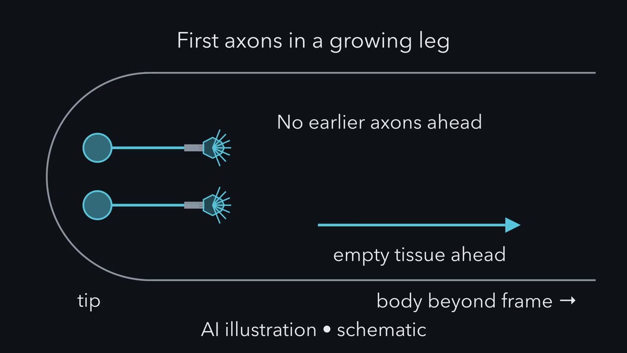 ▶ Watch A Route in Legs · 2:24
Read the transcript of “A Route in Legs”
Film transcript: A Route in Legs
The first axons start out through the far end of a growing leg. No earlier axons lie ahead of them. How do they find the way? The moving tip of a growing axon is the growth cone. It does not carry a map. It is roughly 10 µm across, and its filopodia reach farther. It can touch what lies against it. And it can compare how much of a cue reaches its two sides. Take the simplest case. One cell releases an attractant into uniform tissue. The growth cone heads straight up its gradient. What shape is the path?
straight line to the source. In this ideal case, the cue falls off equally in every direction. So uphill always points at the source. And at the source, the climb ends. Add a second source, and the path can curve. Where it ends depends on where it starts. Either way, an attractant gives a destination, not a list of stops.
Real routes reach a place and go on from it. The first axons out of an insect leg make several sharp turns. A turn like that usually means the growth cone has started reading something new. A cell it touches, a boundary in the tissue, or a different cue. Reach adds a second limit. A secreted cue thins as it spreads. Far enough away, the difference across a growth cone is lost in noise. Simple models with plausible values put that at hundreds of micrometres to a few millimetres. It is an estimate, not a fixed range. So here is the rule. A growth cone reads only what is within its reach. So a long route is usually built as a chain of legs. Each leg ends at a choice point, where the growth cone picks its next path.
There it reads something new, or reads the same thing differently. At many choice points, growth cones pause and spread out before they choose. Not every bend needs a new reading. A pioneer axon can lead followers around long curves.
From the film
- A growth cone reads only what is within its reach.
- So a long route is usually built as a chain of legs.
- Each leg ends at a choice point, where the growth cone reads something new or reads the same thing differently.
In the growing leg of a grasshopper embryo, the first two axons to head for the central nervous system have no earlier axons to follow through the far end of the leg. Kill a particular guidepost cell along their route before they get there, and they lose their way.
That result, from David Bentley and Michael Caudy in 1983, is a clue to how a growth cone navigates. It does not carry a map. It reads what is around it, a short stretch at a time, and some of what it reads is other cells. This lesson answers three questions. What can a growth cone read? Why are long routes built from legs, and what marks the end of each leg? And why can the same signal mean different things to different axons?
The one idea to carry through this lesson. A growth cone reads only what is within its reach. So a long route is usually built as a chain of legs. Each leg ends at a choice point, a place where the growth cone picks its next path by reading something new, or by reading the same thing differently.
“Read something new” means a guidepost cell such as Cx1 below, a tissue boundary, the next bundle of axons, or a different cue that takes over. “Read the same thing differently” means a change in the growth cone’s receptors or internal state, as when netrin-1 attracts one growth cone and repels another (the last section).
What a growth cone can read
The growth cone is the moving tip of a growing axon: a fan of actin with fingers called filopodia, backed by microtubules from the axon shaft. It advances when adhesion grips the substrate, and it turns when that grip differs between its two sides (see the growth cone). The question here is what tells it which side to favour.
Guidance cues sort by two questions. Is the cue fixed to a cell or to the matrix between cells, so that it acts on contact, or is it released and spread through the tissue, so that it acts at a distance? And does it draw the growth cone in or push it away?
| Draws the growth cone in | Pushes it away | |
|---|---|---|
| On contact (fixed to a cell or the matrix) | Contact attraction. Laminin in the extracellular matrix; adhesion molecules such as L1 on other axons. | Contact repulsion. Ephrins on cell surfaces, which make growth cones carrying the matching Eph receptor collapse and retreat. |
| At a distance (secreted, spreads through tissue) | Long-range attraction. Netrin-1, secreted by the floor plate at the ventral midline of the spinal cord, draws commissural axons, the ones that cross the midline, toward it across a gel in culture. | Long-range repulsion. Secreted semaphorins such as Sema3A, which repel growth cones across a collagen gel. Slit, secreted at the same midline, also repels, but it binds the matrix strongly and how far it acts in the embryo is debated. |
Two notes on the table. Netrin-1 was long taught as the classic long-range attractant in the embryo too, but in mice much of its effect turns out to be local, itself a lesson about reach (Going deeper). And contact attraction is a preference, not a speed: growth cones tend to choose the stickier surface, yet often grow more slowly on it.
The cleanest test of whether a growth cone can steer by a secreted cue is a pipette. In 1979 Gundersen and Barrett set a fine pipette releasing nerve growth factor (NGF) beside growing axons of chick sensory neurons, and the growth cones turned toward it within about twenty minutes. The turn was more than faster growth, which is what makes it chemotropism (they called it chemotaxis): steering up a gradient. Close to a pipette tip the gradient is steep, so the assay shows what a growth cone can read near a source, not how far a cue reaches in an embryo. Nor does it show what NGF does there; in the embryo NGF is thought to act mainly on survival and branching.
Why a route needs legs
Start with what a growth cone can sense. It is roughly 10 µm across, and its filopodia reach farther. It can touch what lies against it, and it can compare how much of a diffusible cue reaches its two sides. Those are the two kinds of reading this lesson deals with.
Predict. Take the simplest case. A single cell releases an attractant into uniform tissue, and a growth cone heads straight up the gradient. What shape is the path?
Check your prediction
A straight line to the source, and the climb ends there. Around one source in uniform tissue the cue falls off equally in every direction, so “uphill” always points at the source. Add a second source and the path can curve; where it ends depends on where it starts and how strong each source is, and between two equal sources the pulls can cancel. Either way, an attractant gives a destination, not a list of stops. Real tissue is rarely this tidy.
Real routes are not like this. The first axons out of an insect leg, or out of the spinal cord into a limb, make several sharp turns, reaching a place and going on from it. A turn like that usually means the growth cone has started reading something new: a cell it touches, a boundary in the tissue, a different cue that takes over. Reach adds a second limit. A secreted cue thins as it spreads, and far enough away the difference across a growth cone is lost in noise. Simple diffusion models with plausible values put that range at hundreds of micrometres to a few millimetres; it is an estimate, not a fixed figure (Going deeper).
Hence the legs. The first axons through a territory are pioneers. They grow early, when the tissue is small and distances are short, and they have no earlier axons to follow. Along their route sit guidepost cells, single cells that a growth cone touches at a turn, and intermediate targets, places an axon grows to and then leaves for its next leg, such as the ventral midline that commissural axons cross. Both mark choice points. Later axons, the followers, often grow along the pioneers, riding them around curves, though they still choose which bundle to join and when to leave it.
Where the rule stops.
- Legs are a common strategy, not a law. A contact pathway, such as a laminin track or a pioneer axon, can be long and curved, and a patterned cue can bend a path with no new reading.
- Not every leg ends at a cell. Parts of the grasshopper route in this lesson are read from boundaries in the tissue.
- Graded cues spread across a whole target are a different problem. Maps such as the retinotectal map are built partly by branching and competition; see topographic maps.
Kill a guidepost
Read the transcript of “Kill a Guidepost”
Film transcript: Kill a Guidepost
A growth cone touches a cell just before it turns. Does it need that cell to make the turn? The worked case is the leg of a grasshopper embryo. Near the tip of the leg sits a pair of sensory neurons called Ti1. They send the first axons toward the central nervous system. In the far part of the leg, no earlier axons lie ahead. So they are pioneers there. Nearer the body, they join stretches pioneered by other neurons. Their route has several turns. At some sit young neurons that the growth cones touch just before turning. At one of these turns sits a pair of cells called Cx1. But touching is not needing. The growth cones might touch Cx1 and turn there for some other reason.
David Bentley and Michael Caudy asked the direct question, in 1983. They selectively destroyed the Cx1 cells before the pioneer growth cones arrived. If Cx1 is only on the way, the route should look normal. If it is needed, the route should go wrong at that turn. Which happened?
The pioneers failed to take the normal route. They sent branches in abnormal directions. At this turn, the growth cones need Cx1. The conclusion is narrow, and that is its strength. At this turn, in this preparation, these cells are needed. It does not say whether Cx1 provides adhesion or a signal. Later work by Caudy and Bentley showed that the full route is built from several legs. Some legs are read from guideposts. Others are read from boundaries in the surrounding tissue. So a guidepost is one important cue among several. A choice point is a place where the growth cone picks its next path. A guidepost cell is one way to guide that choice.
Remove one guidepost, and the pioneers lose their normal route at that turn.
From the film
- Removing a guidepost cell can make pioneer axons lose their normal route at that turn.
- A choice point is a place where the growth cone picks its next path.
- A guidepost is one important cue among several, since some legs are read from boundaries in the tissue.
The worked case is the grasshopper leg. Near the tip of the developing leg, a pair of sensory neurons called Ti1 sends the first axons toward the central nervous system. Their route has several turns, and at some of them sit young neurons that the Ti1 growth cones touch just before they turn. At one of these turns sits a pair of cells called Cx1.
Touching is not needing: the growth cones might touch Cx1 and turn there for some other reason. Bentley and Caudy asked the direct question. They selectively destroyed the Cx1 cells before the Ti1 growth cones arrived, then examined the pioneers’ paths.
Predict. If Cx1 is only on the way, what happens to the pioneers? If Cx1 is needed for the turn?
Check your prediction
If Cx1 were only on the way, the route would look normal. Instead the pioneers failed to take the normal route and sent branches in abnormal directions.
The conclusion is narrow, and that is its strength: at this turn, in this preparation, the Cx1 cells are needed. It does not say what Cx1 provides, whether adhesion or a signal. Later work by Caudy and Bentley found that the full Ti1 route is built from several legs, read partly from guideposts and partly from boundaries in the surrounding tissue. A guidepost is one important cue among several.
Followers read the pioneers
Read the transcript of “Followers, Glue and the Reader”
Film transcript: Followers, Glue and the Reader
A growth cone meets many bundles of axons. Its filopodia touch several. Why does it join just one? When axons grow bundled together, they are fasciculated. When one leaves its bundle, it defasciculates. In grasshopper embryos, Raper, Bastiani and Goodman followed a neuron called G. Its growth cone joins one bundle, the A/P fascicle, made of axons from neurons called A and P. It grows along the P axons. Before G arrived, the team killed the P neurons, the neurons, or both. If any bundle would do, G should take a neighbour. What did G do?
Without the P axons, G failed to extend far along its route. It wandered, branched abnormally, and took no other bundle. Without only the axons, G behaved normally. G needs something particular to the P axons. Hence the labeled pathway hypothesis: bundles carry surface labels that growth cones recognize. In the fruit fly, the search found adhesion molecules, not a unique label for each bundle. Fasciclin II can bind Fasciclin II on a neighbouring axon. Without it, axons that travel together in certain pathways drift apart.
In a separate experiment, extra Fasciclin II kept motor axons stuck in their nerve. They bypassed their muscles. Opposite changes, opposite results. The amount of adhesion between axons matters. A second gene, beaten path, makes a protein on motor axons. Without it, motor axons fail to leave their nerve. Now reduce Fasciclin II in a beaten path mutant. What happens at the choice point?
The bypass defect is suppressed. Motor axons can often leave their nerve again. Lowering adhesion makes up for losing Beat. So Beat works at least partly against adhesion between axons. Beat also helps motor axons recognize a pathway cue called Sidestep. Last, the same cue can mean different things. In a simple comparison, netrin-1 can attract growth cones carrying the receptor DCC. With a second receptor, UNC5, beside DCC, it can repel them instead.
The growth cone's internal state, and the surface beneath it, can also flip the response. A cue is a message. The receptors and internal state of the growth cone decide what it means. So followers choose which axons to ride. And the reader decides what a cue means.
From the film
- Growth cones can choose particular bundles of axons to follow, as the labeled pathway hypothesis proposes.
- In the fly, the amount of adhesion between axons matters, and Beat works at least partly against it.
- A cue is a message, and the growth cone's receptors and internal state decide what it means.
Followers do not grow along just any axon. When axons grow bundled together they are fasciculated; when one leaves its bundle it defasciculates.
Jonathan Raper, Michael Bastiani and Corey Goodman showed how selective this is, again in grasshopper embryos, whose central neurons can be named and filled with dye one at a time. A neuron called G grows into a region crossed by many longitudinal axon bundles, joins one of them, the A/P fascicle, named for the identified A and P neurons whose axons form it, and grows forward along the P axons within it. Its filopodia reach other bundles, yet it chooses this one, and electron microscopy showed its growth cone touching the P axons more than their neighbours. Before G arrived, the team killed the P neurons, the A neurons, or both.
Predict. If G needs any bundle to grow on, what should it do when the P neurons are killed? If G recognizes the P axons in particular, what should it do when only the A neurons, in the same bundle, are killed?
Check your prediction
| Neurons killed before G arrives | What G does |
|---|---|
| None | Joins the A/P fascicle and grows forward along the P axons |
| A only | Behaves normally |
| P only | Fails to extend far along its normal route, wanders and branches abnormally, and adopts no other bundle within reach |
| A and P | Fails as when only P is killed |
Other bundles were within reach of G’s filopodia, yet G did not take one, and losing the A axons from the same bundle did not matter. G needs something particular to the P axons.
This led to the labeled pathway hypothesis: each bundle carries surface labels, and each growth cone has receptors for some of them. The ablations do not show whether what the P axons provide tells G which way to go (see the anecdote “What ablation could not show”).
From labels to amounts
The search for the labels moved to the fruit fly, Drosophila. It found adhesion molecules that control which axons bundle together, rather than a unique label for each bundle, which revised the hypothesis. Fasciclin II is a member of the immunoglobulin (Ig) superfamily of adhesion molecules and binds Fasciclin II on a neighbouring axon. Its vertebrate relative is NCAM.
Predict. What should happen to axons that normally travel together if Fasciclin II is removed (loss of function)? And to motor axons given extra Fasciclin II (gain of function), at a branch point where they should leave their nerve?
Check your prediction
Without Fasciclin II, axons that normally travel together in certain pathways drift apart. With extra Fasciclin II in motor neurons, motor axons that should leave their nerve at a branch point stay stuck in it and bypass their muscles. Opposite manipulations give opposite results, so the amount of adhesion between axons matters.
A second fly gene, beaten path (beat), encodes a protein on motor axons (first described as secreted; later work places it on the axon surface). Set the phenotypes side by side:
| Genotype | What the axons do |
|---|---|
| Normal | Bundled axons stay together; motor axons leave their nerve at choice points |
| No Fasciclin II | Axons that normally travel together in certain pathways drift apart |
| Extra Fasciclin II in motor neurons | Motor axons stay stuck in the nerve and bypass their muscles |
| beat mutant | Motor axons fail to leave their nerve at choice points |
| beat with Fasciclin II strongly reduced | Your prediction: the design problem below |
Design problem
A Drosophila motor axon in a beat mutant fails to leave its nerve. Predict what happens at the choice point when Fasciclin II is strongly reduced in a beat mutant, and say what suppression would establish about the balance between bundle adhesion and guidance away from the bundle.
Saved in this browser only.
One way to do it
Consider two simple models. In the first, Beat supplies the only instruction to leave, steering the axon toward its muscle. Then reducing Fasciclin II would not restore that instruction, and axons should still fail to exit. In the second, Beat lets an axon leave by working against the Fasciclin II adhesion that holds it to its neighbours. Then reducing Fasciclin II should restore exit. The two are not cleanly separable: if other guidance inputs remain, lowering adhesion could permit exit even if Beat were purely a directional receptor.
Fambrough and Goodman reported in 1996 that reducing Fasciclin II, using partial-loss alleles rather than a protein null, suppressed the beat phenotype (suppression: a second mutation lessens the defect caused by the first). So lowering adhesion between axons can compensate for losing Beat, and exit depends on a balance between bundle adhesion and guidance away from the bundle. Suppression alone does not distinguish a direct anti-adhesive action of Beat from guidance toward an external pathway, and does not show that Beat acts on Fasciclin II. Later work identified Beat as a receptor component for Sidestep, a cue on the tissues motor axons follow. The gain-of-function result fits too: extra Fasciclin II on motor neurons mimics beat, producing bypass (Lin and Goodman, 1994).
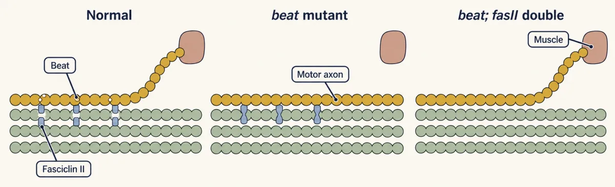
So in the fly, the “label” that lets a motor axon leave its bundle is partly an amount: how much adhesion holds it to its neighbours, and how much Beat works against it at the choice point.
Choice points in vertebrates
Vertebrates show the same logic. Kathryn Tosney and Lynn Landmesser labeled chick motor axons growing into the hindlimb and drew the growth cones one by one. Some stretches of the route are straight. Others are places where routes divide: the plexus, where motor axons sort into dorsal and ventral nerve trunks, and the points where muscle nerves branch off.
Predict. Where along the route would you expect large, complex growth cones, and where small, simple ones?
Check your prediction
At the plexus and where muscle nerves branch off, the growth cones were larger, more lamellipodial and followed more convoluted paths. In straight stretches they were small and simple.
These were fixed embryos, so the study saw shape, not speed; time-lapse work in other systems links such shapes to pausing (Going deeper). Neither shape nor pausing is a test for a choice point, but both often accompany one.
Same cue, different meaning
The second half of the rule is “read the same thing differently”. The same cue can attract one axon and repel another. In a simplified comparison, netrin-1 can attract growth cones that carry the receptor deleted in colorectal cancer (DCC) alone and repel those that carry DCC with a second receptor, UNC5. Inside a single growth cone, the answer can change too. In cultured Xenopus spinal neurons, lowering cAMP signaling turned attraction to netrin-1 into repulsion. Without laminin, Xenopus retinal growth cones are attracted by netrin-1, but laminin turns that response into repulsion.
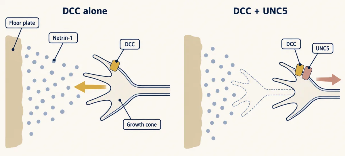
So the four classes in the table describe how a cue behaves in a given experiment, not a fixed property of the molecule. A cue is a message, and the receptors and internal state of the growth cone decide what it means. That is why a few cue families can wire many different axons, though the genome holds far too few genes to name every connection in a brain (the arithmetic is in the wiring problem). Cues act in combination, and each choice point reuses the same few families in a new place and a new combination.
Choice points usually carry several cues: at many of them, a contact cue works beside a long-range one, and an attractant beside a repellent. Overlapping cues make a choice more reliable, though losing a single system can still cause lasting errors, as the knockouts in the next lessons show.
The next lessons take these systems apart: netrin and DCC, Slit and Robo at the midline and semaphorins. For the receptor families themselves, see a field guide to guidance receptors.
Going deeper
How far can a secreted cue reach?
A worked estimate, with its assumptions stated. Take a growth cone about 10 µm across (its width w) that detects a fractional difference in concentration across its width, and receptors that work over about a 100-fold range of concentration around their Kd, the concentration at which half are bound. For a cue whose concentration falls off exponentially with decay length λ, the fractional difference across the cone is about w/λ, the same at every distance. If the cone keeps that sensitivity across the whole 100-fold range, an optimistic assumption, the usable span of the gradient is λ × ln(100), or about 4.6λ.
| Smallest fractional difference the cone can detect | Largest readable decay length λ (the shallowest readable gradient) | Longest usable span |
|---|---|---|
| 2% | 500 µm | 2.3 mm |
| 1% | 1,000 µm | 4.6 mm |
| 0.1% | 10,000 µm | 46 mm |
Detection alone would allow long reach if growth cones were very sensitive. Making a long, shallow gradient may be the harder part. In uniform tissue at steady state, with D the cue’s diffusion coefficient and k its first-order rate of removal, λ = √(D/k), so the cue’s mean lifetime, 1/k, equals λ²/D. With D = 10 to 30 µm²/s, a gradient with λ = 1 mm needs a cue that lasts about 9 to 28 hours on average, in a tissue that holds still; one with λ = 5 mm needs about 10 to 29 days. Source output, source shape and tissue growth matter too. An occupancy-noise estimate (10⁴ receptors split between the cone’s two sides, cue near Kd, signal just equal to noise) puts a single-snapshot threshold near 4%, falling to about 0.4% with 100 independent samples, so a threshold near 1% is plausible but not a hard number. Longer ranges than the body’s estimate would need higher sensitivity and longer-lived cues; this simple model sets no universal upper limit, which is why the rule says “within reach” rather than giving a figure.
Pipette assays sit at the steep end. For a cue that falls off as one over the distance r from a point source, the fractional difference across a cone facing the source is about w/r: 10% at 100 µm and 1% at 1 mm.
Netrin: long range or local?
The floor-plate gradient model is under revision. In mice, netrin-1 laid down by ventricular-zone progenitors along the path carries much of the effect and acts locally, while floor-plate netrin, with Sonic hedgehog, may still act at a distance near the midline. The details are in netrin and DCC.
Adhesion is not speed
The preference for the more adhesive surface comes from culture choice assays (Paul Letourneau, 1975, with chick sensory neurons). It is tempting to say adhesion is what growth cones want, but in several culture studies how sticky a surface is turned out to be a poor predictor of growth rate.
The clutch model explains why. With too little grip, the actin network slips backward and the edge does not advance. With too much, the growth cone cannot let go of old adhesions to move on. On simple charged coatings growth is fastest at intermediate adhesion. On matrix proteins and adhesion molecules, what the surface signals can matter more than how hard it grips.
Laminin is the clearest case. It is a large glycoprotein of basement membranes, and in the early 1980s several laboratories found that it strongly promotes neurite growth in culture. Neurons read laminin through integrins, receptors made of an alpha and a beta chain. In 1986 Kevin Tomaselli, Louis Reichardt and John Bixby blocked integrins with antibodies on dishes where cell bodies could still attach through a second coating. Outgrowth on laminin stopped while the cells stayed down, so integrins were needed for extension, not only to hold the cell on. Integrins link the matrix to actin and signal through Rho-family GTPases (see integrins and Rho GTPases).
Growth cones carry three main families of adhesion molecule:
- Cadherins, such as N-cadherin, which need calcium and bind the same cadherin on another cell.
- Ig-superfamily CAMs, such as NCAM and L1, which bind the same molecule or a different one. Fasciclin II and neuroglian are their fly relatives.
- Integrins, which bind matrix proteins such as laminin. Mammals have 18 alpha and 8 beta chains, forming 24 known pairs.
A uniform coat of laminin permits growth but gives no direction. A border or a gradient can.
Adhesion and other cues
The amount of adhesion between axons may also matter partly because it changes how growth cones respond to other cues. The Fasciclin II experiments in this lesson do not test that idea directly.
Pausing, branching and time-lapse
Later time-lapse imaging, in mouse retinal axons, matched complex growth cone shapes to growth cones that had paused. Growth cone pausing is closely related to axon branching: branches tend to form where a growth cone has stopped to read its surroundings. Much of the evidence for that link comes from cortical neurons, a different system from the ones in this lesson.
Your notes
Select any text in the lesson to highlight it or add a note to it. Your highlights and notes are saved in this browser and collected in your notebook.
This browser is not letting the site save anything (a private window or strict privacy settings), so highlights and notes will be lost when you leave the page.
How we measure it
Killing an identified cell before an axon arrives
In insect embryos individual cells can be recognized and labeled, then destroyed before a growth cone reaches them; methods of this kind include focused light and microelectrodes. Comparing the route with an operated control on a nearby spot asks whether that one cell is needed for a normal turn.
Dye filling of identified neurons
A neuron is injected with a fluorescent dye such as Lucifer Yellow or with horseradish peroxidase, which fills its axon and growth cone. Because the same named neuron can be found in every embryo, you can ask which bundle it chooses and whom its growth cone touches.
Loss-of-function, gain-of-function and double mutants in Drosophila
Remove a gene, express it in extra cells or at higher levels, or combine two mutations, then stain the motor axons with an antibody. Opposite phenotypes from loss and gain point to a quantity the gene sets; a double mutant can show that one gene works by opposing another.
Substrate choice and pipette turning assays
Neurons are grown on dishes coated with patterns of different substrates, or near a pipette releasing a cue. Which surface the growth cones follow, how fast they grow, and whether they turn toward or away from a source each test a different property of the cue.
Practice questions on this lesson (3) → MCAT-style, original, each with an explanation
Where this lesson fits
Before thisReview first
- The Growth Cone: The Tip That Steers the AxonThis course · Lesson 3
You need the growth cone's actin, filopodia and adhesion grip from there to follow how it reads cues and turns.
- Seeing Neurons: Golgi, Cajal and the Neuron DoctrineThis course · Lesson 2
You need the picture of the neuron and its axon from there to follow how single axons are traced and labeled.
After thisStudy next
- Netrin and DCC: One Cue, Two AnswersThis course · Lesson 23
It takes the idea that one cue can attract or repel and shows how DCC and UNC5 decide it.
- Crossing the Midline Once: Slit, Robo and CommThis course · Lesson 24
The midline is the intermediate target described here, and Slit and Robo mark the choice point after crossing.
- Semaphorins and RepulsionThis course · Lesson 25
It takes the long-range repulsion class of cues and shows how it sorts axons at a choice point.
Suggested by an AI model from the lessons themselves, then checked. How we use AI
Finish line
Test yourself
8 questions
Practice and MCAT-style, with explained answers.
Next · Lesson 23
Netrin and DCC: One Cue, Two Answers
The same protein pulls one axon toward the midline of the embryonic spinal cord and pushes another away. What decides?
Continue →