Developmental Neuroscience · Unit 5: Cells on the move
Tangential Migration: Interneurons and the Long Way Round
Rewritten on : new text, figures, flashcards and film. Your notes and highlights stay, but the text around them may have moved.
In a mouse embryo, most of the neurons that will inhibit the cortex are born outside it.
Pause when the reading reaches a film, and offer to play it. Saved on this device.
Watch the film now, or keep listening
Builds on:Radial Migration: Climbing the Glial LadderVentral to Dorsal: Sonic Hedgehog and BMP in the Neural TubeThe Ventricular Zone: Radial Glia as Stem CellsWhy these?
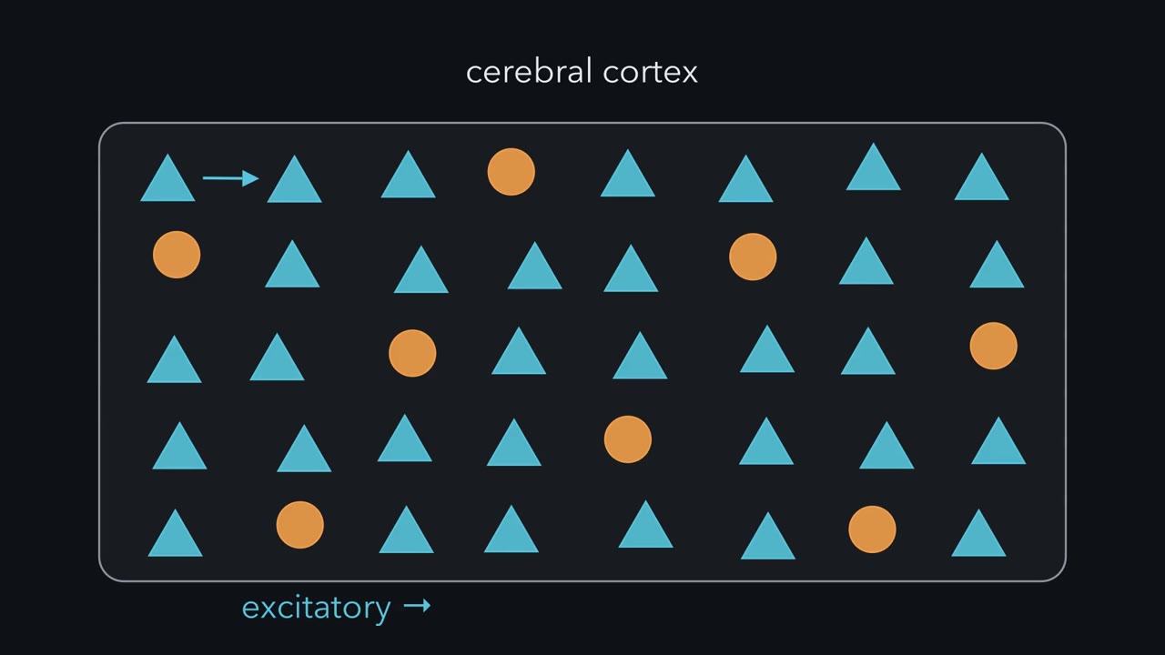 ▶ Watch Born Outside · 2:34
Read the transcript of “Born Outside”
Film transcript: Born Outside
The cerebral cortex needs two kinds of neuron. Some excite their neighbours. Others inhibit them. In rodents, roughly a fifth of cortical neurons are inhibitory interneurons. Yet the cortex's own wall makes the excitatory kind. These pyramidal neurons climb straight out along radial glial fibres, long struts that span the wall. So where do the inhibitory neurons come from?
Start with a map. The dorsal part of the embryonic forebrain, the pallium, forms the cortex. Its ventral part, the subpallium, bulges into the ventricle as three swellings. These are the lateral, medial and caudal ganglionic eminences. Despite the name, they are not ganglia. They are temporary germinal zones, packed with dividing progenitors. Pallial progenitors make neurons that release glutamate. Subpallial progenitors make neurons that release GABA, the main inhibitory transmitter. In rodents, most cortical interneurons are born in the medial and caudal eminences. Then they travel sideways into the cortex. Moving parallel to the brain surface is called tangential migration.
Here is the idea to carry through this lesson. Born outside the cortex, steered by what the cell can read. At the turns studied so far, the road holds a cue. The travelling cell carries what answers it. So a fault can sit in the cell, which cannot answer. Or it can sit in the road, which lacks the cue. Swapping tissue between normal and mutant animals tells you which. And in the cortex, the lane's chemokine signal helps keep a cell in the lane. Hold on to that. The next chapters test it. One limit first. These results come from mice and rats. In humans, the eminences are seen as the main source too. But cortical progenitors also make some inhibitory neurons.
Most cortical interneurons start outside the cortex. What they can read helps decide where they go.
From the film
- In rodents, most cortical inhibitory interneurons are born outside the cortex, in the ventral forebrain, and travel sideways into it.
- Excitatory cortical neurons are made in the cortex's own wall and climb out along radial glial fibres.
- A fault in migration can sit in the cell, which cannot answer a cue, or in the road, which lacks it.
- These results come from mice and rats.
In a mouse embryo, most of the neurons that will inhibit the cerebral cortex are born outside it.
The cortex’s excitatory neurons, the pyramidal neurons, are made in its own wall and climb straight out to their layers, as you saw in radial migration. The inhibitory neurons start in another part of the forebrain and travel sideways, across territories, before they turn and enter the cortex. This lesson asks how that was found, what keeps the travellers on their route, and where else in the brain neurons take the long way round.
Two populations, two birthplaces
Start with a map. The telencephalon is the front-most part of the embryonic brain, which becomes the cerebral hemispheres. Its dorsal part, the pallium, forms the cortex. Its ventral part, the subpallium, bulges into the ventricle as three swellings packed with dividing progenitors: the lateral, medial and caudal ganglionic eminences (LGE, medial ganglionic eminence (MGE) and CGE). Despite the name, they are temporary germinal zones, not ganglia. Much of the LGE becomes the striatum.
The two territories switch on different transcription factors and make different neurons. Pallial progenitors make neurons that release glutamate. Subpallial progenitors make neurons that release GABA, the main inhibitory transmitter of the forebrain. GABA is made from glutamate by glutamic acid decarboxylase (GAD), so antibodies to GABA or GAD mark these cells.
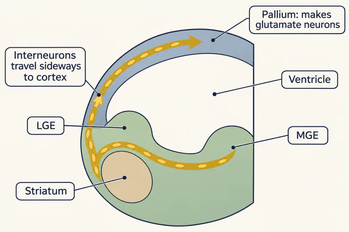
That raises a problem. The cortex needs inhibitory interneurons, local-circuit neurons whose axons stay nearby, roughly a fifth of its neurons in rodents, yet its own wall makes excitatory ones. Either the pallium makes a few GABAergic cells anyway, or they come from somewhere else.
Design problem
You suspect that cortical interneurons come from the ventral telencephalon. Design two independent experiments to show it. Then say what result in a mutant lacking the source, or unable to move cells out of it, would confirm your idea, and what that mutant alone could not tell you.
Saved in this browser only.
One way to do it
First, label the source and follow it. Put a fluorescent dye into the MGE of a cultured embryo or a forebrain slice and look for labelled cells in the cortex a day or two later. Dye shows the route but fades and does not say what the cells become, so a stronger version is a genetic label: a mouse that makes Cre recombinase only in Nkx2.1-expressing cells, most of them in the MGE, crossed to a reporter that every descendant keeps. If labelled cells in the mature cortex contain GABA and not glutamate markers, they came from Nkx2.1-expressing tissue, mainly the MGE.
Second, remove the source or the move. In a mutant whose ventral cells cannot leave, such as the Dlx1/2 double mutant mouse, the cortex should lack most GABAergic interneurons while its pyramidal neurons are made normally. That pattern fits a ventral origin better than a cortical one: interneurons made in the cortical wall without these genes would be left in place.
What the mutant alone cannot tell you: a conventional knockout lacks the gene in every cell, so it cannot say whether the travelling cells or the tissue they cross is at fault, or rule out cortical-wall cells that also need the gene. Grafts between normal and mutant tissue answer the first question. The second needs lineage labels of the cortical wall itself, since an Nkx2.1-Cre map shows where many interneurons come from but not what share comes from elsewhere.
The one idea to carry through this lesson. Born outside the cortex, steered by what the cell can read. In rodents most cortical interneurons are made in the ventral forebrain. At the turns where the cue is known, the striatal semaphorins and the CXCL12 lanes, the tissue holds a cue and the travelling cell carries the receptor that answers it. So a fault can sit in either place: in the cell, which cannot answer, or in the tissue, which lacks the cue. Swapping tissue between normal and mutant animals tells you which. In the cortex, CXCL12-CXCR4 signalling helps keep a cell in its lane; how cells normally leave it is still under study.
These are rodent results; what the idea covers, and what it does not, comes at the end.
Follow, remove, swap
Read the transcript of “Does the Defect Travel with the Cell?”
Film transcript: Does the Defect Travel with the Cell?
How do you work out where a neuron came from, and what it needs for the trip? There are three ways to ask. Follow the cells out. Remove the source. Or swap tissue, and see which one the defect follows. First, follow. In 1996, dye placed in the lateral eminence of cultured rat embryos marked cells that reached the cortex. But dye fades, and it does not say what a cell becomes. A genetic label fixes that. Cells that switch on Nkx2.1, mostly in the medial eminence, turn on a reporter. Every descendant keeps the reporter, even after the gene turns off. In mature mouse cortex, many marked cells are inhibitory interneurons. Second, remove. In 1997, Stewart Anderson, John Rubenstein and colleagues cut mouse cortex away from the ventral forebrain.
The isolated cortex ended up with fewer cells containing GABA. They also used a knockout, a mouse with a gene disrupted in every cell. They disrupted both Dlx1 and Dlx2. These genes encode transcription factors made in the subpallium. In the mutant cortex, which neurons go missing?
The inhibitory ones. No migration from the ventral forebrain into the neocortex was detected, and few cells contained GABA. The pyramidal neurons were still made. Their progenitors do not need these genes. But a knockout lacks the genes in every cell. Is the fault in the travellers, or in the road? In 2001, the same group answered with grafts. They placed labelled pieces of eminence into forebrain slices, crossing normal and mutant tissue both ways. A normal piece in a mutant slice, or a mutant piece in a normal slice. Which fails to migrate?
Normal cells migrated into the cortex of mutant slices. Mutant cells failed to migrate into the cortex of normal slices. The defect travelled with the cells. So whatever these genes do for the trip, they do it inside the travelling cells.
The swap shows where the need lies, not how the genes act. For Dlx, the swap gives a clear answer. The defect travels with the cell.
From the film
- Following cells out, removing the source and swapping tissue are three ways to ask where a neuron came from.
- In mice lacking both Dlx1 and Dlx2, no migration from the ventral forebrain into the neocortex was detected and few cells contained GABA, while pyramidal neurons were still made.
- In graft experiments with slices, the defect travelled with the cells, so these genes act inside the travelling cells.
There are three ways to ask about these cells. Follow them out of the place you suspect, to see where they come from and where they go. Remove the source, or their ability to leave it, to see what goes missing. Swap tissue between normal and mutant animals to see whether a defect lies in the cells or in the tissue they cross.
Follow them
A dye such as DiI dissolves into cell membranes, so any cell that leaves the spot where the dye was placed carries it along. In 1996 J. A. de Carlos, L. López-Mascaraque and F. Valverde injected the dyes DiI or DiA into the LGE region of rat embryos kept alive in culture. Most labelled cells ended up at the surface of the ventrolateral cortex, which lies beside the eminence, reaching it along radial glial fibres. That short trip is not the long sideways route of this lesson; the clue was the other cells, which ran tangentially into the preplate, the first layer of young neurons in the developing cortex. In 1997 Nobuaki Tamamaki and colleagues reported that tangentially oriented neurons in the developing cortical intermediate zone were travelling from the ventral side, with the lateral ganglionic eminence as their likely source. These dye studies showed cells leaving the eminences for the cortex, but the 1996 cells mostly settled in ventrolateral cortex rather than spreading across the neocortex, and neither study identified the cells as mature neocortical GABAergic interneurons. In 1999 Wichterle, Alvarez-Buylla and colleagues showed that medial ganglionic eminence (MGE) cells in embryonic slices migrate extensively toward the cortex, and that grafted MGE cells became neurons.
A dye fades and does not mark identity; the genetic label from the design problem fixes that. Nkx2.1 is a transcription factor made mainly in the MGE and also in the nearby preoptic area, and the Cre switch is a permanent change in the DNA, so the reporter survives long after Nkx2.1 is off. In the mature cortex, many reporter-marked cells are GABAergic interneurons (Fogarty and colleagues, 2007; Xu and colleagues, 2008).
Remove the source, or the move
In 1997 Stewart Anderson, David Eisenstat, Limin Shi and John Rubenstein ran two removal tests in mouse tissue. First, they cut the cortex away from the ventral telencephalon in culture. The isolated cortex ended up with fewer cells containing GABA.
Second, they used a knockout, a mouse in which a gene has been disrupted in every cell. Dlx1 and Dlx2 are homeobox genes. They encode transcription factors, proteins that switch other genes on or off, and are expressed in the subpallium and in the interneurons that come from it.
Predict. In embryos lacking both Dlx1 and Dlx2, what happens to the cortex’s GABAergic interneurons, and to its pyramidal neurons?
Check your prediction
The group detected no migration from the ventral telencephalon into the neocortex, and the neocortex had few GABA-containing cells. Pyramidal neurons were still made, because the pallial progenitors that make them do not need these genes.
The knockout leaves a question open. With the genes missing from every cell, is the fault in the travellers, which might be unable to move, or in the road, which might fail to make something they need?
Swap them: does the defect travel with the cells?
In 2001 Anderson, Oscar Marín, Carrie Horn, Kelly Jennings and Rubenstein answered this with grafts. They placed labelled pieces of ganglionic eminence from one embryo into forebrain slices from another, crossing normal and Dlx1/2 mutant tissue both ways.
Predict. Two ideas compete: Dlx is needed in the migrating cells, or in the tissue they cross. Fill in the last two rows under each idea.
| Donor eminence | Host slice | If Dlx is needed in the migrating cells | If Dlx is needed in the tissue they cross |
|---|---|---|---|
| normal | normal | migrate | migrate |
| normal | Dlx1/2 mutant | ? | ? |
| Dlx1/2 mutant | normal | ? | ? |
Check your prediction
If the need is in the cells, the normal donor migrates into the mutant host and the mutant donor fails in the normal host. If the need is in the tissue, the reverse. If it were needed in both, both mixed grafts would fail.
What happened: normal donor cells migrated into the cortex of mutant slices, and mutant donor cells failed to migrate into the cortex of normal slices. The defect travelled with the cells.
A requirement that travels with the cell like this is called cell-autonomous. Whatever Dlx1 and Dlx2 do for this trip, the requirement lies in the donor cells and not in the tissue they cross. The swap shows where the requirement lies, not how: the genes might control movement itself, or whether the cells become migrating interneurons at all. So it does not show that the cells are reading cues.
Where most of them start
Grafts, fate mapping and further mutants in mice placed most cortical interneurons in the MGE and the CGE, with a smaller share from the preoptic area. MGE identity depends on the transcription factor Nkx2.1, which is switched on by Sonic hedgehog (that lesson covers the patterning). Not every interneuron comes from the MGE: the first dye studies labelled the LGE, and cells from it do reach the cortex.
Moving with no fibre to climb
A pyramidal neuron moves at right angles to the brain surface, from the ventricle outward, mostly by gripping a radial glial fibre, the long process of a progenitor that spans the wall. A tangentially migrating interneuron moves parallel to the surface and crosses regional boundaries with no glial fibre to follow. Think of a climber on a fixed rope versus a traveller walking through streets, choosing at each corner.
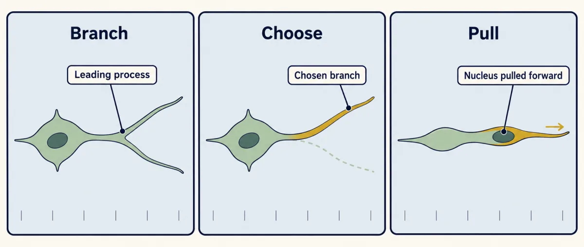
The interneuron’s leading process branches, and the cell advances by choosing one branch and moving its nucleus forward into it. The motors are ones you met in radial migration, dynein and myosin II, but the arrangement need not be the same: in these interneurons, myosin II contraction at the rear of the nucleus is a major push forward (Bellion and colleagues, 2005). Cues along the route make the choices.
Going round the striatum
Leaving the medial ganglionic eminence (MGE), cortex-bound interneurons skirt the striatum. Marín, Rubenstein and colleagues showed in 2001 that these cells carry neuropilin receptors and are repelled by semaphorins made in the striatum, while interneurons that settle in the striatum lack those receptors (see semaphorins).
Predict. Two populations leave the same eminence and pass the same striatum. Take away neuropilin function from the cortex-bound cells. Where do they go?
Check your prediction
Many more of them enter the striatum. In the same 2001 work, loss of neuropilin function let cortex-bound interneurons enter it. The repellent was there for both populations; what decided which cells avoided it was whether the cell could read it.
A map of the cortical wall, and two streams
Once in the cortex, interneurons travel in particular zones of its wall. Here are the ones you need, from the brain surface down to the ventricle, once the cortical plate has formed:
- Meninges. The membranes that cover the brain. Not part of the wall, but a source of signals.
- Marginal zone. The thin outermost layer of the wall, with few cells. The superficial stream runs here.
- Cortical plate. The young neurons that will form the cortical layers.
- Subplate. Early-born neurons just beneath the plate. The arriving plate splits the old preplate into the marginal zone above and the subplate below.
- Intermediate zone. Axons and migrating cells. The deep stream runs in its lower part and in the zone below.
- Subventricular zone. A second layer of dividing progenitors, among them the intermediate progenitors that make projection neurons.
- Ventricular zone. Radial glial progenitors lining the ventricle.
Between the two streams, the young cortical plate holds relatively few interneurons early on. The guides are axons, other cells and diffusible cues, not mainly radial glia (Going deeper has a caveat on axons).
Staying in the lane: CXCL12 and CXCR4
Read the transcript of “Staying in the Lane”
Film transcript: Staying in the Lane
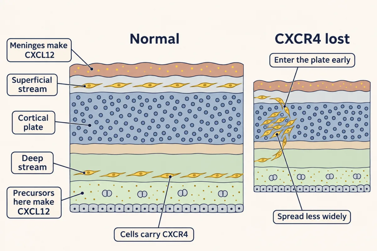
Inside the cortex, why don't interneurons dive into the cortical plate at once? Picture the young cortical wall. Under the meninges, the brain's covering, lies the marginal zone. Below it is the cortical plate. Deeper still are the intermediate zone and the subventricular zone. Interneurons travel in two streams. One runs in the marginal zone, the other deep in the subventricular and lower intermediate zones. Both lanes are lined with a chemokine, a small secreted protein first known for steering immune cells. It is called CXCL12. The meninges make it above. Dividing precursors of projection neurons make it below. The interneurons carry its receptor, CXCR4.
In a chamber assay, precursor cells moved toward CXCL12 placed on one side. Spread evenly on both sides, it gave no extra crossing of the membrane. In this assay, extra crossing needed a difference in concentration. Now take the receptor away. Where do the interneurons end up?
In 2008, two groups blocked or deleted the receptor in mice. The interneurons left their streams and entered the cortical plate early. And they spread less widely across the cortex. Many late-born cells that entered early ended up too deep, though not all did.
Early entry and narrow spread can come from one defect, not two. If cells spent the same time in the lane, moving more slowly would narrow the spread, but they would not enter early. If they leave the lane sooner, they spread less and enter early. That matches the mutants. Normal cells must leave too. Embryonic interneurons gathered near beads soaked in the chemokine. Interneurons from newborn mice did not. So to leave a lane, a cell may stop answering the signal. That compares ages, not single cells. It fits the idea but does not prove it. The cortical plate also makes Neuregulin-3. It attracts interneurons that carry its receptor, ErbB4. How the two are coordinated is still being worked out.
Then the cells turn into the plate, toward their layer. Born outside the cortex, steered by what the cell can read. In the lanes, chemokine signalling helps hold a cell in place.
From the film
- In mice, interneurons travel in two streams lined with the chemokine CXCL12, and they carry its receptor CXCR4.
- Without CXCR4 in mice, interneurons leave their streams and enter the cortical plate early, and they spread less widely.
- To leave a lane, a cell may stop answering the signal; that comparison of ages fits the idea but does not prove it.
A stream is a picture of movement, not an explanation. Why do cells stay in it instead of diving into the cortical plate at once?
Chemokines are small secreted proteins first known for steering immune cells. CXCL12, also called SDF-1, binds the receptor CXCR4, a G-protein-coupled receptor that signals mainly through Gi proteins (that lesson).
In 2003 Ralf Stumm and colleagues found CXCR4 on interneuron precursors travelling from the basal forebrain toward the rat neocortex, and CXCL12 in the meninges.
Precursors dissociated from the striatal region of embryonic day 14.5 mouse brain migrated toward CXCL12 in a chamber assay, and did not migrate more when it was added equally on both sides. Spreading a signal evenly is a standard control, also used in the neurotrophic hypothesis lesson: it separates a response to the signal being there from a response to a difference in its concentration. In this assay, extra crossing needed the difference.
In mice lacking the ligand or the receptor, fewer GABAergic cells sat superficially, where the upper stream runs, and more appeared deeper in the cortex. A stream still entered the cortex, so the signal is not needed for the crossing; its clearest job is positioning cells after entry.
The deep stream has its own source. In 2006 Marie-Catherine Tiveron, Harold Cremer and colleagues found CXCL12 made in the subventricular and intermediate zones by dividing precursors of projection neurons, and in 2015 Abe, Stumm and colleagues removed it selectively from intermediate progenitors and reduced the deep stream. The cortex’s own excitatory precursors help hold the inhibitory immigrants in place.
Predict. Block or delete CXCR4 in mice. Where do the interneurons that have entered the cortex end up, and how widely do they spread?
Check your prediction
In 2008 two groups, López-Bendito, Marín and colleagues, and Li, Pleasure, Rubenstein and colleagues, blocked or deleted CXCR4 in mice. Interneurons left their streams and entered the cortical plate early, and they spread less widely across the cortex. Many late-born interneurons that entered early ended up in the wrong layer, deep layer V instead of upper layers, though not all did.
The chemokine controls retention, timing and spread, and in part final layer.
One defect, two results
Early entry and narrower spread look like two separate facts to learn. A toy model shows they can be one.
Predict. A cell moves along a stream at a steady speed and leaves it at a random moment, after some average time in the stream. Which change gives both early entry into the plate and narrower spread: moving more slowly, or leaving sooner?
Check your prediction
Leaving sooner. Distance travelled is speed times time in the stream.
| Toy model | Speed | Average time in the stream | Average distance before turning in | Enters the plate early? |
|---|---|---|---|---|
| normal | 50 µm per hour | 48 hours | 2.4 mm | no |
| leaves sooner | 50 µm per hour | 12 hours | 0.6 mm | yes |
| moves more slowly | 25 µm per hour | 48 hours | 1.2 mm | no |
Look at the last row: slower cells also spread less but still turn in at the normal time, so only the shorter stay brings cells into the plate early, which is what the CXCR4 mutants showed. The model assumes cells start together, move one way, and leave at a moment that does not depend on their speed. The simplest reading is that losing the receptor mainly shortens the time in the stream, not the speed. The numbers are for illustration, not measurements; 50 µm per hour is the speed in this lesson’s MCAT practice question.
Leaving the lane, and turning in
Normal cells do exit the streams and turn radially or obliquely into the plate toward their layer. Falling responsiveness to CXCL12 is one proposed contributor: Li and colleagues found that interneurons from embryonic mice gathered near beads soaked in CXCL12, while interneurons from newborn mice did not.
That fits the idea but does not prove it, because it compares cells of different ages, not cells leaving a stream with cells staying in it. A direct test would follow single cells in a slice, ask whether CXCR4 signalling falls just before each one turns, and keep the signalling on to see whether the cell stays. Ceasing to answer CXCL12 may not be enough on its own: the cortical plate also pulls. Marín’s laboratory later reported that Neuregulin-3, made in the plate and acting on its receptor ErbB4, attracts ErbB4-carrying interneurons and is needed for their normal invasion of the plate (Bartolini and colleagues, 2017). How the two are coordinated is still under study. All of this was measured in rodents.
Where they settle
Two populations, born in different places and travelling by different routes, end up interleaved in the same layers. In mice, interneurons from the medial ganglionic eminence (MGE) tend to settle in the same layer as pyramidal neurons born at about the same time, while CGE-derived interneurons settle mostly in superficial layers. The cortex the cells reach matters too: when deep-layer projection neurons change identity in Fezf2 mutant mice, the distribution of interneurons shifts with them (Lodato and colleagues, 2011).
Interneurons arrive in excess and their numbers are later trimmed by cell death, the subject of naturally occurring death.
The rostral migratory stream: a tangential route for life
Tangential migration is not only an embryonic event. In adult rodents, the subventricular zone lining the lateral ventricles still contains stem cells, which make dividing transit-amplifying “C cells”, which make young neurons. Those neurons travel forward along the rostral migratory stream to the olfactory bulb and become inhibitory interneurons there.
Marla Luskin traced this route from the anterior subventricular zone in newborn rats in 1993. In 1994 Carlos Lois and Arturo Alvarez-Buylla showed that it runs in adult mice: marked subventricular cells travelled to the bulb and became neurons. In 1996, with José Manuel García-Verdugo, they showed the mode of movement. The young neurons move in chains, sliding along one another inside tunnels formed by astrocytes, with no radial glia and no axon scaffold. This is chain migration, a third style next to glia-guided radial movement and the stream migration of cortical interneurons.
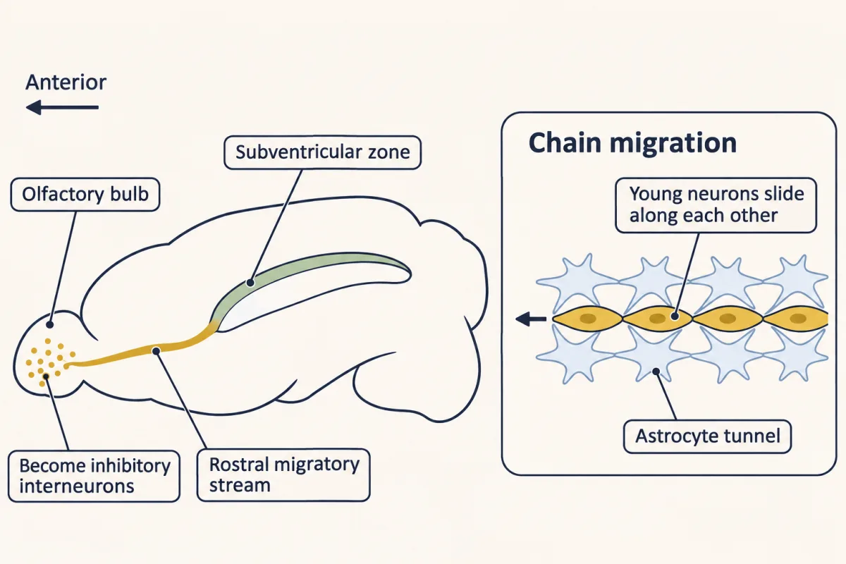
In humans, a comparable stream is seen in infants, declines sharply over the first 18 months of life and is nearly extinct by adulthood (Sanai and colleagues, 2011). Whether adult humans add neurons at all is taken up in adult neurogenesis.
Putting it together
| Experiment | Animal | Observed result | What it supports | What it does not establish |
|---|---|---|---|---|
| Following cells out of the eminences (1996, 1997, 1999) | rodent embryos and slices | labelled cells reach the cortex, some travelling tangentially | cells leave the eminences for the cortex | what a dye-labelled cell becomes |
| Nkx2.1-Cre fate map | mouse | many marked cells in the mature cortex are GABAergic interneurons | many interneurons come from Nkx2.1-expressing tissue, mainly the MGE | the share from other sources |
| Cortex cut from the ventral telencephalon (1997) | mouse tissue in culture | fewer GABA-containing cells in the cortex | the cortex depends on ventral tissue for these cells | that cells, rather than a signal, came from there |
| Dlx1/2 double knockout (1997) | mouse embryo | no detected ventral-to-neocortical migration; few neocortical GABA-containing cells; pyramidal neurons still made | Dlx1/2 are needed for normal interneuron development and migration; with the tracing, a ventral origin | migration versus differentiation; cells versus tissue |
| Reciprocal grafts (2001) | mouse slices | migration follows the donor’s genotype, not the host’s | the Dlx requirement lies in the migrating cells | how Dlx genes act |
| Neuropilin function lost (2001) | mouse | many more cortex-bound interneurons enter the striatum | a cue only some cells can read sorts two populations | — |
| Chamber assay (2003) | embryonic day 14.5 mouse cells | migrate toward CXCL12; no extra crossing when it is spread evenly | in this assay, the cells follow a concentration difference | what the chemokine does in the streams |
| CXCL12 or CXCR4 null (2003) | mouse | fewer GABAergic interneurons superficially, more in deeper cortex; ventral-to-cortical migration still occurs | positioning inside the cortex, not the initial entry | the timing and mechanism of stream exit |
| CXCR4 blocked or deleted (2008) | mouse | early plate entry, narrower spread, many late-born cells too deep | retention, timing, spread, and in part layer | why normal cells leave |
| Beads soaked in CXCL12 (2008) | embryonic and newborn mouse interneurons | embryonic cells gather, newborn cells do not | responsiveness falls with age | that the fall causes the exit |
| Fezf2 mutant (2011) | mouse | interneuron distribution shifts with the projection neurons | the destination helps set the layer | — |
| CXCL12 removed from intermediate progenitors (2015) | mouse | deep stream reduced | the lane’s cue must be present | — |
For the route through the rodent forebrain, the evidence fits the guiding idea: born outside the cortex, steered by what the cell can read. Here is what it covers, and what it does not.
- Which cells. Cortical GABAergic interneurons of mouse and rat embryos, from the medial ganglionic eminence (MGE), the CGE and the preoptic area. Not pyramidal neurons, which are made in the cortical wall. “Born outside” is shown for rodents and probably holds in outline for humans, where the size of a cortical share is still under study (Going deeper).
- Which decisions. The route, and when cells enter the cortical plate: leaving the eminence, going round the striatum, staying in a stream, and turning into the plate. Not the cells’ subtype, which is set largely by where and when they are born. Not their number, which cell death trims later. And not all of their final layer, because the pyramidal neurons they meet shift where they settle.
- Cell or tissue. Either side can carry a defect. The Dlx1/2 defect travels with the cells; the deep-stream defect after CXCL12 removal stays with the tissue; and two populations leaving one eminence part ways because only one can read the striatal repellent.
- Leaving the lane. Falling CXCL12 responsiveness, untested in single cells, and a Neuregulin-3 pull from the plate seem to contribute; how they are coordinated is still under study.
Design problem
You suspect that the need for CXCR4 lies in the interneurons and not in the tissue they cross. Design the swap, and say what result would support your idea.
Saved in this browser only.
One way to do it
Run the Dlx logic again. Graft labelled MGE pieces from CXCR4-mutant embryos into normal forebrain slices, and normal pieces into mutant slices, and follow the cells into the cortex. If the need is in the interneurons, mutant cells should leave the streams early even in a normal host, and normal cells should stay in the streams of a mutant host. A second route is to delete the receptor only in the interneurons and ask whether that alone reproduces the interneuron migration defect; the 2008 work included conditional mutants of this kind. Either way, the question is whether the defect travels with the cells.
Going deeper
Where human interneurons come from
In human fetal tissue, a 2002 study by Letinic, Zoncu and Rakic argued that many interneurons arise in the cortex’s own proliferative zones. Studies published in 2013 (Hansen and colleagues; Ma and colleagues) found most human interneuron progenitors in the ganglionic eminences, with a larger share from the caudal eminence than in rodents. The ganglionic eminences are now seen as the main human source. Later lineage studies of human fetal cortex also found cortical progenitors making some GABAergic neurons, mostly from midgestation, and how large that share is remains under study.
Axons as guides
Some deep-stream cells travel in contact with bundles of descending cortical axons that carry the adhesion molecule TAG-1, a finding from Denaxa and colleagues in 2001. Contact is not the same as need: mice lacking TAG-1 were later reported to keep normal cortical interneuron migration (Denaxa and colleagues, 2005). Later in mouse development a third stream runs through the subplate.
A second receptor for CXCL12
A second receptor for CXCL12, CXCR7 (also called ACKR3), shapes the response as well.
Why a chemokine stays near its source
Like many chemokines, CXCL12 binds sugar chains on cell surfaces and matrix, which tends to keep it near where it is made, one way a secreted cue can act over short range (that lesson).
What the Dlx genes might do
One report suggests the Dlx genes help cells migrate partly by restraining the growth of their axons and dendrites (Cobos and colleagues, 2007).
Is the layer fixed at birth?
Heterochronic transplants, which place medial ganglionic eminence (MGE) cells of one age into a host of another age, indicate that an interneuron’s future layer is not fixed by its birthday alone: it can still change in response to cues around the progenitor’s last division, and those cues are already present in the MGE before migration begins (Valcanis and Tan, 2003).
Interneuron death does not follow the NGF rule
The rule of the neurotrophic hypothesis lesson, let the target keep what it can feed, was worked out for NGF-dependent sympathetic and sensory neurons. Southwell and colleagues (2012) found that developmental death of cortical interneurons needs Bax and is timed roughly by the cells’ own age. In their transplants, interneurons lacking the neurotrophin receptor TrkB survived as well as control cells. Together these argue against a simple competition for a target neurotrophin. Later work implicated activity from pyramidal neurons in deciding which interneurons survive (Wong and colleagues, 2018).
Your notes
Select any text in the lesson to highlight it or add a note to it. Your highlights and notes are saved in this browser and collected in your notebook.
This browser is not letting the site save anything (a private window or strict privacy settings), so highlights and notes will be lost when you leave the page.
How we measure it
Lipophilic dye tracing in embryos and slices
A fluorescent dye such as DiI, which dissolves into membranes, is placed in one small region of a living embryo or brain slice. Cells that leave that region carry the dye with them, so the route they take can be seen hours later. It shows direction and route, not what the labelled cells become.
Gene knockout
A gene is disrupted in the mouse germ line so that every cell lacks it. If cortical interneurons are missing in Dlx1/2 double mutants while pyramidal neurons are present, the genes are needed somewhere in the making or moving of interneurons.
Tissue transplantation
A piece of tissue from a marked donor is grafted into a host slice or embryo. Swapping mutant and normal donors and hosts shows whether a defect lies in the migrating cells or in the territory they cross.
Cre-based genetic fate mapping
Cre recombinase is made only in cells that express a chosen gene, such as Nkx2.1 in the medial ganglionic eminence, and it switches on a reporter that every descendant keeps. The reporter then marks where cells from that source end up, long after the source gene is off.
Practice questions on this lesson (3) → MCAT-style, original, each with an explanation
Where this lesson fits
Before thisReview first
- Radial Migration: Climbing the Glial LadderThis course · Lesson 17
Pyramidal neurons climb glial fibres there, and this lesson contrasts that with interneurons moving sideways with no fibre.
- Ventral to Dorsal: Sonic Hedgehog and BMP in the Neural TubeThis course · Lesson 10
Sonic hedgehog patterning gives the ventral forebrain, including MGE identity through Nkx2.1, that these interneurons come from.
- The Ventricular Zone: Radial Glia as Stem CellsThis course · Lesson 13
You need radial glia as the cortex's own progenitors to see why excitatory neurons are made in its wall.
After thisStudy next
- Too Many Neurons: Naturally Occurring Cell DeathThis course · Lesson 28
Interneurons arrive in excess, and this lesson takes up the cell death that trims their numbers afterwards.
- Two Places the Adult Brain Still Makes NeuronsThis course · Lesson 47
It takes up the rostral migratory stream and asks whether adult humans add neurons.
- Opening and Closing a Critical PeriodThis course · Lesson 40
Once interneurons settle in cortex, GABA circuits help open and close the critical period.
Suggested by an AI model from the lessons themselves, then checked. How we use AI
Finish line
Test yourself
8 questions
Practice and MCAT-style, with explained answers.
Next · Lesson 20
The Neural Crest: Cells That Leave Home
Why can one mutation leave a child with part of the colon without gut neurons, a white forelock and deafness?
Continue →