Developmental Neuroscience · Skim version
The Neural Crest: Cells That Leave Home
Read the full lesson → Skim the whole course →
The guiding idea
Why can one mutation leave a child with part of the colon without gut neurons, a white forelock and deafness?
One border, many destinations: where along the body neural crest cells start biases what they can become, and their route helps decide which fates appear.
From the film: One Border, Many Destinations
- Where along the body a crest population starts biases what it can become; the signals on its route, and where it stops, help decide which fates appear.
- Crest cells form at the border between the neural plate and future skin, then change from epithelial cells into free mesenchymal cells in an epithelial-to-mesenchymal transition.
- Because many tissues share that start and many need the same genes, one failed gene can strike tissues that look unrelated.
From the film: Mark It, Move It
- Quail nuclei carry a built-in mark that neither fades nor dilutes as cells divide, so quail-into-chick grafts show where each level's crest goes.
- Moving cells to a different level changes what the population makes: adrenal-level trunk crest at the vagal level formed enteric ganglia, some with cholinergic neurons.
- A dish shows what a cell can make, tracing in the embryo shows what it did make, and whether the typical crest cell is multipotent is still debated.
From the film: Leave, Steer, Arrive: The Puzzle Solved
- In chick, Snail2 is needed for crest cells to leave and probably lifts the Cadherin-6B brake, at least partly.
- In the chick trunk, crest cells pass only through the front half of each sclerotome because the back half repels them; ephrin-B in chick and semaphorin 3F in mouse are repellents, and neither is known to be the only one.
- Tissues that fail together often share a requirement, and which tissues fail depends on where the gene is needed, the variant and each tissue's sensitivity.
-
A border population that leaves home
-
How do you follow a cell that leaves?
-
Leaving home: an identity first, then a release
-
Steering: a forbidden half and cells that back away
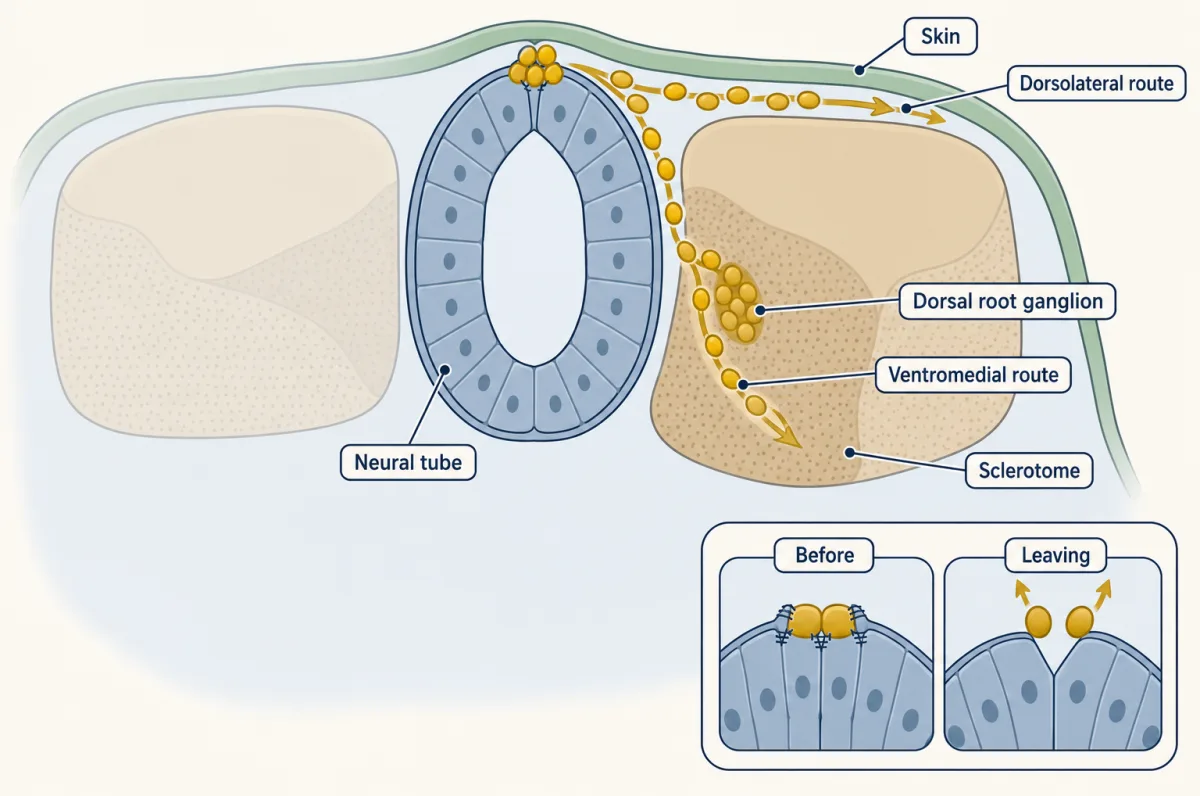
Crest cells leave the top of the neural tube and take two routes in the chick trunk. The ventromedial route, through the rostral half of each sclerotome, gives dorsal root ganglia, sympathetic ganglia and adrenal medulla; the later dorsolateral route, under the skin, gives mostly melanocytes. AI-generated illustration 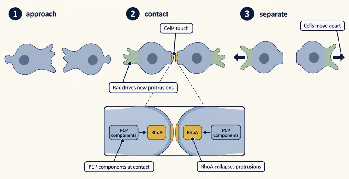
Contact inhibition of locomotion, shown in frog and zebrafish cranial neural crest. When two crest cells touch, planar cell polarity signaling activates RhoA at the contact, collapsing protrusions there, while Rac drives protrusions at the free edges, and the cells head off in new directions. How far this applies to trunk crest in birds and mammals is less clear. AI-generated illustration -
Stopping and gathering
-
Neurocristopathies: tissues that fail together often share a requirement
-
Putting it together
-
Going deeper
Where this lesson fits
Before thisReview first
- Neural Induction: The Default ModelThis course · Lesson 6
The border between neural plate and future skin, where crest forms, comes from the BMP gradient set up there.
- Neurulation and Neural Tube DefectsThis course · Lesson 7
You need the rolling up of the plate, which puts border cells at the dorsal tube, before they can leave.
- Tools for Following Cells and GenesThis course · Lesson 4
The lesson uses grafts, lineage tracing and clonal logic for fate mapping, which that lesson introduces.
After thisStudy next
- Pioneers, Guideposts and the Logic of Guidance CuesThis course · Lesson 22
Repellent cues like ephrin-B that fence off the caudal sclerotome are developed there as a general logic of guidance.
- Semaphorins and RepulsionThis course · Lesson 25
Semaphorin 3F and neuropilin-2, the mouse caudal-half repellent for crest, are treated there as repulsive cues.
- Too Many Neurons: Naturally Occurring Cell DeathThis course · Lesson 28
Crest-derived sensory and sympathetic neurons that arrive at their targets then face heavy cell death, as covered there.
Elsewhere on the siteFilms, simulations and method films in other courses
- Cre-lox and binary expression systemsMethod film
Genetic lineage tracing of crest derivatives in the lesson's deeper section uses recombinase-based marking of crest cells.
- Wnt Plugs the DrainFilm · Cell Signaling
Wnt signaling helps specify the crest at the plate border, so this film shows that signal in detail.
Suggested by an AI model from the lessons themselves, then checked. How we use AI