Developmental Neuroscience · Unit 1: The problem and the tools
Tools for Following Cells and Genes
A neuron has no label saying when it was born or where it came from. So how do you find out?
Pause when the reading reaches a film, and offer to play it. Saved on this device.
Watch the film now, or keep listening
Builds on:The Wiring Problem: How a Brain Gets BuiltSeeing Neurons: Golgi, Cajal and the Neuron DoctrineWhy these?
 ▶ Watch What Can a Label Prove? · 3:21
Read the transcript of “What Can a Label Prove?”
Film transcript: What Can a Label Prove?
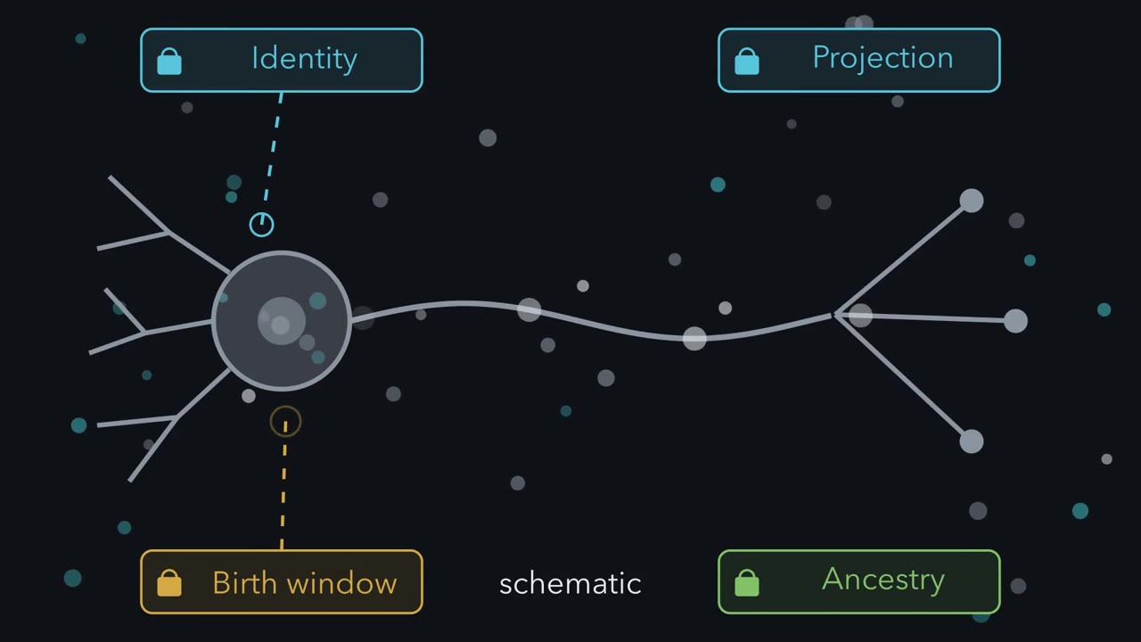
A finished neuron hides its past. Which label could reveal the part you need in that tissue? Its shape alone cannot show its birth time, ancestry, or distant projection. Each question needs an event that can carry that information forward.
Start with identity. A Nissl stain reveals many cell bodies. A Golgi stain fills a few cells and their branches. An antibody binds one chosen protein. These marks report what they detect, not when the cells formed. For a projection, use the neuron's own transport. Put a tracer near cell bodies, and anterograde transport carries it toward terminals. Put a retrograde tracer at a target, and terminals carry it back. The result maps a sampled population, not every synapse. Birth-dating follows a different event. During a short pulse, tritiated thymidine or BrdU enters DNA being synthesized. In a dividing cell, that means S phase. The pulse marks a cohort copying DNA during that window.
After the pulse comes the chase. Each further division usually dilutes label among the descendants. Heavy label later supports few additional divisions and an approximate last S phase. Weak label fits continued cycling, although uptake and detection also vary. Ancestry needs a mark that survives division. A grafted quail nucleus stays recognizable in chick tissue. A retroviral insert enters a dividing cell's DNA.
Cre can permanently switch on a reporter. In each case, descendants inherit a durable difference. Predict the next frame. One founder carries a permanent DNA change, then divides twice. Which cells should carry that change?
Every descendant inherits the changed DNA. Yet one colour across many founders gives a population fate map, not separate clones. Sparse induction, multiple colours, or distinct barcodes can separate neighbouring families. Here is the rule. A tool supports only what its mark records.
Protein binding can report target detection. Transport can report a projection. DNA synthesis can date a cohort approximately. An inherited DNA change can report ancestry. The rule also tells you when to stop. A tracer does not prove every synapse. A weak birth label is not a precise birthday. A shared lineage colour may hide many founders. State the strongest claim, then name the limit.
From the film
- A method can reveal only what its mark records.
- Transport reveals a projection, not every synapse.
- DNA synthesis approximately dates a cohort.
- An inherited DNA change can reveal ancestry.
A neuron carries no label saying when it was born, which cell it descends from, or which genes it needs. Developmental neuroscientists make those hidden histories readable, then ask only what the mark can justify.
The guiding idea. Within the methods here, the event a tool records or changes sets the strongest claim it can support: protein binding reports detection of a molecular target, transport reports a projection, DNA synthesis approximately dates a cohort, inherited DNA change reports ancestry, deletion tests necessity, and imposed activity tests sufficiency.
Scope: this rule chooses and interprets the methods in this lesson. “Strongest claim” means the direct claim under the tested conditions, not a complete account of the biology.
| Question | Event carrying the information | Strongest direct claim |
|---|---|---|
| What is the cell? | A stain or antibody binds its target | The target was detected there |
| Where does it project? | A tracer is transported along an axon | Labelled cells project to, or labelled terminals arise from, the injected region at population scale |
| When did it stop dividing? | Label enters DNA during synthesis, then dilutes with division | Heavy residual label approximately dates the last S phase |
| Whose descendant is it? | A durable mark passes to daughter cells | Marked cells descend from the marked founder or population |
| What does a gene or activity do? | An intervention removes or imposes it | The outcome tests necessity or sufficiency under those conditions |
What is this cell, and where does it connect?
Three staining strategies, three uses. A Nissl stain binds the RNA-rich cytoplasm around a nucleus, so it shows cell bodies across a section, along with glial nuclei and some proximal dendrite: good for counting cells and finding the boundaries of layers. A Golgi stain fills a small, unpredictable fraction of cells completely, including their dendrites, so you see the whole shape of a few neurons against a clear background. Immunostaining uses an antibody against one chosen protein, so it reports whether that molecular target was detected. The cost is specificity in the other direction: an antibody raised against a mouse protein may not recognize the chick version, and a monoclonal antibody that sees one site can fail when that site is masked. The logic of antibodies as detection reagents is covered in detecting a signal.
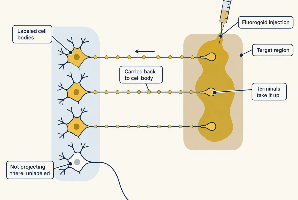
Connections use the neuron’s transport machinery. Inject an anterograde tracer such as biotinylated dextran amine into a region; neurons there carry it toward their terminals, reporting where those cells project. Cholera toxin B subunit travels both ways and is used mainly retrogradely, so direction must be established for each tracer. Inject Fluorogold into a target instead, and terminals carry it back to cell bodies. That labels neurons whose terminals take up enough tracer there. Either dye injection gives population wiring, not one cell’s synapses. Genetically targeted viruses go further: a rabies virus restricted to one synaptic step labels cells directly presynaptic to a defined starter population, or even one starter cell.
Predict. An antibody gives no signal in a chick section, although it detects the mouse version of the protein. Does the negative result prove that chick cells lack the protein?
Check your prediction
No. The antibody may not recognize the chick version, or its single recognized site may be masked. A negative detection result is not automatically evidence that the target is absent.
When was this cell born?
Tritiated thymidine, and later the thymidine analog BrdU, use DNA replication as a clock. They are incorporated wherever DNA is being synthesized, which in a dividing cell means S phase. Give a pulse, wait through a chase, and look for label.
Predict. A BrdU-positive nucleus appears in injured tissue whose cells are thought to be non-dividing. Must that cell have passed through S phase?
Check your prediction
No. Thymidine analogues can also enter DNA during repair. In a dividing cell the main interpretation is S phase, but BrdU alone does not distinguish repair from replication in injured tissue. Cell-cycle evidence and the experimental context are needed.
After a pulse in proliferating tissue, each further division usually dilutes label among descendants. Heavy residual label supports few additional divisions; weak label is consistent with continued cycling, although uptake and detection also vary. The inferred “birthday” is therefore an approximate last S phase, not an exact timestamp or a readout of cell identity.
The main model has now done its work. The human cases and their limits are in Going deeper.
Whose descendant is this cell?
A birth date marks a cohort; a lineage is a family. Classical methods lead to modern multicolour reporters and genetic barcodes that separate one founder’s family from its neighbours. Reconstructing somatic mutations is the main route to lineage in the human brain.
Grafting with a nuclear marker is the classic approach. Quail nuclei carry a distinctive condensed-chromatin pattern, so quail cells grafted into a chick embryo stay recognizable in sections after they migrate and differentiate. Le Douarin built an experimental program on that difference, following grafted populations into ganglia, gut and head mesenchyme.
Retroviral labeling works inside one animal. A retrovirus integrates its genome into the DNA of a dividing cell, so every descendant of that cell inherits the insert and the marker gene it carries. Keep the dose low enough that labeled clones do not overlap, and a cluster of marked cells is one progenitor’s family. That assumption is a sampling argument, not an observation, and it is where this method can fail.
The genetic version is the one you will use. A Cre-dependent reporter commonly places a loxP-flanked transcriptional stop before a fluorescent protein gene; other designs leave the gene inverted until Cre flips it. When Cre recombinase deletes the stop, the recombined allele passes to descendants. They stay labelled as long as the reporter’s promoter is active. The DNA marking is permanent and heritable through division.
Predict. Quail-derived cells later appear in a chick peripheral ganglion. Does the marker show that the host contributed no ganglion cells?
Check your prediction
No. The marker shows that the marked cells descend from the graft. Unmarked host cells may also contribute, and the graft does not by itself reveal which signals were required or the potential of any single founder. Reporters and actuators themselves are taught in hacking the cell; here the point is recombination.
A cell-type Cre that recombines many founders with one colour gives a population fate map, not separate clones. Following one family requires sparse or low-dose induction, multicolour reporters, or founder-specific barcodes.
The same logic can now change a gene rather than merely preserve a history.
Read the transcript of “Build a Conditional Test”
Film transcript: Build a Conditional Test
Chapter one asked what a label can prove. How can we test what a gene does in one cell type, after birth? We need to control three things: the target, the place, and the time. First, build the target. Place loxP sites around an important gene segment. This floxed allele is designed to work normally until Cre recombinase removes that segment. A Cre-free control must test whether the allele really does work normally. Next, control the place. A cell-type promoter supplies Cre only where that driver is active. Cre deletes the bracketed segment largely in those cells. But a driver's actual range and completeness must be measured, not assumed from its name.
Then control the time. CreER is designed to keep Cre outside the nucleus until tamoxifen is given. A gold time gate opens after birth. Recombination is concentrated after dosing, but its actual timing must be measured. Now read the cross one locus at a time. AvilCre/+ supplies one Cre driver allele.
TrkAfl/− supplies one floxed allele and one null allele. The semicolon joins the two instructions in one mouse. A matched control changes one causal piece. Keep the floxed and null TrkA alleles, but remove Cre. That mouse preserves the target genotype without triggering deletion. A second control can retain Cre while keeping one working TrkA copy. Now predict carefully. A cell glows green from a Cre-dependent reporter. Does that glow prove the separate floxed target gene was deleted?
No. The reporter and target are separate lox-flanked loci, and they can recombine differently. Old target protein can also persist after deletion. Reporter light shows reporter recombination. Measure the target protein itself in those cells. Only then interpret the outcome. Removing a gene tests whether it is necessary under those conditions. Imposing activity with an actuator tests whether activity is sufficient. A reporter observes a state; it does not impose that state. A conditional experiment is a chain. Match the genotype to the perturbation, the perturbation to the measured readout, and the readout to the claim. Check every link with a control in practice.
One weak link limits the conclusion.
From the film
- Cre changes a floxed allele only where recombination occurs.
- An inducible driver adds control over timing.
- Read each locus separately before interpreting a cross.
- Reporter light does not prove target-gene deletion.
The second film began with a floxed allele; what it did not show is how that engineered allele reaches a mouse or what a real driver name can hide.
How to make a mouse
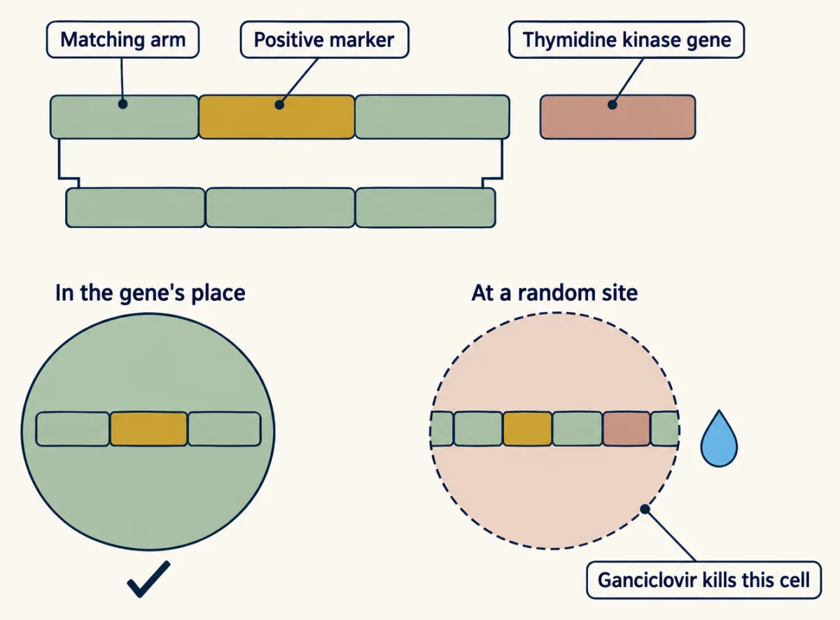
To ask what a gene does, change it. In the classical route, mouse embryonic stem cells from an early embryo can still contribute to any tissue of the body, including the germ line. Supply DNA matching the target gene on both sides of an intended change. In rare cells, homologous recombination swaps the new version into the gene’s own place.
Because targeting is rare, select twice. A positive marker inside the construct retains cells that took up DNA. A negative marker outside the matching arms, classically a thymidine kinase gene that makes ganciclovir lethal, kills most cells that inserted the whole construct randomly and kept that marker. Survivors still require genotyping. Destroying a gene is a knockout; writing in a sequence such as a fluorescent reporter is a knock-in. Selected cells enter an early embryo. The pup is a chimera, and the allele is heritable if those cells made its gametes.
A straight knockout can kill an embryo before the question matters, and whole-body deletion cannot identify the tissue that mattered. The loxP sites introduced in the film are each 34 base pairs long. A doxycycline-driven tetracycline-controlled system provides an alternative to CreER for timing.
Keep the reminder short: the changed event limits the claim. A superscript names what is written at a locus; a slash separates its alleles. Thus p75NTR-/- is a homozygous knockout, DR6+/- a heterozygote, and p75NTR-/-;TNFR1-/- a double knockout.
Now go beyond the chapter’s notation. In AvilCre/+;TrkAfl/-, Advillin controls the Cre allele. This driver has long been used for sensory neurons, but advillin and Avil-Cre recombination also occur in sympathetic, parasympathetic and enteric neurons. The floxed TrkA copy is the only potentially functional copy available to lose. The genotype therefore deletes TrkA wherever this Avil-Cre is active, including sensory and some autonomic neurons.
Predict. The full CreER, floxed-gene and reporter genotype receives vehicle instead of tamoxifen. Which failure does that control test?
Check your prediction
It tests ligand-independent CreER activity: recombination when the inducing drug was withheld. A Cre-negative TrkAfl/- littermate instead preserves the floxed and null alleles without Cre. AvilCre/+;TrkAfl/+ retains a wild-type TrkA copy and is a further control for Cre activity itself.
The schematic cannot tell how often a real driver is leaky, recombining where or when it should not, or mosaic, recombining in only some target cells. Nor can reporter light quantify residual target protein. Measure the driver’s range and the target’s loss in the tissue, ages and animals used.
Intersectional targeting adds a second recombinase, Flp, with its own sites and runs two independent switches. Requiring both switches in the same cell increases specificity.
A reporter cross tells you where Cre acted, not where the floxed gene was lost: the two lox-flanked targets recombine independently. This is a sketch of the logic, not a count from a real cross; how many and which cells keep the gene is arbitrary here.
Design problem
Gene X is needed for the embryo to survive, so a straight knockout dies before birth. You want to know what X does in sensory neurons after birth. Design the mouse: list the alleles, the drug, the reporter, and three controls.
Saved in this browser only.
One way to do it
Alleles: a sensory-neuron CreER driver, such as an Advillin-CreER line; X floxed over floxed; and a Cre-dependent fluorescent reporter. Map recombination in autonomic ganglia too, because advillin is not sensory-specific after birth. Give tamoxifen after birth to preserve X through embryonic development and trigger deletion later; the vehicle control tests whether ligand-independent recombination undermines that timing. The reporter marks cells in which Cre was active.
Three controls: CreER with no floxed allele, given tamoxifen, tests whether Cre plus tamoxifen harms neurons by itself. Floxed over floxed with no CreER, given tamoxifen, tests whether the floxed allele is hypomorphic, or partly defective without Cre. The full experimental genotype given vehicle tests ligand-independent CreER activity. In the same animals, measure how much X protein is actually gone in reporter-positive cells, because phenotype size depends on it.
Necessary, sufficient, reporter, actuator
Two pairs of words now finish the experimental logic.
The first pair is about inference. Sufficient means you added the thing and the outcome followed. Necessary means you removed it and the outcome failed. Each can hold without the other.
Predict. Overexpressing protein A makes structure B expand, but knocking out A leaves B unchanged. Is A necessary, sufficient under the tested conditions, both, or neither?
Check your prediction
A is sufficient under those conditions but not necessary for this outcome. The gain-of-function may place A at levels or locations it never normally reaches, or a redundant relative may cover its loss. The reverse pattern—necessity without sufficiency—would mean A is one requirement among several.
A reporter reads out a state: a fluorescent protein marks where a cell is, a tagged protein shows where it sits, and a calcium sensor brightens when calcium rises. An actuator imposes a state. Blue light opens channelrhodopsin, a cation channel, to drive firing; yellow light drives halorhodopsin to move chloride and suppress activity. Light-gated channels are introduced in ion channels as receptors.
Chemical actuators use the same logic. Put transient receptor potential vanilloid 1 (TRPV1), a capsaicin-gated channel, into chosen neurons of a mouse whose own TRPV1 is knocked out; capsaicin then activates those neurons and not the rest. An activity reporter alone shows that activity goes with an outcome, not that it caused it. An actuator can ask whether that activity is enough.
Going deeper
Human birth-dating
Giving a thymidine analog to people only to study the brain is not done. The classic exception, Eriksson and colleagues in 1998, used postmortem tissue from patients already treated with BrdU during cancer care. For humans generally, atmospheric nuclear weapons testing raised carbon-14 sharply before the 1963 test ban treaty, after which it fell. A cell’s genomic-DNA carbon-14 therefore reflects the year its DNA was last made. Spalding and colleagues described matching genomic-DNA carbon-14 to the atmospheric record in 2005. The method is the point here; findings about turnover in particular human brain regions are contested and belong to adult neurogenesis.
From classical targeting to newer routes
Capecchi’s laboratory, together with Smithies’s and Evans’s, established classical gene targeting. CRISPR shortened that path: a guide RNA brings Cas9 nuclease to a matching sequence in a fertilized egg, so many alleles can now be built without stem cells or chimeras.
Your notes
Select any text in the lesson to highlight it or add a note to it. Your highlights and notes are saved in this browser and collected in your notebook.
This browser is not letting the site save anything (a private window or strict privacy settings), so highlights and notes will be lost when you leave the page.
How we measure it
Thymidine-analog birth-dating
Give a pregnant animal tritiated thymidine or BrdU, which cells incorporate while they synthesize DNA, principally in S phase but also during repair. Later, cells that are still heavily labeled divided few times after the pulse, so the pulse approximately dates their last S phase. Autoradiography or an anti-BrdU antibody reveals them.
Axonal tract tracing
Inject a tracer that neurons take up and transport. An anterograde tracer such as biotinylated dextran amine or PHA-L is carried from cell body toward terminals; a retrograde tracer such as Fluorogold or cholera toxin B subunit moves from terminals back to cell bodies. Cholera toxin B travels both ways but is used mainly retrogradely. The pattern of labeled cells maps a connection.
Interspecies grafting with a nuclear marker
Transplant tissue between two species whose nuclei can be told apart, classically quail into chick. Donor descendants stay recognizable after they migrate and differentiate, so you learn where a cell population goes rather than only where it starts.
Conditional gene targeting with Cre and lox
Flank part of a gene with loxP sites and supply Cre recombinase from a cell-type-specific promoter. Cre deletes the flanked segment in cells that make Cre, as completely and as exclusively as that driver turns out to be. Fusing Cre to a modified estrogen receptor concentrates recombination after tamoxifen, although ligand-independent activity must be tested.
Method films · the methods library
Practice questions on this lesson (3) → MCAT-style, original, each with an explanation
Where this lesson fits
Before thisReview first
- The Wiring Problem: How a Brain Gets BuiltThis course · Lesson 1
It sets out why the genome cannot list the wiring, which is the question these tools exist to answer.
- Seeing Neurons: Golgi, Cajal and the Neuron DoctrineThis course · Lesson 2
Dye-filling and Golgi-style staining here build on the way single neurons were first seen and counted as separate cells.
After thisStudy next
- The Organizer: Spemann and MangoldThis course · Lesson 5
Graft experiments are the classic use of transplanted tissue to follow cells and test what a signal does.
- The Ventricular Zone: Radial Glia as Stem CellsThis course · Lesson 13
Lineage tracing and birth-dating are the tools that showed radial glia make most cortical neurons.
- Birth Order and Fate: Temporal Identity and the Inside-Out CortexThis course · Lesson 16
It uses birth-dating labels to show that the first-born cortical neurons sit deepest.
Elsewhere on the siteFilms, simulations and method films in other courses
- Engineered mutants: site-directed, dominant-negative and constitutively activeMethod film
Making a protein constitutively active or dominant-negative is the same sort of tool as the conditional gene changes covered here.
- Targeted cell ablationMethod film
Removing chosen cells is one of the necessity tests this lesson sorts, using genetically targeted toxins.
Suggested by an AI model from the lessons themselves, then checked. How we use AI
Finish line
Test yourself
8 questions
Practice and MCAT-style, with explained answers.
Next · Lesson 5
The Organizer: Spemann and Mangold
How can a small graft make its host build a second spinal cord from cells meant for skin?
Continue →