Neuroscience · Unit 2: Synapses and plasticity
Transmitter Systems and Drugs
Rewritten on : new text, figures, flashcards and film. Your notes and highlights stay, but the text around them may have moved.
Four drugs that act on GABA push inhibition in opposite directions. What decides which way a drug pushes?
Pause when the reading reaches a film, and offer to play it. Saved on this device.
Watch the film now, or keep listening
Builds on:The Action Potential and ConductionThe Resting Membrane and Passive SignalsSynaptic Transmission and Quantal ReleaseWhy these?
 ▶ Watch Three Steps, One Drug · 3:01
Read the transcript of “Three Steps, One Drug”
Film transcript: Three Steps, One Drug
Four drugs act on one transmitter, with opposite effects. What decides which way each one pushes? In 1921, Otto Loewi reported slowing a frog heart by stimulating its vagus nerve. He moved the fluid around it onto a second heart, and that heart slowed too. Its own nerve was untouched. The first nerve had released a chemical. If the message is a chemical, a drug can imitate it, block it, or make it last. Follow one molecule across a synapse. Three things happen to it. First, it is released from the terminal, mostly when a spike arrives. Second, it is read by a receptor, a protein on the target cell. Some receptors are ion channels that open within milliseconds.
Others are G-protein-coupled receptors. They act through messengers inside the cell, over tens of milliseconds to minutes. Third, it is removed, by an enzyme or by a transporter that pulls it back into a cell. Source, readers, off switch. Every transmitter fills these three slots. Think of a key, a lock, and a cleaner that sweeps keys away. A copy of the key that turns the lock is an agonist. A key that fits but will not turn is a competitive antagonist. Oil in the lock, so the real key turns more easily, is a positive allosteric modulator. Stopping the cleaner is a reuptake blocker or an enzyme inhibitor. GABA is the brain's main fast inhibitory transmitter.
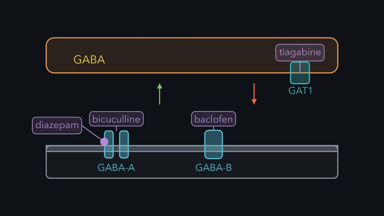
Here are four drugs that act on its system. Baclofen activates GABA-B receptors. Bicuculline sits in the GABA site of GABA-A, without opening it. Diazepam binds a different site on GABA-A and strengthens GABA's effect. Tiagabine blocks GAT1, a GABA transporter. Picture the receiving cell at a mature synapse, where GABA inhibits. Which drugs make inhibition stronger, and which weaker?
Baclofen, diazepam and tiagabine make it stronger. Bicuculline weakens it, and in animals it causes seizures. On terminals, baclofen can also cut GABA release, so circuits can differ. Same transmitter, opposite effects. What decides is which step a drug changes, and in which direction. The idea: classify a drug by the main step it changes. Release, reading, or removal. How much it changes that step depends on how much of its target it fills. What that does to a disease is a separate question. The takeaway: find the step a drug changes, and the direction it pushes.
From the film
- Every transmitter is released, read by receptors and removed, and a drug mainly changes one of these steps.
- Agonists imitate the transmitter, competitive antagonists block it, modulators change its effect and reuptake blockers or enzyme inhibitors make it last.
- Four drugs on one transmitter, GABA, can push inhibition in opposite directions depending on which step they change and in which direction.
In 1921 Otto Loewi reported an experiment with two isolated frog hearts. He stimulated the vagus nerve of one, which slows its beat, and then moved fluid that had bathed that heart onto the second. The second heart slowed too, although its own vagus had not been touched.
The nerve had acted by releasing a chemical, later identified as acetylcholine. If a nerve’s message is a chemical, another chemical can imitate it, block it or make it last. That is why so many drugs act at synapses, and it raises three puzzles.
A chemical message and three puzzles
- Opposite effects. Four drugs that act on GABA, the brain’s main fast inhibitory transmitter, push inhibition in opposite directions. How can one transmitter be pushed both ways?
- How much. A drug that fills nine in ten of its targets looks nearly done. Filling ninety-nine in a hundred takes eleven times the concentration that filled nine in ten. Why would those last few sites matter?
- Mechanism and cause. Drugs that block the serotonin transporter help many people with depression. Does that show that depression is caused by too little serotonin?
One idea answers all three. It needs one term: a drug’s target is the protein it binds, such as a receptor, a transporter or an enzyme.
The one idea to carry through this lesson. Classify a drug by the main step of a transmitter system it changes: release, reading or removal. How much it changes that step depends on how much of its target it fills. For a drug that competes with the natural molecule for its site, the size has a simple form, worked out below: the natural molecule needs 1 + L/K times its usual concentration to fill the same share of sites, where L is the free drug concentration and K is the drug’s dissociation constant. What that does to a disease is a separate question, which the mechanism alone cannot answer.
The idea: three steps, and a drug changes mainly one
Follow a transmitter’s signal across a typical synapse. It has three steps, in order.
- It is released. A nerve terminal stores the transmitter in vesicles and releases it, mostly when a spike arrives; synaptic transmission covers how. Small transmitters are usually made in the terminal, peptides are made in the cell body and shipped out, and some terminals take transmitter up instead. The neurons that release a given transmitter are its source.
- It is read. Across the cleft, the transmitter binds a receptor, a protein that the transmitter activates and that then changes the target cell. Receptors come in two kinds.
- An ionotropic receptor is itself an ion channel. Binding opens it, and current flows within milliseconds.
- A metabotropic receptor is a G-protein-coupled receptor, or GPCR. It has no pore of its own. It starts a chain of messengers inside the cell, so its effects take tens of milliseconds to minutes.
- It is removed. Each signal has to be cut short, so that the next change can be told apart. Something clears the transmitter: an enzyme that breaks it down, a transporter, a membrane carrier that pulls it back into a cell, or, for peptides, peptidases. This is the off switch.
Source, readers, off switch: together they make a transmitter system. Every transmitter in this lesson fills the same three slots. Only the contents differ. The receptor field guide and GPCRs and G proteins survey the receptor classes.
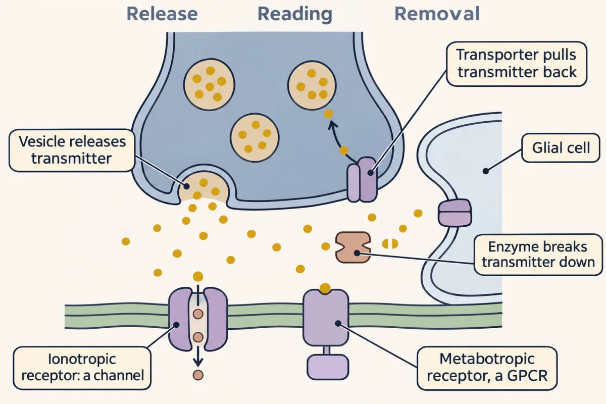
A lock and key helps you hold the steps. The transmitter is a key, the receptor is a lock it turns, and a cleaner sweeps used keys away. A drug can be:
- a copy of the key that also turns the lock: an agonist;
- a key that fits but will not turn, so the real key cannot get in: a competitive antagonist;
- oil in the lock, so the real key turns more easily: a positive allosteric modulator (allosteric means acting from another site);
- a way of stopping the cleaner, so keys pile up and keep turning locks: a reuptake blocker or an enzyme inhibitor.
The analogy breaks down in one place. Many real receptors need two transmitter molecules bound before they open, and a modulator’s site can sit far from the transmitter’s own site. The four roles still hold.
Predict. Here are four drugs that act on the GABA system:
- Baclofen activates GABA-B receptors.
- Bicuculline sits in the GABA site of GABA-A receptors without opening them.
- Diazepam binds a different site on GABA-A receptors and strengthens GABA’s effect.
- Tiagabine blocks GAT1, a transporter that clears GABA.
Which of the four make inhibition stronger, and which make it weaker?
Check your prediction
Picture the cell that receives GABA at a mature synapse, where GABA inhibits. There, three make inhibition stronger. Baclofen fakes the signal at one reader, diazepam strengthens it at another, and tiagabine keeps GABA around longer. Bicuculline weakens inhibition, and in animals it causes seizures. It is the same transmitter each time, with opposite effects. What decides the effect is which step a drug changes, and in which direction. Circuits can differ: baclofen also acts on GABA-B receptors on terminals, where it can cut GABA release.
The reader decides the sign
A transmitter is not excitatory or inhibitory by nature. The receptor and the ion gradient decide.
Use the rule from the action potential: an open channel pulls the membrane potential, Vm, toward that ion’s equilibrium potential, the voltage at which the ion has no net push in or out. GABA-A channels pass mainly chloride, with some bicarbonate, so they pull Vm toward a voltage near chloride’s equilibrium potential, ECl. This lesson treats them as chloride channels.
Picture a number line with rest at −70 mV and threshold at −50 mV. Where ECl sits on it sets what GABA does.
- ECl below Vm. Opening the channels pulls Vm down, away from threshold.
- ECl between rest and threshold, say −60 mV. Opening the channels depolarizes the cell. With the chloride and resting conductances equal and no other input, Vm settles at the balance point, halfway: (−70 + −60)/2 = −65 mV. Yet any excitation that pushes Vm past −60 mV now meets an opposing chloride current, so GABA usually blunts excitation that arrives at the same time, an effect called shunting. Whether it helps or hinders a spike still depends on its size, timing and location.
In most mature neurons ECl sits near or below rest, which is why GABA usually inhibits.
Predict. GABA opens GABA-A channels in two neurons. In the first, ECl is −75 mV, below rest. The second is an immature neuron, and suppose its ECl is −45 mV, above its threshold. What does GABA do to each?
Check your prediction
In the first neuron, the channels pull Vm down toward −75 mV, away from threshold: inhibition. In the second, they pull Vm up toward −45 mV, which lies past threshold, so GABA can excite. Same transmitter, same receptor; the chloride gradient decided the sign. That switch during development is in CNS synapses.
Walking the systems
Now fill the three slots for each transmitter, and attach one drug to one step. How most of these transmitters are made belongs to amino acids in the biochemistry course.
Glutamate
- Source. Glutamate is the brain’s main excitatory transmitter. In the textbook account nearly all excitatory neurons in the central nervous system release it, so its source is everywhere rather than one nucleus.
- Readers. Three ionotropic receptors (AMPA, kainate and N-methyl-D-aspartate (NMDA)) and metabotropic glutamate receptors. NMDA receptors open more slowly than the other channels. The NMDA receptor has two kinds of binding site: glutamate sites and separate co-agonist sites. It opens only when both kinds are filled. At normal extracellular magnesium, magnesium ions strongly block its pore at negative membrane potentials; depolarization relieves much of that block and allows a larger current. Glycine or D-serine fills the co-agonist sites. The NMDA receptor’s role as a coincidence detector is in ion channels as receptors.
- Off switch. High-affinity transporters in glia and presynaptic terminals clear it from the cleft. There is no glutamate-destroying enzyme in the cleft.
GABA
- Source. GABA is made by local interneurons, neurons whose axons stay within a region, and also by projection neurons: cerebellar Purkinje cells and the spiny projection neurons of the striatum are classic examples.
- Readers. GABA-A receptors are ligand-gated chloride channels. GABA-B receptors are G-protein-coupled receptors (GPCRs), built from two different subunits, GB1 and GB2.
- Off switch. Neurons and glia both take GABA back up through high-affinity transporters.
- Drugs. Of the four you met, three act at readers and one at removal. Baclofen is an agonist at GABA-B receptors. Bicuculline is a competitive antagonist at GABA-A receptors. Benzodiazepines such as diazepam are positive allosteric modulators: they bind at an interface of GABA-A subunits separate from the GABA sites. Tiagabine is a reuptake blocker at GAT1.
Glycine
- Source and readers. Glycine carries much of the inhibition in the spinal cord, through strychnine-sensitive chloride channels.
- Off switch. Transporters remove it.
- One drug. Strychnine blocks these inhibitory glycine receptors.
Acetylcholine and the amines
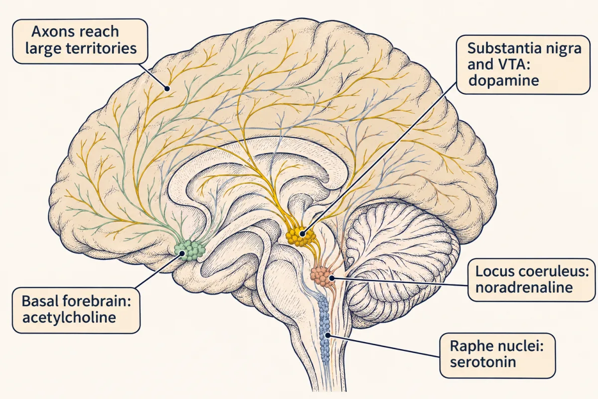
Most of these come from a nucleus, a cluster of neuron cell bodies inside the brain, whose axons reach large territories. That is why a drug acting on one can change arousal, mood or movement at once. Acetylcholine also comes from neurons that do not fit that pattern, such as striatal interneurons and motor neurons.
| Transmitter | Major source | Receptors | Off switch |
|---|---|---|---|
| Acetylcholine | Basal forebrain; laterodorsal and pedunculopontine tegmental nuclei; striatal interneurons; motor neurons | Nicotinic channels; muscarinic GPCRs | Hydrolysis by acetylcholinesterase |
| Dopamine | Substantia nigra and ventral tegmental area (midbrain) | D1-like (D1, D5) and D2-like (D2, D3, D4) GPCRs | Uptake by the dopamine transporter, DAT |
| Noradrenaline | Locus coeruleus (pons) | α1, α2 and β GPCRs | Uptake by the noradrenaline transporter, NET |
| Serotonin | Dorsal and median raphe nuclei (brainstem) | GPCRs, plus the 5-HT3 channel | Uptake by the serotonin transporter, SERT |
The sources are the major ones, not the only ones, taken from review syntheses drawn largely from rodent anatomy. Note the exception among the receptors: the 5-HT3 receptor is a ligand-gated channel.
Acetylcholine is the one in the table ended by an enzyme: acetylcholinesterase hydrolyses it at many cholinergic synapses, most famously the neuromuscular junction. One drug: donepezil is an enzyme inhibitor that slows that breakdown. With the enzyme slowed, acetylcholine stays in the cleft longer and keeps reaching receptors, so each release acts for longer. In patients with probable Alzheimer disease, PET showed donepezil reducing cortical acetylcholinesterase activity: a measure of mechanism, not of clinical benefit.
The amines are ended by transporters that use the sodium gradient to carry them back into the terminal; SERT’s uptake depends on sodium and chloride. In prefrontal cortex, dopamine is also taken up by NET. One drug: paroxetine is a reuptake blocker. It and S-citalopram sit in the central site of SERT, where serotonin binds; the structure on this page shows paroxetine there.
Dopamine deserves one more caution, since you will meet it again in motivation and reward and the basal ganglia. Popular accounts reduce it to a signal for pleasure. Recordings and manipulations in animals tie its signals instead to reward prediction, movement and motivation, among other roles, and those lessons give the evidence.
Peptides and co-transmission
- Source. Neuropeptides are short chains of amino acids packaged in large dense-core vesicles. Neuropeptide Y, for example, is made by neurons spread throughout the brain.
- Readers. Most neuropeptides act at GPCRs.
- Off switch. Peptidases usually cut them into inactive fragments (some fragments stay active), so reuptake is not their main off switch as it is for the amines.
A neuron can release more than one transmitter. In adult mouse slices, some dopamine neurons of the substantia nigra also inhibit striatal projection neurons by releasing GABA, and Melani and Tritsch (2022) found that this GABA depends on uptake through the transporter Gat1 (see the anecdote “Where does a dopamine neuron get its GABA?”). So “a dopamine neuron” names a cell’s main transmitter, not its only one, and a drug aimed at one transmitter of a neuron may leave its partner untouched.
How much: occupancy and the factor 1 + L/K
Read the transcript of “How Much a Drug Changes a Step”
Film transcript: How Much a Drug Changes a Step
A drug that fills nine in ten of its targets looks nearly done. Is it? Take paroxetine. It blocks the transporter that clears serotonin. Picture the transporters as a field of dots. As the drug's concentration rises, the dots fill. Half fill at a very low concentration. That concentration is the dissociation constant, KD. Nine in ten takes nine times that concentration. Ninety-nine in a hundred takes eleven times the 90% % concentration. The curve flattens at the top, so the last few sites cost far more drug. Do those last few sites matter? Build a simple model to find out. Suppose the transporter is the only way out, and is far from busy. Block half, and serotonin lingers twice as long.
Block nine in ten, and it lingers ten times as long. Block ninety-nine in a hundred, and it lingers about a hundred times as long. That stretch is the factor 1 + L/K. L is the free drug concentration. K is the drug's half-fill concentration. Real tissue has other routes out. They make all three numbers smaller, and limit the stretch most when the block is nearly complete. The same factor appears at a receptor. Picture GABA's dose-response curve. A competitive antagonist slides the curve right. You need more GABA, by that same factor, but you still reach the top. Diazepam, a modulator at a separate site, slides the curve to the left instead. Keep adding bicuculline.
Then, separately, keep adding diazepam. What happens to each curve? The antagonist keeps pushing the curve right, for as long as you add more. The modulator stops. Once its own sites are full, it can do no more. That is its ceiling. One warning. This rule is for blockers that compete, not for agonists. An agonist is different. With spare receptors, filling a tenth of them can give about half the full effect. The takeaway: if the transporter is the only way out, the last few percent blocked stretch the signal most.
From the film
- The share of targets a drug fills depends on its concentration relative to its dissociation constant, and the last few percent cost far more drug.
- In a simple model where a transporter is the only way out, blocking it stretches the signal by the factor 1 plus L over K.
- A competitive antagonist shifts a dose-response curve right without lowering the top, while a modulator at a separate site has a ceiling.
The quantitative side of binding is in binding and dose-response. Here is the version you need, starting with three pictures.
- The occupancy curve. Drug concentration runs along the bottom on a log scale (each equal step multiplies the concentration by the same factor; see log scales), and the share of targets filled runs up the side. The curve rises steeply through the middle and flattens near the top.
- A signal that lingers. Serotonin in the space around a synapse is plotted against time after one release. It falls as transporters clear it. Block more of the transporters and the fall stretches out.
- Two drugs at a receptor. GABA concentration runs along the bottom on a log scale, and current up the side. The control curve rises from no current to a maximum. With bicuculline, the same curve slides to the right and still reaches the same top. With diazepam, it slides to the left.
Predict. Look at the first two pictures. If the transporter is the only route out, does going from 90% to 99% occupancy stretch the serotonin signal a little or a lot?
Check your prediction
A lot. Each transporter still working clears serotonin, so blocking most of the few that remain cuts clearance far more than 9 points of occupancy suggest. Step 4 below puts numbers on it.
Worked problem: how much transporter does a drug occupy, and what does that do? Coleman, Green and Gouaux (2016) reported paroxetine binding to wild-type human SERT in HEK293 cell membranes and reported a dissociation constant, KD, of 0.08 nM. The dissociation constant is the free drug concentration at which half the binding sites are filled. Assume one class of site, equilibrium, and so little binding that free drug equals added drug.
The first equation turns a drug concentration into the share of targets it fills:
In words The fraction occupied is the drug concentration divided by the dissociation constant plus the drug concentration.
- f“f”
- the fraction of transporters occupied, from 0 to 1
- L“L”
- the free drug concentrationnM
- KD“K D”
- the dissociation constant: the concentration that fills half the sites, 0.08 nM for paroxetine at SERTnM
Sanity check With no drug (L = 0), f = 0. When L equals KD, f = 1/2. When L is far above KD, f creeps toward 1 but never reaches it, which is why the last few sites are so costly (see Step 3).
Try one number
Take L = 3 × KD, whatever KD is.
f = 3KD / (KD + 3KD) = 3KD / 4KD = 0.75.
Three times the dissociation constant fills three quarters of the sites.
Need a refresher? rates and “per”
Step 1. At L = 0.08 nM: f = 0.08 / (0.08 + 0.08) = 0.50. Half the transporters are occupied.
Step 2. What L gives 90%? Rearrange the equation to solve for L: multiply both sides by (KD + L) to get f × KD + f × L = L; gather the L terms, f × KD = L × (1 − f); divide by (1 − f): L = f × KD / (1 − f). So L = 0.90 × 0.08 nM / 0.10 = 0.72 nM, nine times KD. Check: 0.72 / (0.08 + 0.72) = 0.72 / 0.80 = 0.90.
Step 3. For 99%: L = 0.99 × 0.08 nM / 0.01 = 7.92 nM. That is 11 times the concentration needed for 90%, all to fill the last 9% of sites.
Step 4. What does blocking do to how long serotonin lingers? Build a toy model. Suppose the transporter is the only route out and works far below saturation, so serotonin is too scarce to keep the free carriers busy. Suppose too that paroxetine binding stays at equilibrium, a filled transporter cannot transport, and the KD measured in membranes also describes the block. Then only the free transporters clear serotonin, and the free share is 1 − f = KD / (KD + L). Uptake runs slower, and serotonin lingers longer, by the factor
In words The stretch is one over the free share of transporters, which works out to one plus the drug concentration over the dissociation constant.
- f“f”
- the fraction of transporters occupied by the drug
- L“L”
- the free drug concentrationnM
- KD“K D”
- the drug's dissociation constantnM
Sanity check With no drug, f = 0 and the factor is 1: no stretch. At half occupancy (L = KD) the signal lasts twice as long. In this model the factor keeps growing as L rises; Going deeper shows how other routes out put a ceiling on it.
Try one number
Continue with L = 3 × KD, which fills f = 0.75.
Free share: 1 − 0.75 = 0.25, so clearance runs at a quarter speed.
Stretch: 1 / 0.25 = 4, and the other side agrees: 1 + 3 = 4.
Where it comes from optional
Step 1, the free share is 1 − f = 1 − L / (KD + L) = KD / (KD + L).
Step 2, take the reciprocal: 1 / (1 − f) = (KD + L) / KD.
Step 3, split the fraction: KD / KD + L / KD = 1 + L / KD.
Need a refresher? rates and “per”
At 0.08 nM, half occupancy, it is 1 + 0.08/0.08 = 2: the signal lasts twice as long. At 0.72 nM it is 1 + 0.72/0.08 = 10. At 7.92 nM it is 1 + 7.92/0.08 = 100. So the costly last 9% of sites stretch the signal from ten times to a hundred times its normal length. In this model, the flat top of the occupancy curve is where the action is for a blocker.
This is the factor 1 + L/K from the one idea.
Step 5. The same factor appears at a receptor. Contrast two drugs, using made-up round numbers. Suppose GABA gives half its maximal current at a GABA-A receptor at 10 µM. That concentration is its half-maximal effective concentration (EC50). A competitive antagonist with a dissociation constant KB = 1 µM, present at [B] = 3 µM, shifts the curve right by the dose ratio:
In words The dose ratio is one plus the antagonist concentration divided by its dissociation constant.
- dose ratio“dose ratio”
- the agonist concentration needed with the antagonist, divided by that needed without it, for the same response
- [B]“B concentration”
- the antagonist concentration (square brackets mean concentration)µM
- KB“K B”
- the antagonist's dissociation constantµM
Sanity check With no antagonist, the dose ratio is 1: no shift. With [B] equal to KB, the agonist needs twice its usual concentration.
Try one number
[B] = 3 µM and KB = 1 µM: dose ratio = 1 + 3/1 = 4.
GABA’s EC50 of 10 µM becomes 4 × 10 = 40 µM.
Need a refresher? rates and “per”
Why that factor? The antagonist makes each GABA site 1 + [B]/KB times harder to fill, so GABA must be that many times more concentrated to fill the same share. It is the transporter factor again: on its own, this antagonist would fill 3 / (1 + 3) = 75% of the sites, and 1 / (1 − 0.75) = 4.
The apparent EC50 becomes 40 µM, and enough GABA still reaches the same maximum. A benzodiazepine does the opposite kind of thing: it shifts the GABA curve to the left and opens few or no channels by itself.
A key to the constants: KD is any drug’s dissociation constant; KB names it for an antagonist at a receptor, and Ki for an inhibitor at a transporter or enzyme. Km is different: the serotonin concentration at which a transporter runs at half its maximum.
Predict. Keep adding bicuculline, then, in a separate experiment, keep adding diazepam. What happens to each curve?
Check your prediction
The bicuculline curve keeps sliding right, by 1 + [B]/KB, for as long as you add more. The diazepam curve stops moving once diazepam’s own sites are full: a positive allosteric modulator has a ceiling set by its own saturation.
Where the factor holds. It describes the immediate action of a drug at one target, at equilibrium, when the drug competes for the natural molecule’s site: a competitive antagonist at a receptor, or a competitive inhibitor at a transporter or enzyme. At a receptor it gives the dose ratio. At the off switch it gives the slowing of uptake only if the carrier works far below saturation, and the stretch of the whole signal only if the blocked route is the only way out and the drug’s occupancy stays fixed. The numbers come from a model and from SERT in cell membranes: they are not a measured change in synaptic serotonin and not a dose for a person. The factor does not predict:
- what an agonist does, because a cell with spare receptors, more than it needs for a full response, can respond strongly with few of them filled;
- the ceiling of an allosteric modulator;
- chronic adaptation, which the anecdote about the dopamine transporter knockout shows;
- drugs that hit several targets, or releasers such as amphetamine, which change both removal and release;
- clinical benefit or the cause of a disease.
- Occupancy at your L
- 50.0%
- L needed for the target
- 0.72 nM
- times the L for 90% (above 90% only)
- 1.00
In this sketch: one class of site, equilibrium, free drug equal to added drug; paroxetine at wild-type human SERT in HEK293 membranes, reported KD = 0.08 nM (Coleman, Green and Gouaux, 2016).
Occupancy f = L / (KD + L) climbs steeply at first and flattens at the top, so filling the last sites takes far more drug than filling the first.
In the simulation, set the target to 90% and then to 99%, read off the concentration each needs, and work out 1 + L/KD for each.
A mechanism is not a cause
Read the transcript of “A Mechanism Is Not a Cause”
Film transcript: A Mechanism Is Not a Cause
If a drug that blocks serotonin uptake helps depression, was low serotonin the cause? Start with what is established. SSRI drugs block the serotonin transporter, in the pocket where serotonin binds. In 1990, Pedro Delgado and colleagues ran a blinded experiment. They studied 21 patients whose depression had remitted on antidepressants. Each received a drink with no tryptophan, serotonin's building block, and on another occasion a control drink. Neither the patient nor the rater knew which drink was which. After the tryptophan-free drink, 14 of the 21 relapsed. They returned to remission within one to two days on normal food. The control drink had no significant effect.
Does this show that low serotonin causes depression? It does not. These patients were chosen because an antidepressant had helped them. The result suggests that some of them needed serotonin to stay well. A treatment can depend on a transmitter that was never low. Pooled depletion studies split people by risk. Healthy volunteers without a family history showed no significant mood drop. Those with one dropped slightly. Drug-free people recovered from depression dropped more. A 2022 review found no consistent support for a low-serotonin cause. Other researchers dispute its methods. Meanwhile, a 2018 analysis of trials found 21 antidepressants better than placebo, for acute major depression in adults.
Certainty ranged from moderate to very low. Both can be true. Aspirin relieves a headache, yet headaches are not caused by a lack of aspirin. Still, the tryptophan experiment suggests that some treated patients need serotonin to stay well. The takeaway: a drug's mechanism is evidence about the drug. It is not proof of what causes the disease.
From the film
- In a blinded tryptophan-depletion study, 14 of 21 patients in remission on antidepressants relapsed, which suggests some treated patients need serotonin to stay well.
- A treatment can depend on a transmitter that was never low.
- A drug's mechanism is evidence about the drug, not proof of what causes the disease.
SSRIs, the selective serotonin reuptake inhibitors such as paroxetine, block SERT; that is established, down to a structure of the drug in its pocket. The step from there to “depression is caused by low serotonin” is a separate claim. One blinded experiment shows how to keep the two apart.
Design problem
Neurons make serotonin from the amino acid tryptophan, which they take up from the blood. Delgado and colleagues (1990) gave 21 patients whose depression had remitted on antidepressants a tryptophan-free amino-acid challenge, which lowers the precursor of serotonin, and a control challenge, double-blind and in crossover: neither patient nor rater knew which was which. Fourteen of the 21 relapsed after tryptophan depletion and returned to remission over 24 to 48 hours on normal food; the control challenge had no significant effect. Does this show that low serotonin causes depression? If not, what would you need to test that claim?
Saved in this browser only.
One way to do it
It does not. The patients were selected because they had responded to antidepressants, most of which act on serotonin or noradrenaline systems. The result suggests that remission in some treated patients depends on serotonin being available. A treatment can depend on a transmitter that was never low to begin with.
To test whether low serotonin causes depression, you would study people who are not on these drugs. Deplete tryptophan, with a balanced control drink that contains tryptophan, in people who have never been depressed and in untreated people with depression, double-blind and in crossover. If this acute drop in serotonin synthesis were enough by itself to lower mood in the people tested, over the hours observed, mood should fall relative to the control session. Analyse the two groups separately: further worsening in people already depressed is not guaranteed. A null result would not rule out a chronic or vulnerability-dependent role for serotonin. Depletion studies in such groups have been pooled. A 2007 meta-analysis found no significant mood drop in healthy volunteers without a family history of depression, a slight drop in healthy volunteers with one, and a moderate drop in drug-free people who had recovered from depression, so the answer depends on who is vulnerable. Check that blood tryptophan actually fell, and report how many participants changed, not only group means.
Then weigh the result against the wider evidence below, keeping apart whether a drug helps and what caused the illness.
The wider evidence
A 2022 umbrella review by Moncrieff and colleagues found no consistent support for depression being caused by lowered serotonin concentration or activity. Other researchers dispute its methods and argue the serotonin system is still implicated. A separate question is whether the drugs help: a 2018 network meta-analysis by Cipriani and colleagues found all 21 antidepressants it included more effective than placebo for acute major depression in adults, with certainty of evidence from moderate to very low.
Both findings can be true at once. Aspirin relieves a headache, and headaches are not caused by a lack of aspirin. The comparison has a limit, though: Delgado’s result suggests that serotonin may be part of how the drug works in some treated patients. It still does not show that serotonin was low before treatment.
Going deeper
Telling a competitor from a modulator
The dose ratio 1 + [B]/KB is the Schild relation. It holds at equilibrium for an antagonist that competes for the agonist’s site, even when several agonist molecules must bind. So the dose ratio minus 1 grows in direct proportion to the antagonist’s concentration. A negative allosteric modulator, which lowers the agonist’s affinity from a separate site, can mimic one shift. In a model check (an affinity-only model with a cooperativity factor of 0.05), such a modulator gives a 2.73-fold shift where a competitor at the same [B]/KB = 2 gives 3-fold. At [B]/KB = 8 it gives about 6.4-fold against the competitor’s 9-fold, because its effect levels off as its own sites fill, in this model at 20-fold. One concentration cannot tell the two apart. Several can help, and binding or structural experiments add evidence on where the drug sits.
Where the factor 1 + L/K stops working
These are model calculations, made to show the scope conditions, not measurements.
- Other routes out. In a well-mixed model with fixed parallel first-order clearance, suppose diffusion and other drug-insensitive routes supply 5% of the baseline clearance rate constant. Then blocking the transporter can lengthen serotonin’s decay time constant at most 1/0.05 = 20-fold, however much drug you add: at 50%, 90% and 99% block the stretch is about 1.9, 6.9 and 16.8 times, against 2, 10 and 100 with no other route. The bound need not hold if clearance is nonlinear or changes across space.
- A busy carrier. Take a competitive inhibitor at nine times its Ki and compare uptake at the same serotonin concentration. With serotonin far below the transporter’s Km, uptake runs about 10 times slower. With serotonin at Km, it runs only about 5.5 times slower. During a falling serotonin transient the ratio changes as serotonin falls.
- Spare receptors. In the operational model with a transducer ratio of 10, filling 10% of the receptors gives half the largest response the system can give; with a ratio of 100, about 1% does.
How the 5-HT3 receptor was shown to be a channel
In 1989 Derkach, Surprenant and North recorded single-channel currents opened by serotonin in outside-out patches from guinea-pig enteric neurons, in the submucous plexus of the gut wall. The channels had apparent conductances of 15 and 9 pS. A response in an excised patch, without the cell’s cytoplasm, showed that the 5-HT3 receptor is a ligand-gated channel.
Glycine’s second job, and D-serine
In cultured mouse brain neurons, Johnson and Ascher found that glycine potentiated N-methyl-D-aspartate (NMDA) receptor responses by a mechanism separate from the inhibitory glycine receptor (see the anecdote “Glycine has a second job”). Whether glycine or D-serine fills the NMDA receptor’s co-agonist site depends on the synapse and the stage of development.
Your notes
Select any text in the lesson to highlight it or add a note to it. Your highlights and notes are saved in this browser and collected in your notebook.
This browser is not letting the site save anything (a private window or strict privacy settings), so highlights and notes will be lost when you leave the page.
How we measure it
Outside-out patch clamp
A pipette sealed onto a neuron is pulled away so that a small patch of membrane closes over its tip with the outer face toward the bath. You apply a transmitter to the bath and record single-channel currents. A response that survives without the cytoplasm points to a receptor that gates its own channel.
Radioligand binding
Membranes carrying a target, such as SERT expressed in HEK293 cells, are incubated with a radioactive drug at a range of concentrations, and bound drug is separated and counted. A fit to a binding model gives the dissociation constant, KD. It measures affinity, not what the drug does once bound.
X-ray crystallography and cryo-electron microscopy
Purified receptors or transporters are imaged with a drug bound, and the map shows which residues line the drug's pocket. Each structure is one state, so it must be paired with functional recordings before you say whether binding opens, blocks or modulates.
Blinded crossover depletion challenge
Each participant receives a challenge that lowers a transmitter's precursor on one occasion and a matched control on another, with neither participant nor rater knowing which. Each person serves as their own control, and the result applies to the population that was enrolled.
Practice questions on this lesson (3) → MCAT-style, original, each with an explanation
Where this lesson fits
Before thisReview first
- The Action Potential and ConductionThis course · Lesson 4
You need equilibrium potentials and channels pulling Vm toward them to see why GABA's sign depends on E_Cl.
- The Resting Membrane and Passive SignalsThis course · Lesson 3
Resting potential and conductance explain the balance-point calculation for chloride channels opening near rest.
- Synaptic Transmission and Quantal ReleaseThis course · Lesson 5
The lesson uses vesicular release and the cleft as the starting point and does not re-teach them.
After thisStudy next
- Integration, Inhibition and NeuromodulationThis course · Lesson 7
It takes the chloride shunting and inhibition-by-conductance idea from here and shows how inhibitory inputs shape firing.
- Feeding, Motivation and RewardThis course · Lesson 19
It builds on dopamine's source, receptors and uptake here to test what dopamine signals actually do.
- Descending Control and the Basal GangliaThis course · Lesson 16
Dopamine and GABA projection neurons introduced here are placed into movement circuits there.
Elsewhere on the siteFilms, simulations and method films in other courses
- Spectrophotometry and enzyme kineticsMethod film
Competitive inhibition raising the apparent Km by 1 + I/Ki is the same factor used here.
- Dose–response explorerSimulation · Cell Signaling
Occupancy from a dissociation constant and shifted dose-response curves under a competitive antagonist are worked through there.
- The type of receptor sets the speed of the responseFilm · Cell Signaling
It shows how the receptor type, not the transmitter, sets the response, as with ionotropic and metabotropic readers here.
- The Mark and the ReaderFilm · Cell Signaling
It shows how a signal is ended at the receptor and by degradation, the removal step here.
Suggested by an AI model from the lessons themselves, then checked. How we use AI
Finish line
Test yourself
9 questions
Practice and MCAT-style, with explained answers.
Next · Lesson 7
Integration, Inhibition and Neuromodulation
An inhibitory input can leave a resting cell's voltage unchanged and still stop it from firing. How?
Continue →