Organismal Biology · Skim version
Nervous Control: A First Look
Read the full lesson → Skim the whole course →
The guiding idea
A doctor taps the tendon below your kneecap and your leg kicks before you decide anything. What route does the signal take, and how fast does it run?
A nervous response runs a route, receptor to sensory neuron to integrating centre to motor neuron to effector, and in a simple model its time is the sum of its legs: each axon's length over its speed, about a millisecond per synapse in the cord, and short delays at the ends.
From the film: Every Response Has a Running Time
- A nervous response runs a route: receptor, sensory neuron, integrating centre, motor neuron, effector.
- Along an axon the signal is an all-or-none impulse; at most synapses it crosses as a released chemical.
- Helmholtz timed a frog nerve from two points; subtracting the two delays removed the junction and muscle, leaving the nerve's own speed.
- In a simple model, a response's time is the sum of its legs: each axon's length over its speed, about a millisecond per synapse in the cord, and short delays at the ends.
- In the knee-jerk model, nearly all of the bar is time on the two axons, and its legs add up to just under the measured time at which the thigh muscle's electrical signal starts.
From the film: One Synapse or Several
- The knee jerk's direct path has one synapse in the spinal cord; it is monosynaptic.
- Interneurons in the cord also quiet the opposing muscle, so even the knee jerk is not a single wire.
- The withdrawal reflex runs through interneurons from the start, exciting flexors and inhibiting extensors.
- In this example, the withdrawal is slower mostly because its pain fibres conduct slowly; each extra synapse adds only about a millisecond.
- Both reflexes are somatic, because their effector is skeletal muscle, not because they are voluntary.
-
One neuron: what the first film left out
-
How fast? What Helmholtz’s subtraction can tell you
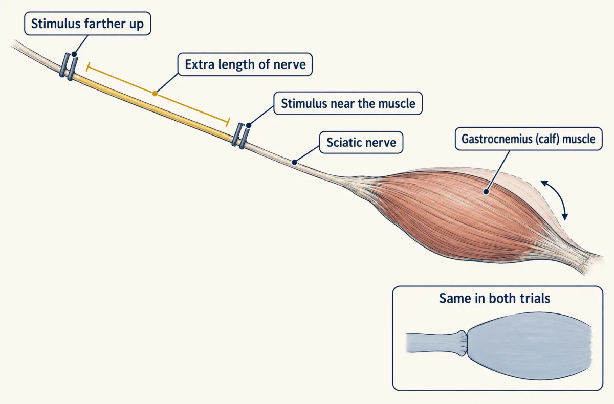
Helmholtz stimulated a frog's sciatic nerve near the gastrocnemius and then farther up, and timed the delay to the twitch each time. The junction and muscle respond in the same time in both trials, so the extra delay belongs only to the extra length of nerve. AI-generated illustration -
The reflex arc: what the second film left out
-
Two autonomic divisions and their exceptions
-
Four layouts of neurons
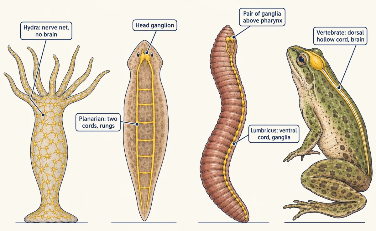
Every layout links sensing to acting, but they differ: a net with no brain, a ladder, a segmented ventral cord, a dorsal hollow cord with a brain. None is a step toward another. AI-generated illustration -
Going deeper
Where this lesson fits
Before thisReview first
- Tissues, Organs and HomeostasisThis course · Lesson 6
It introduces receptor, control centre and effector, the same route this lesson times leg by leg.
- Homeostasis in ActionBiology · High school
It shows a feedback loop with sensor, control centre and effector, which this lesson treats as a timed route.
After thisStudy next
- Hormones and the Endocrine SystemThis course · Lesson 8
Hormones are the second kind of control, so you can compare their slow delivery with the nerve route timed here.
- Muscle, Bone and MovementThis course · Lesson 19
The effector end of the reflex arc is a muscle, and this lesson shows how its fibres shorten.
- Heat Balance and Body TemperatureThis course · Lesson 9
Heat balance uses sensors, a control centre and autonomic effectors such as sweat glands and blood vessels.
Suggested by an AI model from the lessons themselves, then checked. How we use AI