Organismal Biology · Unit 3: Supply and exchange
Gas Exchange and Breathing
Rewritten on : new text, flashcards and film. Your notes and highlights stay, but the text around them may have moved.
Water holds far less oxygen than air, yet a trout's gill can send its blood out at a higher oxygen partial pressure than the water leaving it. Your lung cannot do that with air. Why not?
Pause when the reading reaches a film, and offer to play it. Saved on this device.
Watch the film now, or keep listening
Builds on:Size, Scale and SurfaceWhat an Organism Has to DoTissues, Organs and HomeostasisWhy these?
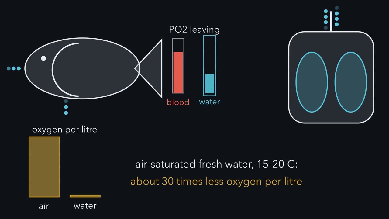 ▶ Watch The Last Gas the Blood Touches · 3:15
Read the transcript of “The Last Gas the Blood Touches”
Film transcript: The Last Gas the Blood Touches
Water holds far less oxygen than air. Yet a trout's gill can send its blood out at a higher oxygen partial pressure than the water leaving it. Your lung cannot do that with air. Why not? Each gas in a mixture has a partial pressure, its share of the total pressure.
Dry air at sea level is about one fifth oxygen. But partial pressure is not amount. Water left touching air reaches the same oxygen partial pressure. Yet each litre holds far less oxygen. Oxygen crosses an exchange surface only by diffusion. Molecules cross both ways, but the net flow runs from higher partial pressure to lower. So here, richer means higher partial pressure, not more oxygen. Fick's law gives three dials for speed. More area, a steeper partial pressure difference and a thinner barrier all speed transfer. That is why gills and lungs are large and thin. They are moist because gases must dissolve before crossing cells. Here, all along the surface, the water or air stays richer than the blood beside it.
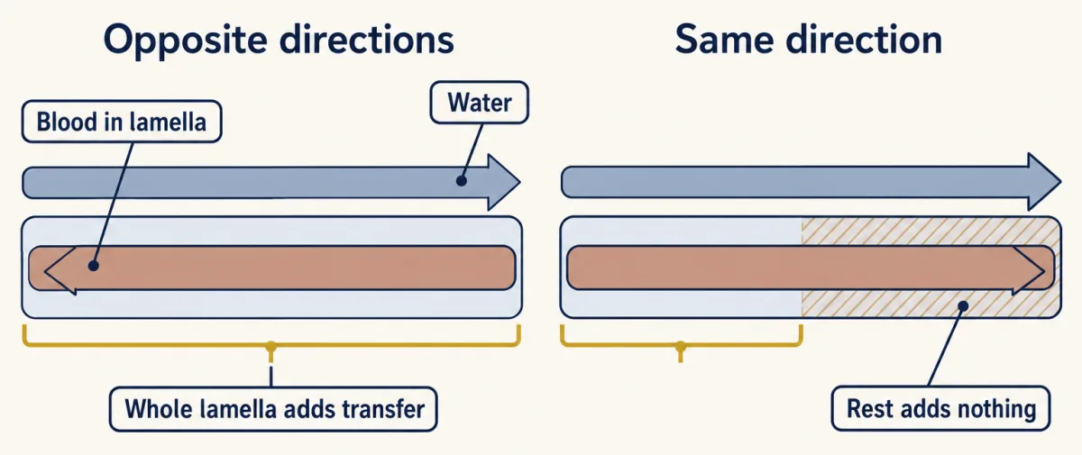
Diffusion brings the blood toward it, never past. So the leaving blood can at best reach the partial pressure it last touched. Run water and blood the same way along a thin gill plate, a lamella. The water starts rich and the blood starts poor. Partway along, their partial pressures close in on each other. The rest of the plate adds little. Now reverse the blood, so it runs against the water. Where will the leaving blood end up?
The blood about to leave meets water that has just arrived. All along the plate, the water stays a little richer than the blood beside it. This is countercurrent exchange. With a large, thin surface, the leaving blood can beat the water leaving. Running the same way, it never could. Fold the surface into a dead-ended sac, an alveolus in a mammal lung. Air goes in and out by the same tube. Each breath only partly renews the gas inside. Blood keeps taking oxygen out. So the sac's gas sits well below the inhaled air. Every capillary touches that same mixed gas. Blood can at best match it. That answers the trout. In its gill, the leaving blood last touches fresh, incoming water.
In your lung, it last touches stale, mixed gas. Fick's law and the flows of blood and water or air set how close the blood gets. The flow arrangement sets what it last touches. Breathing keeps that water or air fresh.
From the film
- Oxygen and carbon dioxide cross every exchange surface by diffusion, a net flow from higher partial pressure to lower.
- Partial pressure is not amount: water at the same PO2 as air holds far less oxygen per litre.
- Fick's law: more area, a thinner barrier and a steeper partial-pressure difference all speed transfer.
- While the water or air stays richer, blood can at best reach the PO2 it last touched; the surface and the flows set how close it gets.
- In a teleost gill the leaving blood last touches incoming water; in a mammal lung it can at best match the alveolar gas.
Water holds far less oxygen than air, yet a trout’s gill can send its blood out at a higher oxygen partial pressure than the water leaving it. Your lung cannot do that with air. Why not?
The first film built the answer from diffusion, the net movement of a gas from higher partial pressure to lower (the pressure one gas in a mixture would exert alone: PO2 for oxygen, PCO2 for carbon dioxide), and showed that partial pressure is not amount. Vertebrate blood widens that gap: most of its oxygen is bound to haemoglobin, which adds to the amount without raising the PO2. In this lesson “richer” always means higher PO2.
The one idea. Oxygen and carbon dioxide cross every exchange surface only by diffusion, which pulls the blood’s partial pressure toward that of the water or air beside it. So blood that takes up oxygen all along the surface can leave with no higher PO2 than the water or air it last touched, and blood that gives up carbon dioxide all along, with no lower PCO2. Fick’s law (area, thickness) and the flows of blood and of water or air set how close the blood gets; how those flows run past each other sets what it last touches; breathing keeps that water or air fresh.
Scope: a ceiling on partial pressure, not total gas, for passive exchange at steady state (flows and rates constant), one gas at a time, while the water or air stays richer than the blood beside it all along the contact (lower in PCO2, for carbon dioxide), as in every case here. A small or thick surface, short contact time, mismatched air and blood flow, or blood that bypasses the surface leave real blood below it. When streams that touched different gas mix, as in birds, the ceiling holds for each stream, not the mix.
Fick’s law: how fast gas crosses
Predict. These numbers are invented. Barrier 1 is 50 m2 and 0.5 micrometres thick; barrier 2 is 30 m2 and 1.0 micrometre thick. With the same gas, partial-pressure difference and material, does barrier 2 manage more or less than half of barrier 1’s rate?
Check your prediction
Less than half: 30%, because the lost area and the added thickness multiply.
The rate across a sheet of tissue follows Fick’s law (see also size and scaling):
rate of gas transfer = k x A x (P1 - P2) / T
A is the area, P1 - P2 the partial-pressure difference (ΔP), T the barrier thickness, and k a constant for that gas and barrier. For scale, dry air at sea level (about 21% oxygen at about 760 mmHg, 101 kPa) has a PO2 of roughly 160 mmHg (21 kPa).
Worked problem.
- Ratio. Rate 2 / Rate 1 = (A2 / A1) x (T1 / T2), since k and ΔP cancel; thickness flips because it sits below the line.
- Area. 30 m2 / 50 m2 = 0.60.
- Thickness. 0.5 micrometres / 1.0 micrometre = 0.50.
- Multiply. 0.60 x 0.50 = 0.30, so 30% remains.
- Area ratio, A2 / A1
- Thickness ratio, T1 / T2 (flipped: T is below the line)
- Pressure-difference ratio, ΔP2 / ΔP1
- Rate 2 / Rate 1 (k cancels)
- A / T, barrier 2
- A / T, barrier 1
- Check: (A2 / T2) / (A1 / T1) × ΔP ratio
Rule to keep: rate ∝ A × (P1 − P2) / T. More area, a steeper difference and a thinner barrier all raise the rate; lost area and added thickness multiply. In this sketch: invented numbers; same gas, same k, steady diffusion across a uniform sheet; not to scale.
Water or air: skin, gills and tracheae
Water is hard to breathe: air-saturated fresh water at 15 to 20 degrees C holds roughly 30 times less oxygen per litre than air, and it is about 800 times denser and far more viscous, so a gill does more work for less oxygen.
Skin. An earthworm exchanges gases across its moist skin. Frogs combine skin and lungs, and adult lungless salamanders (family Plethodontidae) breathe through the skin and mouth lining alone. Skin breathing works where skin stays wet and oxygen demand per unit of surface is modest.
Gills. In teleost fish, the large group of bony fishes that includes trout, each gill is folded into filaments carrying thin plates called lamellae: large area, thin barrier. Most teleosts move water one way, in through the mouth and out under the gill cover; tunas ram it through an open mouth as they swim. Blood runs opposite to the water inside each lamella, the countercurrent exchange the first film built, which raises the ceiling to the PO2 of the incoming water. Reaching it is another matter.
Predict. A trout’s lamellae have plenty of area, but suppose the fish pumps only a trickle of water, carrying less oxygen per minute than the blood could take up. Does countercurrent flow still bring the leaving blood close to the incoming water?
Check your prediction
No. The ceiling is unchanged, but the trickle loses much of its oxygen on the way, so its PO2 falls toward the blood’s before the blood is full. Getting near the incoming water takes both a large, thin surface and a water flow whose oxygen-carrying capacity per minute is at least the blood’s.
Blood from a teleost gill goes on to the body in a single circuit, as in circulation and the heart.
Tracheae. Most insects carry oxygen in air, not blood: air enters openings called spiracles into tracheae, branching tubes whose finest branches reach the tissues. Many larger insects pump the abdomen, and in 2003 Mark Westneat and colleagues used synchrotron X-ray imaging to watch tracheae in live beetles, crickets and ants compress and expand rhythmically.
A leaf, by contrast, exchanges gases by diffusion through pores called stomata, with no pump; see moving water and sugar in plants.
Three lungs: mammal, bird, alligator
A lung is an internal, air-filled exchange organ.
Mammals: tidal. Air flows in and out through the same airways, tidal ventilation, into tiny dead-ended sacs, the alveoli. Their PO2 sits near 100 mmHg in a resting adult human, below the roughly 150 mmHg of warmed, humidified inhaled air, for the reasons the first film gave.
Birds: one way, crosscurrent. Birds have no diaphragm, and their stiff lungs keep a nearly constant volume and exchange gas in narrow tubes called parabronchi. Thin-walled air sacs in front of and behind the lungs work as bellows as the sternum and ribs move. Through most parabronchi, air flows the same way during inspiration and expiration, so fresh air keeps arriving at one end.
Capillaries cross the parabronchus at right angles, each at a different point along it: crosscurrent exchange. Each capillary can at best match the air at its own point: nearly fresh air near the inflow end, only drained air near the outflow end. When the streams join, their oxygen contents combine, so the mixed blood lands between its richest and poorest streams and can carry a higher PO2 than the air leaving the parabronchi.
Predict. Take idealized gill, bird-lung and mammal-lung arrangements. Give each the same incoming PO2 in medium and blood, the same ratio of the medium’s oxygen-carrying capacity per minute to the blood’s, even flow everywhere and no diffusion limit. The first film put the gill above the mammal lung. Where does the bird lung fall, and why?
Check your prediction
Between them. The gill’s leaving blood last touches incoming water; the bird’s, a spread of air from fresh to drained; the mammal’s, mixed alveolar gas. This ranks idealized shapes, not real animals (see Going deeper).
Alligators: one way, no air sacs. In 2010 C. G. Farmer and Kent Sanders reported one-way airflow in the bronchi they measured in the American alligator, Alligator mississippiensis, which has no air sacs. Birds and crocodilians are both archosaurs, the reptile group that holds them and their extinct relatives, and the shared trait fits inheritance from a common ancestor without proving it. Later work found one-way flow in monitor lizards and green iguanas too, so the trait may be older than archosaurs, or may have evolved more than once; see reading phylogenetic trees.
So air moves one way through most bird parabronchi and the alligator bronchi measured so far, while a mammal’s alveoli are dead ends whose gas depends on how much fresh air each breath delivers. One reminder of the rule: blood can at best match the gas it last touched, so a surface works only while that gas is renewed. The second film follows one breath in.
Read the transcript of “Keeping the Gas Fresh”
Film transcript: Keeping the Gas Fresh
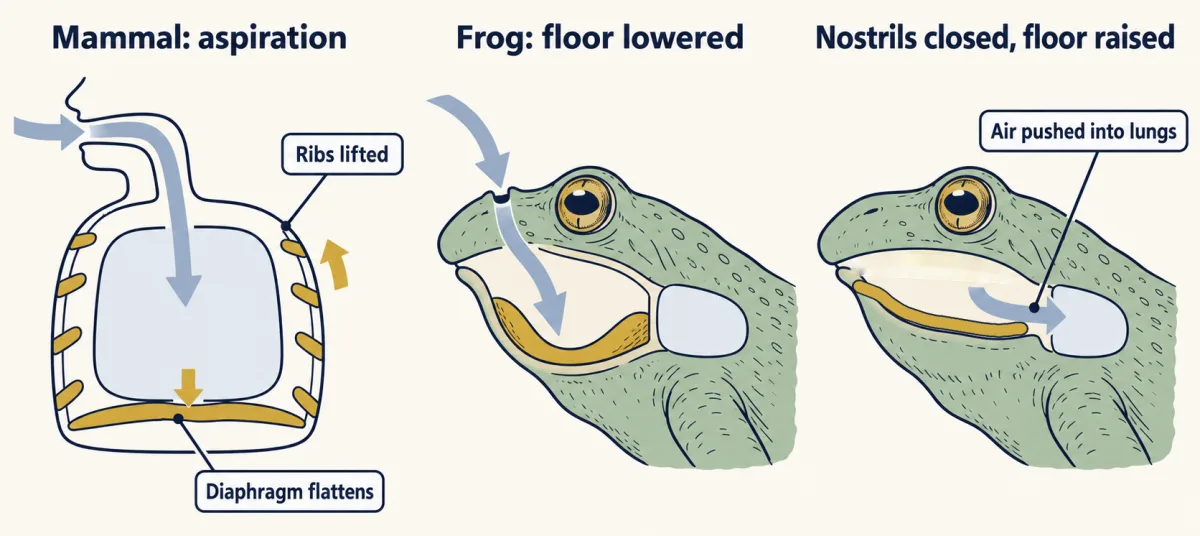
Two people move the same total air each minute. So why is one of them building up carbon dioxide while the other is not? The first film ended at a dead-ended sac, the alveolus. Blood can at best match the gas inside it. Blood flow keeps bringing new blood. But the gas must be renewed too, through the airways. So what keeps moving fresh air in? A mammal pulls air in. The diaphragm, a dome of muscle under the lungs, contracts and flattens. The chest enlarges, pressure in the lungs falls, and air flows in. This is aspiration. But a tube leads to the sac. The airways, from nose and mouth down to the smallest bronchioles with no alveoli, exchange nothing. They are dead space.
So the first air into the sac each breath is stale air left in the tube. And fresh air still in the tube when you stop breathing in is breathed out unused. Take slow, deep breaths. Each breath clears the dead space and sends a large fresh portion into the sac. The total air moved each minute is minute ventilation. The fresh part reaching the alveoli is alveolar ventilation. Now keep the total air per minute the same, but take smaller, faster breaths. Does carbon dioxide in the sac rise, fall or stay the same?
It rises. Each small breath barely clears the dead space. Less fresh air reaches the sac, while the body makes carbon dioxide as fast as before. Here the first film's rule runs in reverse. Blood giving up carbon dioxide leaves with no lower partial pressure than the gas it last touched. So the blood's carbon dioxide climbs with the sac's. The brainstem watches that result. Carbon dioxide crosses into the brain's fluid and lowers its pH.
Sensors near the brainstem surface detect the fall, and the drive to breathe rises. Haldane and Priestley sampled the last of a forced breath out. In resting subjects, a small rise in alveolar carbon dioxide greatly increased breathing. Lowering oxygen did little until it fell far. So breathing keeps the gas the blood touches fresh. At rest, a mammal's controller mostly reads carbon dioxide. That level is the result of its own breathing, set against the body's carbon dioxide output. Too little fresh air, and carbon dioxide climbs. The sensors then push breathing back up.
From the film
- Breathing renews the air or water at the exchange surface and blood flow renews the blood; together they keep the partial-pressure difference up.
- In a mammal, the first air of each breath into the alveoli is stale air from the dead space, the airways where no exchange happens.
- At steady state, breathing ordinary air with carbon dioxide production unchanged, alveolar ventilation, not total air moved, sets alveolar PCO2.
- In resting mammals, carbon dioxide, sensed largely through brain pH, sets most of the drive to breathe.
Moving the gas: aspiration and the frog’s pump
The second film showed aspiration, the diaphragm flattening to lower chest pressure, but not the ribs or the breath out. The external intercostal muscles lift the ribs as the diaphragm contracts, and the lungs follow the enlarging chest. Quiet expiration in adult humans is normally passive recoil of lungs and chest wall once the muscles relax; forceful expiration adds the internal intercostals and abdominal muscles.
The wet lining of the alveoli has surface tension, pulling inward like a soap bubble. Surfactant, a lipid and protein mixture secreted by alveolar cells, lowers it, raising compliance (how much the lung stretches for a given pressure change), so the lung inflates with less effort.
A frog works the other way. In buccal pumping it lowers the floor of its mouth with the nostrils open, drawing in air, then closes the nostrils, opens the glottis (the opening into the airway) and raises the floor, pushing air into the lungs under positive pressure. Mammals and birds both draw air in by lowering pressure: mammals in the chest, birds in the air sacs. Buccal pumping is thought to be the older method, from which aspiration in amniotes (reptiles, birds and mammals) evolved, but a living frog is its own lineage, not an ancestor of mammals or birds.
Carbon dioxide runs the controller
Breathing rhythm comes from brainstem neurons (see the nervous system map and the hypothalamus and autonomic system). The second film gave J. S. Haldane and J. G. Priestley’s 1905 result, which made carbon dioxide the main resting stimulus in these men. Their method: because windpipe gas mixes fresh and stale air, they took the end of a forced breath out as alveolar air, with themselves and colleagues as resting subjects breathing added carbon dioxide or less oxygen. Alveolar PCO2 also stayed nearly constant for each man at rest.
Later work found two kinds of sensor: the central chemoreceptors the second film showed, which read the pH of brain extracellular fluid, and the carotid bodies, sensory clusters where each carotid artery forks, which respond mainly to low arterial oxygen and are also affected by carbon dioxide. In adult humans, ventilation rises steeply with hypoxia, too little oxygen, only once arterial PO2 falls well below normal. In teleosts, low oxygen in the water is a major ventilatory stimulus.
Predict. A resting person breathes a gas mixture low enough in oxygen to drop arterial PO2 well below normal, and ventilation rises steeply. Carbon dioxide production stays the same. Which sensor is mainly driving the rise, and what happens to alveolar PCO2 while it does?
Check your prediction
Mainly the carotid bodies, which respond to low arterial oxygen; nothing here raised carbon dioxide. With production unchanged, the extra breathing is more than production requires, so alveolar PCO2 falls, a state called hypocapnia.
Worked problem. The second film showed the direction; here are the numbers. Illustrative values for an adult human at rest: tidal volume (one breath) 500 mL, 12 breaths per minute, and 150 mL of dead space, the conducting airways, which have no alveoli and exchange no gas.
- Minute ventilation, total air moved, = 12 x 500 mL = 6,000 mL per minute.
- Alveolar ventilation, fresh air reaching alveoli, = 12 x (500 - 150) = 4,200 mL per minute.
- Breathe rapidly and shallowly, 24 breaths of 250 mL: minute ventilation = 24 x 250 = 6,000 mL per minute, unchanged.
- Alveolar ventilation = 24 x (250 - 150) = 2,400 mL per minute, 57% of before.
- At steady state, breathing ordinary air, alveolar PCO2 is about 0.863 x carbon dioxide production (mL per minute) / alveolar ventilation (L per minute). With production held at an illustrative 200 mL per minute, alveolar PCO2 would rise from about 41 to about 72 mmHg.
Gas carriage in blood is in carrying oxygen and carbon dioxide, and acid-base adjustment in the kidney.
Design problem
Birds have one-way airflow driven by air sacs. Alligators have none. How would you find out whether air moves one way through an alligator’s lung, and what would the result tell you?
Saved in this browser only.
One way to do it
Put directional probes, such as heated thermistors, in identified bronchi of live alligators: flow the same way during inspiration and expiration means one-way flow there. This is the approach Farmer and Sanders reported in 2010. Then take the brain and breathing muscles out of the question by pumping air in and out of an excised lung through the trachea. If one-way flow persists and the airways have no flap valves, the lung’s own structure, most likely its airway geometry, produces it. As a control, a mammal lung on the same pump should show flow reversing with each breath; if not, the probes or pump are at fault.
Limits: Farmer and Sanders’s pump tests used four lungs, one live animal had a direction-sensing probe, and only some bronchi were measured.
Going deeper
Ranking flow arrangements, and a tracer aside
In 1972 Johannes Piiper and Peter Scheid compared idealized models of the three arrangements under the matched conditions of the Predict above: countercurrent transferred the most gas by their measure, crosscurrent next, the mixed pool of a mammal lung least. When the surface is small or thick, the arrangements barely differ. The textbook rule that air takes two breaths to pass through the bird lung is a schematic; helium tracer work in songbirds found some inhaled gas reaching the front air sacs within one breath.
What Haldane and Priestley could not yet see
They argued that carbon dioxide reached the brain’s respiratory centre through the arterial blood; separate sensor cells had not yet been found.
Sensing carbon dioxide in the brainstem
In mice, retrotrapezoid nucleus neurons carry two pH sensors that work in parallel, the proton-sensing receptor GPR4 and the potassium channel TASK-2; losing GPR4 alone removes much of this response (see GPCRs and G proteins).
Your notes
Select any text in the lesson to highlight it or add a note to it. Your highlights and notes are saved in this browser and collected in your notebook.
This browser is not letting the site save anything (a private window or strict privacy settings), so highlights and notes will be lost when you leave the page.
How we measure it
End-expiratory sampling of alveolar air
The subject breathes out suddenly and deeply through a tube, and the last part of the breath is sampled and analysed. That last portion comes from the gas-exchanging part of the lung, so its carbon dioxide and oxygen give an estimate of alveolar partial pressures.
Directional flow probes in the airways
A heated thermistor placed in a bronchus is cooled by moving air, and a paired arrangement tells which way the air is going. Recording in live animals and in excised lungs ventilated by a pump separates the lung's own structure from the animal's breathing muscles and brain.
Synchrotron X-ray imaging of live animals
A synchrotron produces X-rays bright enough to film air-filled tubes inside a small living animal. It shows tubes changing shape; pressure and gas measurements are still needed to say how much air those changes move.
Spirometry and whole-body plethysmography
A spirometer records the volume of air moved into and out of the lungs, giving tidal volume and breathing rate. In small animals such as mice, a sealed chamber records the pressure changes caused by breathing, so ventilation can be measured in a conscious animal breathing test gases.
Practice questions on this lesson (4) → MCAT-style, original, each with an explanation
Where this lesson fits
Before thisReview first
- Size, Scale and SurfaceThis course · Lesson 2
Fick's law and surface area against volume from there are used here without being re-taught.
- What an Organism Has to DoThis course · Lesson 1
It sets up the trade-off of taking in gas across a surface while losing water, which this lesson builds on.
- Tissues, Organs and HomeostasisThis course · Lesson 6
Breathing here is a controlled loop with sensors and set points, as built up there.
After thisStudy next
- Carrying Oxygen and Carbon DioxideThis course · Lesson 13
It takes the blood leaving the exchange surface and shows how haemoglobin carries oxygen and carbon dioxide.
- Osmoregulation and Nitrogenous WasteThis course · Lesson 15
Gills are also the fish's salt and water surface, so the same large thin surface now faces a water-balance problem.
- Moving Water and Sugar in PlantsThis course · Lesson 23
It treats plant gas exchange through stomata and the water lost alongside it.
Elsewhere on the siteFilms, simulations and method films in other courses
- A cell’s energy budgetSimulation · Biology
It shows why cells need a steady oxygen supply and produce carbon dioxide.
Suggested by an AI model from the lessons themselves, then checked. How we use AI
Finish line
Test yourself
9 questions
Practice and MCAT-style, with explained answers.
Next · Lesson 13
Carrying Oxygen and Carbon Dioxide
A litre of your arterial blood carries about 200 mL of oxygen. If that blood were plasma alone, it would carry about 3 mL. What makes up the difference, and how does carbon dioxide get back to the lungs?
Continue →