Biochemistry · Nitrogen Metabolism
Protein Turnover and Amino Acid Catabolism: Tag, Recognize, Destroy, Recycle
Rewritten on : new text, figures and flashcards. Your notes and highlights stay, but the text around them may have moved.
A protein's lifespan runs from minutes to a lifetime, set in large part by tags the cell reads and acts on.
Pause when the reading reaches a film, and offer to play it. Saved on this device.
Watch the film now, or keep listening
Builds on:The Citric Acid Cycle: Harvesting ElectronsGluconeogenesis: Pushing Glucose Back UphillThe Pyruvate Dehydrogenase Complex: The One-Way Gate to the CycleWhy these?
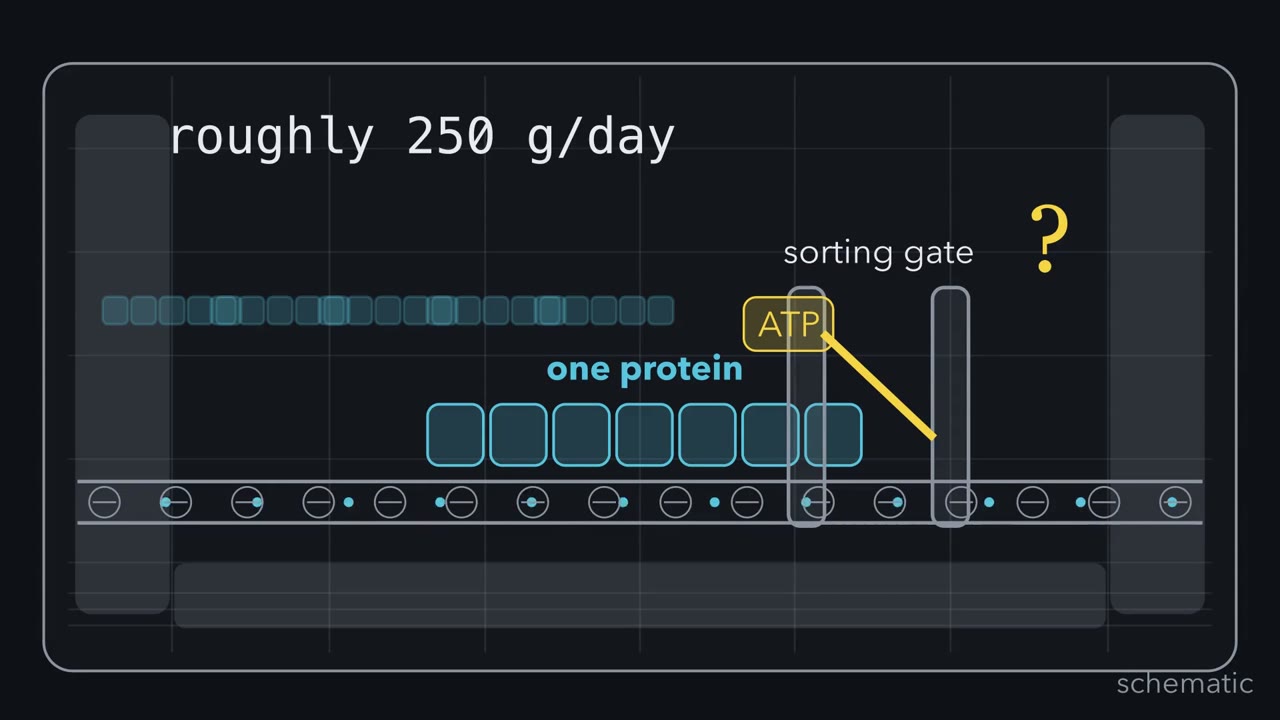 ▶ Watch Choose, Recycle, Split · 3:27
Read the transcript of “Choose, Recycle, Split”
Film transcript: Choose, Recycle, Split
Your body dismantles roughly 250 grams of protein daily. Why spend ATP to destroy what the cell already built? Follow one protein through a recycling center, where selection matters more than demolition. Proteins have different expiration dates. Some last minutes; others last months or a lifetime. Turnover removes damaged proteins and changes signals. Yet peptide-bond hydrolysis is downhill. What is ATP buying? Ubiquitin is the address label. E1 spends ATP to activate it. E2 carries it. An E3 ligase recognizes the target and applies the tag.
Hundreds of E3s create selectivity. A classic K48 chain sends the protein toward the proteasome. Predict where energy goes. Does ATP power peptide-bond cutting, or pay for something else? Hold your answer as the tagged protein reaches the machine.
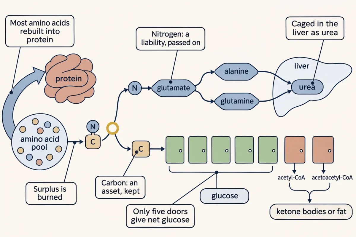
Not to cutting. The proteasome's regulatory cap recognizes the tag, then uses ATP to unfold and thread the protein. The core cuts it into short peptides without ATP. Deubiquitinases recover the tag. Energy buys choice and controlled handling, not favorable bond hydrolysis. Peptidases release amino acids into a shared pool. In an adult at nitrogen balance, most return to protein. The surplus is burned. The cell selects proteins, reuses most products, then separates surplus amino acids by atom. In this simplest adult human picture, each burned amino acid poses two problems. Nitrogen can be toxic; carbon remains valuable. Nitrogen passes onto glutamate and often through alanine or glutamine in blood.
The carbon skeleton moves toward central metabolism. In the liver, most waste nitrogen is packaged as urea. One urea nitrogen comes from ammonium; the other comes from aspartate. The cycle spends four high-energy phosphate bonds per urea. It converts a dangerous load into an excretable product. Carbon skeletons enter through seven metabolic doors. Five can supply net glucose because they can become oxaloacetate. The acetyl-CoA and acetoacetyl-CoA doors cannot. Only leucine and lysine are purely ketogenic. Now the recycling center's logic is visible. Select the protein before dismantling it. Reuse most amino acids. When surplus is burned, cage its nitrogen and keep its carbon in metabolism.
Choose what dies, recycle most, then split surplus amino acids by atom.
From the film
- In proteasomal degradation, ATP pays for tagging and handling, not peptide-bond cleavage.
- E3 ligases give ubiquitin tagging its target specificity.
- In an adult at nitrogen balance, most released amino acids return to protein.
- In adult humans, most waste nitrogen leaves as urea.
- Amino-acid carbon enters seven metabolic doors, but only five can yield net glucose.
The film above shows the choose–recycle–split logic; the text below adds mechanisms, evidence, exceptions and clinical cases.
Right now, inside you, proteins are being destroyed on purpose. Your body turns over roughly 250 grams of protein a day, tearing down and rebuilding what looks like a stable adult. Rudolf Schoenheimer showed this in the 1930s: rats fed nitrogen-labeled amino acids built the heavy tag into proteins throughout the body. He called it “the dynamic state of body constituents.”
The clocks differ wildly. Ornithine decarboxylase can be gone in about 10 minutes. Hemoglobin lasts as long as its red blood cell, about 120 days. The crystallins at the core of your eye’s lens are as old as you are, never replaced. Cells set many of these clocks deliberately.
The film above poses Irwin Rose’s puzzle: peptide-bond hydrolysis is downhill, and a protease such as trypsin removes the kinetic barrier without ATP. Yet the cell spends ATP on protein destruction.
The one idea to carry through this lesson: choose, recycle, then split by atom. The film above follows that logic; the sections below explain how each choice and handoff works.
This is the adult human picture, liver and muscle, fed or fasting. The split applies only to amino acids that are burned. Urea carries most waste nitrogen, not all: the kidney excretes some as ammonium, and some leaves as creatinine and uric acid. Lysine and threonine bypass the usual first step of nitrogen removal, and birds and many reptiles excrete uric acid, not urea.
Choosing: the ubiquitin tag
Rose, Avram Hershko and Aaron Ciechanover found the answer in reticulocyte extracts, and it earned the 2004 Nobel Prize in Chemistry. The tag is ubiquitin, a 76-amino-acid protein about 96% identical from yeast to humans. Its C-terminal Gly-Gly forms an isopeptide bond (an amide bond to a side-chain amine, not to the backbone) with a lysine on the doomed protein.
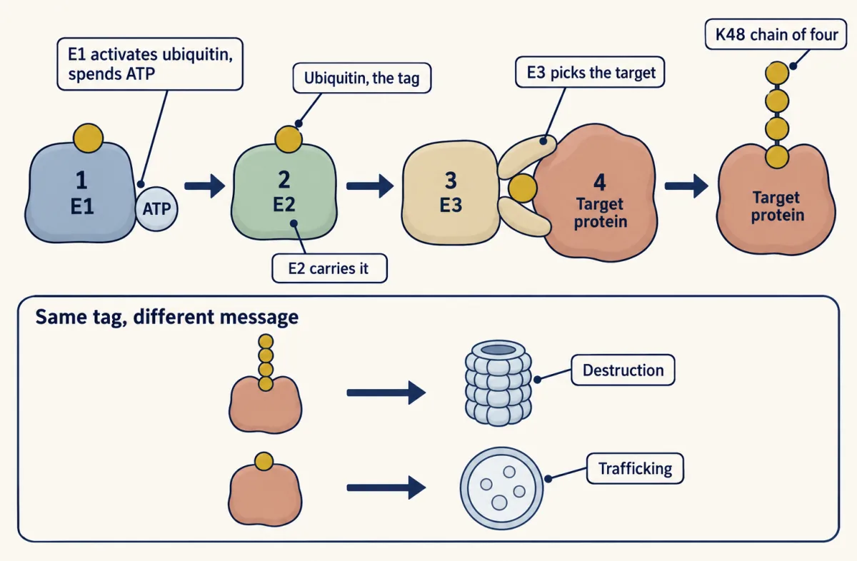
Three enzymes write the tag. E1 activates ubiquitin, spending ATP (to AMP plus pyrophosphate) to form a thioester to its own cysteine. E2, the conjugating enzyme, carries the activated ubiquitin. E3, the ligase, picks the address: it chooses the target and gets the ubiquitin onto it. Count them: roughly 2 E1 enzymes, about 40 E2s and more than 600 E3s. Specificity lives in the E3s. PROTAC drugs exploit this: they recruit an E3 ligase to a protein it would normally ignore, condemning that protein to the proteasome.
What does an E3 read? A degron, a feature that marks the target for destruction. The cleanest example is the N-end rule: half-life set by the first residue. A yeast test protein starting with methionine, glycine or alanine lasted more than 20 hours; the same protein starting with arginine or phenylalanine was gone in about two or three minutes. One residue, hundreds of fold.
The chain is a code. Ubiquitin has lysines of its own, so ubiquitins can be chained; K48 means a link through ubiquitin’s lysine 48. A chain of four or more K48-linked ubiquitins is the classic destruction signal. A K63 chain usually means “signal, do not destroy”: DNA repair, trafficking, immune signaling. Other linkages and single ubiquitins carry other messages.
Destroying: the proteasome
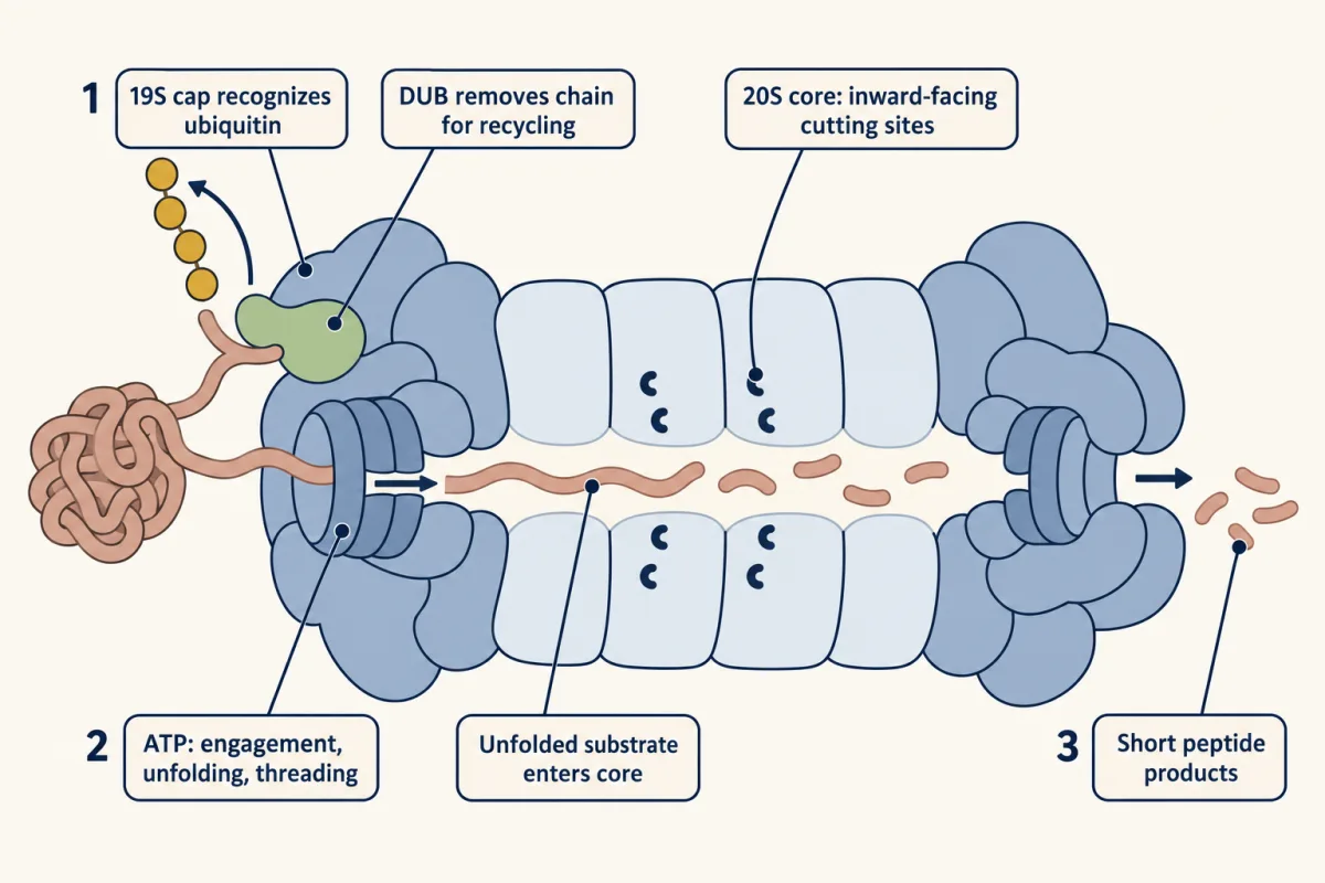
Tagged proteins go to the 26S proteasome, one or two 19S caps on a 20S core. The caps recognize the K48 chain, unfold the protein and thread it in, using ATP. The core’s active sites face strictly inward, so bystanders are rarely chewed up. Short peptides come out, and deubiquitinases (DUBs) recycle the ubiquitin.
Predict. Trypsin cuts with no ATP, so where does this system’s ATP go?
Check your prediction
Not to cutting: the 20S core cuts without it. At E1, ATP charges ubiquitin so the E3 can mark the target. At the 19S cap, ATP pays for mechanical work: unfolding the protein and threading it in. The ATP buys specificity (the right target) and processivity (once a protein enters, it is cut all the way down to short peptides).
Destruction as a switch
NF-κB sits trapped by its inhibitor IκB. When a signal arrives, IκB is phosphorylated, tagged with a K48 chain and destroyed, which frees NF-κB to enter the nucleus and switch on inflammatory and immune genes. Destroying the inhibitor is the activation.
Oxygen sensing, the subject of the 2019 Nobel Prize in Medicine, also runs on regulated destruction. In normal oxygen, a prolyl hydroxylase uses O₂ and α-ketoglutarate to hydroxylate HIF-1α, and the VHL E3 ligase recognizes the hydroxyproline and sends HIF-1α to the proteasome.
Predict. Oxygen falls in a tissue. What happens to HIF-1α, and why does that switch genes on?
Check your prediction
The hydroxylase needs O₂, so hydroxylation slows. Without the hydroxyproline, VHL cannot recognize HIF-1α, and HIF-1α survives. It switches on VEGF and glycolytic genes, while its sibling HIF-2α drives EPO. Nothing was added to switch the response on; the destruction that kept it off stopped.
Multiple myeloma plasma cells churn out so much antibody that they are addicted to clearing misfolded protein. Block the proteasome with bortezomib (a reversible inhibitor) and misfolded protein piles up, killing these cells first.
From destruction to fuel: most amino acids are reused
The film above shows the broad reuse loop. Peptidases release amino acids from proteasomal peptides into the same pool as food. The roughly 250 g of body protein broken down each day is rebuilt, and an adult in nitrogen balance burns about as much as they eat, typically around 80 g, so about three-quarters of the amino acids leaving the pool go back into protein (250 of about 330 g). The surplus is burned after a protein-rich meal or during a fast. Turnover by itself is nearly a closed loop; urea comes mainly from burning the surplus (plus a small obligatory loss), and that is where nitrogen and carbon part ways.
The nitrogen road
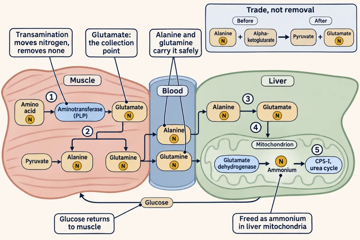
Free ammonia is neurotoxic. At body pH nearly all of it is ammonium ion (NH₄⁺); only about 1 to 3% is NH₃, and here “ammonia” means both. So the cell does not let the amino group fall off; it hands it from carrier to carrier first.
Predict. Alanine carrying a heavy-nitrogen (¹⁵N) label meets alanine aminotransferase (ALT) and α-ketoglutarate. Afterward, how much of that nitrogen has left the amino-acid pool?
Check your prediction
None. The enzyme only trades:
alanine + α-ketoglutarate ⇌ pyruvate + glutamate
The ¹⁵N is now on glutamate, still an amino acid. Transamination moves nitrogen; it removes none. It is reversible, so the same enzymes also help build several non-essential amino acids, such as alanine and aspartate.
For most amino acids, aminotransferases collect the amino group onto α-ketoglutarate, so glutamate is the collection point. They all use pyridoxal phosphate (PLP, vitamin B6). The main way to release the nitrogen is a second move, oxidative deamination by glutamate dehydrogenase in liver mitochondria (glutaminase and a few other enzymes free some ammonium directly):
glutamate + NAD⁺ (or NADP⁺) + H₂O ⇌ α-ketoglutarate + NH₄⁺ + NAD(P)H + H⁺
The reaction can run both ways; in liver the urea cycle drains the ammonium, so it runs toward release.
The film above animates how muscle and other tissues ship nitrogen to the liver in two nontoxic carriers. In the glucose-alanine cycle, muscle puts nitrogen onto pyruvate from glycolysis, making alanine; the liver takes it off and turns the pyruvate into glucose for muscle and brain. In a fast this route carries the nitrogen of muscle protein breakdown, and some of the liver’s new glucose carbon comes from muscle amino acids. Glutamine, the most abundant amino acid in blood, carries two nitrogens; glutaminase in liver and kidney releases one as ammonium.
Caging the nitrogen: the urea cycle
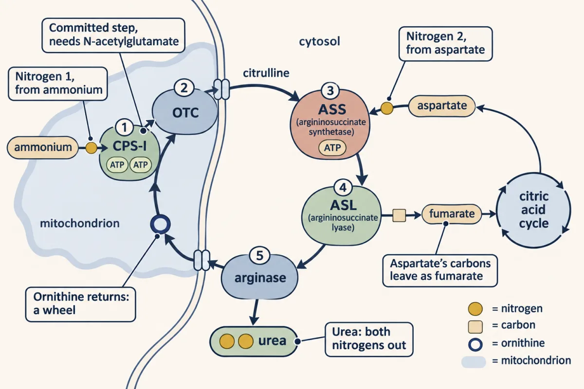
The liver now holds waste nitrogen in two forms: free ammonium, and the amino group of aspartate, made when glutamate hands its nitrogen to oxaloacetate. Urea, H₂N–CO–NH₂, has two nitrogens, and the cycle Hans Krebs and Kurt Henseleit found in 1932 takes one from each source. It starts in the mitochondrion, where much of the liver’s ammonium is made and where the rest arrives, so free ammonium meets carbamoyl phosphate synthetase I (CPS-I) without leaving the compartment.
Step 1, the first nitrogen (mitochondrion). Carbamoyl phosphate synthetase I (CPS-I) joins ammonium to bicarbonate, spending two ATP, to make carbamoyl phosphate: the committed, rate-limiting step. CPS-I works only with N-acetylglutamate bound, which the liver makes from acetyl-CoA and glutamate faster when amino acids are plentiful. So the cycle speeds up when nitrogen arrives.
Step 2, onto the carrier (mitochondrion). Ornithine transcarbamylase (OTC) hands the carbamoyl group to ornithine, making citrulline, which leaves for the cytosol.
Predict. Ornithine has just taken on the carbamoyl group. Does the liver need a fresh ornithine for every urea it makes?
Check your prediction
No. Ornithine comes back out at step 5. Krebs and Henseleit spotted this in liver slices: a little ornithine supported far more urea than it could have made itself. Used at one step and returned at another, ornithine is a wheel, not a fuel. This was the first metabolic cycle ever described.
Step 3, the second nitrogen (cytosol). Argininosuccinate synthetase joins citrulline to aspartate, making argininosuccinate. Aspartate’s amino group is urea’s second nitrogen. This step spends one ATP but cuts it to AMP and pyrophosphate, and the pyrophosphate is then split, so it costs two high-energy bonds.
Step 4, the carbon leaves (cytosol). Argininosuccinate lyase cuts argininosuccinate into arginine and fumarate. Aspartate’s nitrogen stays on arginine; its four carbons leave as fumarate.
Step 5, urea out, ornithine back (cytosol). Arginase adds water to arginine, releasing urea and regenerating ornithine, which returns to the mitochondrion.
| What | Enters at | Leaves as |
|---|---|---|
| Nitrogen 1 | Ammonium, step 1 | Urea |
| Nitrogen 2 | Aspartate, step 3 | Urea |
| Urea’s carbon | Bicarbonate, step 1 | Urea |
| Aspartate’s four carbons | Step 3 | Fumarate, step 4 |
| Ornithine | Step 2 | Ornithine again, step 5 |
| Energy | 2 ATP at step 1; 1 ATP to AMP at step 3 | Four high-energy bonds per urea (gross; Going deeper) |
regulated step − used up
Mitochondrial. The committed, rate-limiting step: free ammonia and bicarbonate are condensed into carbamoyl phosphate at the cost of two ATP, the first two of the four high-energy bonds per urea. Two ATP, two bonds.
Regulation. Activated only when N-acetylglutamate (NAG) is present — a feedforward signal, made from acetyl-CoA + glutamate and amplified by arginine, that says nitrogen is arriving.
Clinical. Severe CPS-I deficiency causes neonatal hyperammonemia; milder forms can present later.
Mitochondrial. The carbamoyl group is handed to ornithine, building citrulline, which is exported to the cytosol. Ornithine is the carrier regenerated at the end — Krebs's catalytic wheel.
Clinical. OTC deficiency is the most common urea cycle disorder (~1/56,000 in the US) and X-linked. Lab triad: high ammonia, low citrulline, and high urinary orotic acid (excess carbamoyl phosphate spills into pyrimidine synthesis).
Cytosolic. Aspartate donates the second nitrogen of urea. ATP is cleaved to AMP + PPᵢ and the pyrophosphate is hydrolyzed, so this single step costs two high-energy bonds, completing the four per urea.
Clinical. Deficiency causes citrullinemia: citrulline backs up because it cannot be condensed with aspartate.
Cytosolic. The molecule is cleaved into arginine and fumarate. The fumarate is the gear that meshes the urea cycle with the TCA cycle (the Krebs bicycle): fumarate → malate → OAA → aspartate returns the carbon aspartate brought in, so the loop recycles carbon rather than adding new carbon for glucose.
Clinical. Deficiency (argininosuccinic aciduria) accumulates argininosuccinate; managed with arginine plus nitrogen scavengers.
Cytosolic. Arginine is hydrolyzed to release urea, the final nitrogen waste, and regenerate ornithine, which re-enters the mitochondrion to turn the wheel again. Ornithine is a true catalyst: consumed at step 2, returned here.
Clinical. Arginase deficiency (argininemia) is a rare urea cycle disorder (NAGS deficiency is rarer) and causes spastic diplegia more than acute hyperammonemic crisis.
Where the fumarate goes: the Krebs bicycle. Fumarate becomes malate, then oxaloacetate, which takes an amino group from glutamate and becomes aspartate again for step 3. So the urea cycle and the citric acid cycle are two wheels on one chain. Although oxaloacetate can become glucose, this loop only returns the carbon aspartate brought in. A fasting liver’s net new glucose carbon comes from elsewhere: the skeletons of glucogenic amino acids from broken-down protein, and glycerol from fat.
When the cycle breaks
When the cycle fails, ammonia rises and astrocytes swell as they trap it in glutamine, causing brain edema. OTC deficiency, the most common urea cycle disorder, shows a triad: high ammonia, low citrulline (OTC makes it) and high urinary orotic acid. Why orotic acid? The cytosol builds the ring of the pyrimidine bases C, T and U from carbamoyl phosphate and aspartate. Carbamoyl phosphate that OTC cannot use spills out of the mitochondrion into that pathway, and its intermediate, orotic acid, reaches the urine. Treatment: restrict protein, give scavengers (sodium benzoate, sodium phenylbutyrate) that escort nitrogen out by other routes, supply arginine (except in arginase deficiency), and dialyze in a crisis.
The carbon road: seven doors
The film above shows the seven-door carbon map; the table below supplies all 20 assignments, including amino acids that use more than one door.
| Door | Net glucose? | Who enters here |
|---|---|---|
| Pyruvate | Yes | Alanine, serine, glycine, cysteine; part of tryptophan |
| α-Ketoglutarate | Yes | Glutamate, glutamine, proline, arginine, histidine |
| Succinyl-CoA | Yes | Valine, isoleucine, methionine, threonine |
| Fumarate | Yes | Part of phenylalanine and tyrosine; aspartate, when it gives its nitrogen to urea |
| Oxaloacetate | Yes | Aspartate, asparagine |
| Acetyl-CoA | No | Leucine; part of isoleucine, threonine and tryptophan |
| Acetoacetyl-CoA | No | Leucine, lysine; part of phenylalanine, tyrosine and tryptophan |
Acetoacetyl-CoA here stands for acetoacetate and its activated form.
The rule: the film’s five-versus-two split follows from carbon accounting. The first five doors can become oxaloacetate, the starting point for glucose. Pyruvate dehydrogenase is irreversible, so acetyl-CoA cannot return to pyruvate. Each turn of the citric acid cycle takes in two acetyl carbons and gives off two as CO₂, making no extra oxaloacetate. Acetoacetyl-CoA is two acetyl units, so the same count applies.
Only two amino acids are purely ketogenic: leucine (acetyl-CoA and acetoacetyl-CoA) and lysine (acetoacetyl-CoA). Five are mixed: isoleucine, phenylalanine, threonine, tryptophan and tyrosine. The other 13 are purely glucogenic.
The succinyl-CoA door explains two vitamins. Its four amino acids are broken down to propionyl-CoA. A biotin enzyme carboxylates it to methylmalonyl-CoA, and methylmalonyl-CoA mutase, a B12 enzyme, rearranges that into succinyl-CoA. Cofactors fingerprint the step: PLP for amino-group transfer, biotin for carboxylation, B12 for the rearrangement.
Predict. A liver is fed a diet whose only amino acid is leucine. Can it make net glucose from it?
Check your prediction
No. Leucine’s carbon enters only at acetyl-CoA and acetoacetyl-CoA: two in, two out, no extra oxaloacetate. (A labeled leucine carbon can still turn up in glucose, but not as net glucose; see Going deeper.)
Only five doors give net glucose, because each can become oxaloacetate. Acetyl-CoA cannot: pyruvate dehydrogenase is irreversible, and each turn of the citric acid cycle takes in two carbons and gives off two as CO₂, so it never makes extra oxaloacetate. Acetoacetyl-CoA is two acetyl units, so the same count applies. That is why leucine and lysine can feed ketone bodies or fat but never net glucose.
Break one of these enzymes and you get an inborn error of metabolism; each is rare, but as a group they are not. In 1902 Archibald Garrod argued that alkaptonuria, which turns urine black and stains cartilage, is inherited: the first recognized inborn error. Phenylketonuria (PKU) (defective phenylalanine hydroxylase) is caught by newborn screening and managed by a low-phenylalanine diet. In MSUD (defective branched-chain keto-acid dehydrogenase), branched-chain amino acids and their keto acids build up, the urine smells of maple syrup, and the excess leucine is neurotoxic.
The whole lesson in one bird
The bar-tailed godwit flies 11,000 km nonstop in about eight days. It burns fat for energy and metabolic water, and it breaks down some of its own muscle protein, probably for glucose, for anaplerosis (refilling citric acid cycle intermediates) and for more water. The nitrogen leaves as uric acid, which costs almost no water. Choose, then split by atom: the E3s pick what dies, the nitrogen is caged, the carbon is kept.
Going deeper
The main line above does not depend on these details.
The rest of the ubiquitin code
Ubiquitin has seven internal lysines (K6, K11, K27, K29, K33, K48, K63). K11 chains, made by the cell-cycle ligase APC/C, also send proteins to the proteasome. Linear (M1) chains, built through ubiquitin’s N-terminal methionine, help switch on NF-κB. A single ubiquitin often directs trafficking or changes a protein’s partners, as on histones. K48 chains of four or more are the classic signal, not the only one: shorter or mixed chains, and several single ubiquitins, can also target a protein.
More on the N-end rule
The rule was found in 1986 by Andreas Bachmair, Daniel Finley and Alexander Varshavsky. Later work added a layer: many of the “stable” first residues become degradation signals once they are N-terminally acetylated.
More on the proteasome
The 20S core is a barrel of 28 subunits in four stacked rings, and its peptides are about 3 to 22 residues long. A second route, autophagy into lysosomes, handles much bulk turnover, whole organelles, and aggregates the proteasome cannot take. Besides bortezomib (reversible), myeloma is treated with carfilzomib (irreversible) and oral ixazomib.
Transaminases on a liver panel
Alanine aminotransferase (ALT) and AST leak into the blood when liver cells are injured, which is why these transaminases sit on standard liver panels. AST also comes from muscle and heart.
What urea really costs
Four high-energy bonds per urea is the textbook gross cost. The NADH made at malate dehydrogenase in the bicycle (and at glutamate dehydrogenase) offsets part of it, so the net cost is lower; textbooks differ on the exact figure.
Threonine in humans
The textbook classes threonine as mixed, and this lesson and the sorter follow that. In adult humans the route that yields acetyl-CoA (threonine dehydrogenase) is minor, and most threonine goes to propionyl-CoA and succinyl-CoA.
A label is not net glucose
The two CO₂ that leave in a turn of the citric acid cycle are not the same two carbons that entered. So an acetyl-CoA carbon can turn up in glucose as an isotope label, because over later turns it trades places with oxaloacetate carbons. What matters is the count: two carbons leave as CO₂ for every two that enter, so no net glucose is made.
Treating PKU
Besides the low-phenylalanine diet, phenylketonuria (PKU) can be managed with sapropterin, a synthetic form of the enzyme’s cofactor, tetrahydrobiopterin (BH4), or with pegvaliase, an injected enzyme that breaks down phenylalanine.
Habits from this course
This lesson closes the course’s main sequence; supplements on nucleotide metabolism and the integration of metabolism follow. Five habits to carry forward: ask what the ATP is buying; compartmentalization is regulation; cofactors fingerprint mechanism; cells run in cycles, not arrows; and when something breaks, ask which step, which cofactor, which compartment.
Your notes
Select any text in the lesson to highlight it or add a note to it. Your highlights and notes are saved in this browser and collected in your notebook.
This browser is not letting the site save anything (a private window or strict privacy settings), so highlights and notes will be lost when you leave the page.
How we measure it
Isotope tracing plus mass spectrometry
Schoenheimer fed rats 15N-labeled amino acids and tracked the heavy tag by mass spectrometry. Much of the label turned up in tissue proteins, showing the adult body is in constant turnover rather than a fixed frame. Non-radioactive heavy atoms let you thread a label into a molecule, feed it, and follow it without poisoning anything; this is still the central tool of metabolic biochemistry.
Cell-free fractionation with reticulocyte lysate
Hershko, Ciechanover, and Rose worked in reticulocyte extracts because mature red cell precursors lack lysosomes, isolating the ATP-dependent degradation system. Splitting the extract on a column killed the activity; only recombining two fractions plus ATP restored it. One fraction held a small heat-stable protein, APF-1, soon recognized as ubiquitin. Reconstitution from purified fractions is how a multi-enzyme cascade is taken apart and rebuilt.
The Warburg manometer (gas measurement of cycles)
Krebs worked with the Warburg manometer, a sealed flask whose pressure change reports gas exchange in living tissue slices, and measured the urea the slices made. Finding that small amounts of ornithine supported much more urea than could be built from the ornithine itself is what revealed a catalytic cycle rather than a linear consumption. The trick of spotting a molecule that is simultaneously consumed and regenerated defines a metabolic cycle.
Newborn metabolic screening (Guthrie and tandem MS)
The Guthrie bacterial-inhibition assay, and now tandem mass spectrometry on a dried blood spot, screens newborns in many countries for selected inborn errors of amino acid metabolism. A high blood phenylalanine flags PKU before brain damage occurs; raised leucine/isoleucine and valine flag MSUD. Catching a defective enzyme by its accumulating metabolite, days after birth, turns a catastrophic disease into one managed largely by diet, with lifelong monitoring.
Practice questions on this lesson (3) → MCAT-style, original, each with an explanation
Where this lesson fits
Before thisReview first
- The Citric Acid Cycle: Harvesting ElectronsThis course · Lesson 19
Fumarate, oxaloacetate, alpha-ketoglutarate and succinyl-CoA are the cycle intermediates where amino-acid carbon enters and the urea cycle meshes.
- Gluconeogenesis: Pushing Glucose Back UphillThis course · Lesson 17
You need to know how oxaloacetate becomes glucose to see which amino-acid doors give net glucose.
- The Pyruvate Dehydrogenase Complex: The One-Way Gate to the CycleThis course · Lesson 18
Its irreversibility is why acetyl-CoA cannot return to pyruvate, so ketogenic amino acids give no net glucose.
After thisStudy next
- Integration of Metabolism: How the Body Switches FuelsThis course · Lesson 30
It uses muscle protein breakdown, urea and amino-acid carbon for glucose to show how the body switches fuels between fed and fasting.
- Nucleotide Metabolism: Build It, Recycle It, or Pay in Uric AcidThis course · Lesson 29
It takes up aspartate, glutamine and carbamoyl phosphate, which also feed the pyrimidine pathway where orotic acid appears.
Elsewhere on the siteFilms, simulations and method films in other courses
- HIF and oxygenSimulation · Cell Signaling
It shows how oxygen-controlled ubiquitin tagging destroys HIF, the same PHD and VHL switch used here.
- Filter Far More, Take Most BackFilm · Organismal Biology
It follows the urea that the liver makes out of waste nitrogen into the kidney and urine.
- Take It Apart, Build It AgainFilm · Biology
It shows how proteins are broken into amino acids by adding water, the first step of the recycling here.
Suggested by an AI model from the lessons themselves, then checked. How we use AI
Finish line
Test yourself
8 questions
Practice and MCAT-style, with explained answers.
Play it · about 4 min
Metabolic Autopsy: Urea cycle
A baby with high ammonia. Find the broken urea-cycle enzyme.
Play →Next · Lesson 29
Nucleotide Metabolism: Build It, Recycle It, or Pay in Uric Acid
Nucleotides are costly to build — Lesch-Nyhan lives where recycling breaks, and gout and allopurinol live at the uric-acid end.
Continue →Unit review · Fuels, Fat & Nitrogen
MCAT Gauntlet: Fuels, Fat & Nitrogen
15 timed questions across the pentose phosphate pathway, fatty acid oxidation, ketone bodies, fatty acid synthesis, amino acid catabolism and the urea cycle. Three lives.
Also in the arcade for this lesson: Ledger Rush: Amino acids
← Lesson 27 · Lipid Synthesis: Building Cholesterol from Acetyl-CoA