Sensory Systems · Unit 5: Vision
Melanopsin Cells: Light for the Clock and the Pupil
Rewritten on : new text, flashcards and film. Your notes and highlights stay, but the text around them may have moved.
A mouse with no working rods or cones still sets its body clock to the day and still narrows its pupils in bright light. Which cells are measuring the light?
Pause when the reading reaches a film, and offer to play it. Saved on this device.
Watch the film now, or keep listening
Builds on:Parallel Channels Out of the EyeFive Ways to Turn a Stimulus into a CurrentWhy these?
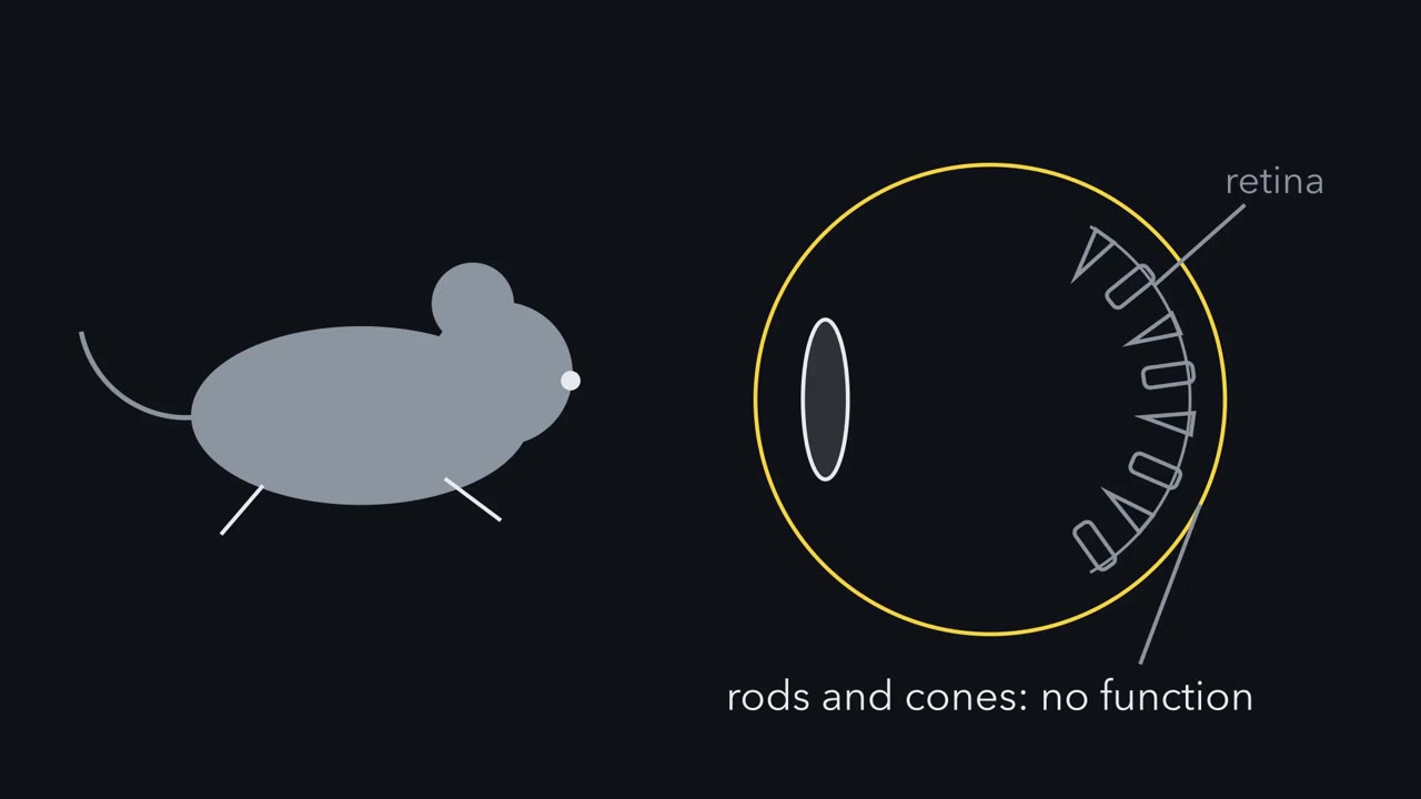 ▶ Watch A Blind Mouse That Knows It Is Day · 2:29
Read the transcript of “A Blind Mouse That Knows It Is Day”
Film transcript: A Blind Mouse That Knows It Is Day
Take a mouse with no working rods and no working cones. Can it still tell day from night? Normally, rods and cones catch the light. Bipolar cells pass their signals on to ganglion cells. Ganglion cells are the retina's output neurons. Their axons carry its signals to the brain. In 1999, two papers studied mice that lack both rods and cones. How do you see a clock in a mouse? Watch its running wheel. A mouse is nocturnal, so it runs in the dark. Each line of the record is one day. Stack the days. If running starts at the same time each day, near lights-off, the clock looks locked to the light cycle. In these rodless, coneless mice, light still shifted the timing of their daily running.
Light still suppressed melatonin from their pineal gland. Work on the pupil followed two years later. So something in the eye, other than rods and cones, was still measuring light. Responses like these use light without forming an image. They are called non-image-forming. Two are best established. The first is setting the body clock. The second is narrowing the pupil. The brain's master clock is the suprachiasmatic nucleus, or SCN. Light reaching it keeps the clock aligned with day and night. That is called photoentrainment. The pupil reflex is driven from the olivary pretectal nucleus. Which cells were measuring the light? In these mice, the answer was a small set of retinal ganglion cells.
They are called intrinsically photosensitive retinal ganglion cells. For short, ipRGCs. Each makes its own light response with a pigment called melanopsin. It also relays signals from rods and cones. In mice, these cells are the main route by which light reaches the clock and the pupil. Three questions come next. What does the cells' own light response look like? How does light become a voltage inside them? And how do we know the clock and pupil depend on them? Hold one picture in mind. Two light signals arrive at one cell, and leave by one exit.
From the film
- A small set of retinal ganglion cells, the ipRGCs, make their own light response with melanopsin and also relay rod and cone signals.
- In mice, these cells are the main route by which light reaches the clock and the pupil.
- Each ipRGC pools two light signals into one output.
A mouse with no working rods or cones still sets its body clock to the day and still narrows its pupils in bright light. Which cells are measuring the light, and what pigment do they use?
Those mice were real. In 1999 two papers in Science studied mice that lack both rods and cones, each with a different mouse model. Freedman and colleagues found that light still shifted the timing of the mice’s daily running, and Lucas and colleagues found that light still suppressed melatonin from their pineal gland. Work on the pupil followed in 2001. Something in the eye other than rods and cones was still measuring light.
Responses like these use light without forming an image, so they are called non-image-forming. The two best established are setting the clock and narrowing the pupil.
- The clock. The suprachiasmatic nucleus (SCN) is the brain’s master clock. Light reaching the SCN shifts the clock so that it stays aligned with day and night. This is photoentrainment. To watch it in a mouse, watch its running wheel. A mouse is nocturnal, so it runs in the dark. A running record has one line per day. Stacked, the days show whether running starts at a fixed time relative to lights-off, as it does when the clock is locked to the light cycle.
- The pupil. The olivary pretectal nucleus (OPN) drives the pupil light reflex.
In those mice, the light for both turned out to be measured by a small set of retinal ganglion cells. Ganglion cells are the retina’s output neurons; their axons carry its signals to the brain. An intrinsically photosensitive retinal ganglion cell (ipRGC) is a ganglion cell that makes its own light response with the pigment melanopsin and also relays signals that rods and cones send through bipolar cells. The core lesson on sleep and rhythms introduced melanopsin as the light input to the clock, and the retina described the photoreceptors without mentioning these cells at all.
Three questions come first: what the cells’ own light response looks like, how light becomes a voltage inside them, and how we know that the clock and the pupil depend on them. Then come their separate outputs, their subtypes, mood and learning in mice, what people show and, more modestly, pattern vision.
A light response that survives the block
Read the transcript of “The Light Response That Survived the Block”
Film transcript: The Light Response That Survived the Block
Cut a ganglion cell off from rods and cones. If it still responds to light, whose response is it? Here is the logic. Rods and cones reach ganglion cells through synapses. Block those synapses with drugs. If a ganglion cell still responds once the block is verified, the response is most likely its own. In 2002, Berson, Dunn and Takao did this in rat retina. First they labelled ganglion cells from the SCN. That told them which cells project to the clock. Then they recorded from those labelled cells, while the blockers were applied. Predict. Suppose the rod and cone relay were the cell's only light signal. What should the recording show after the block?
There should be no light-evoked change. But one remained. The labelled cells still depolarized to light. So the response was intrinsic, made within the ganglion cell itself. They also recorded from a cell body removed from the retina. That is a stronger test, with no synapses left at all. So one cell can give two kinds of light response. Put both on one time axis. The relayed response, from rods and cones, usually starts fast and works in dim light. It can have both brief and lasting parts. The cell’s own response is usually slow to start and needs brighter light. It is often long-lasting, though it adapts too. When the light goes off, it decays slowly.
The cell stays depolarized for a while. In people, these cells drive the post-illumination pupil response. The pupil stays small for a while after light goes off. Why would slow and sustained be useful? The clock and the pupil need to know how bright it is. A camera must follow fast changes. A light meter needs a steady reading. The cells that serve the clock and the pupil. are mostly a type called M1. They report how bright the world is, not what is in it. The rat response fitted a pigment that peaks near 484 nanometres. That is a rat estimate, not a human one. In the same volume of the journal, Hattar and colleagues showed that these rat cells carried melanopsin.
And melanopsin-marked mouse axons reached the SCN. A response, a pigment and a target now pointed to one set of cells. Their signature is slow, insensitive and sustained.
From the film
- A ganglion cell that still responds to light once rod and cone synapses are blocked is most likely making its own response.
- The cell's own response is usually slow to start, needs brighter light and is long-lasting.
- Slow and sustained suits a cell that reports how bright the world is, not what is in it.
The first step was to show that the response is made inside the ganglion cell, not handed on by rods and cones. Block synaptic transmission from the photoreceptors with drugs. If a ganglion cell still responds to light once that block is verified, the response is its own.
In 2002 Berson, Dunn and Takao did this in isolated, flat-mounted adult rat retina. They labelled ganglion cells from the suprachiasmatic nucleus (SCN), so they knew which cells project to the clock, and recorded from those cells while the blockers were applied.
Predict. Before the block, a labelled cell responds to light through rods, cones and bipolar cells. If that relay were the cell’s only source of light information, what would the recording show once synaptic transmission is blocked?
Check your prediction
No light-evoked change would remain, though baseline activity and noise could. But one remained: the labelled cells still depolarized to light, so the response was intrinsic, made within the ganglion cell. Berson and colleagues also recorded from a cell body removed from the retina, a stronger test, because a cell on its own has no synapses left to block.
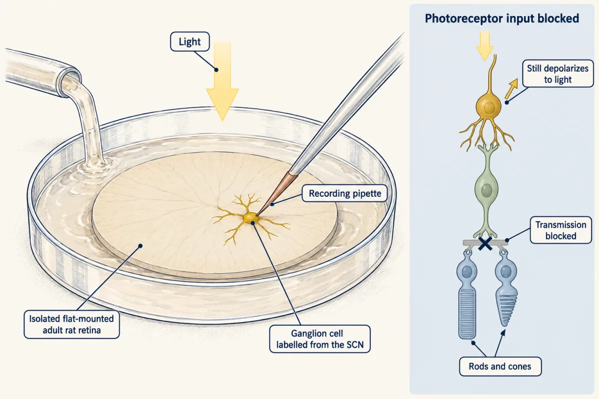
The signature response
So one cell can give two kinds of light response. Picture both on one time axis, with a steady light switched on and, later, off.
| Relayed from rods and cones | The cell’s own melanopsin response | |
|---|---|---|
| Start | fast | slow to start |
| Sensitivity | high: works in dim light | low: needs brighter light |
| In steady light | can have brief and strongly sustained parts, depending on the circuit and the light | often sustained, though it adapts too; depends on subtype and light |
| After the light goes off | falls back quickly | decays slowly, so the cell stays depolarized for a while |
These are typical differences under the same light, not absolute rules. The melanopsin column, above all its slow decay, is the signature of these cells. In people the same cells drive the post-illumination pupil response, in which the pupil stays small for a while after a light goes off; it is being studied as a clinical test.
Why would a slow, insensitive, sustained response be useful? The clock and the pupil need to know how bright it is, and how bright it has stayed, not where the edges are. A camera must follow fast changes; a light meter needs a steady reading. In that sense the ipRGCs that serve the clock and the pupil, mostly a subtype called M1, are the retina’s light meter: they report how bright the world is, not what is in it.
The action spectrum of the rat response, its sensitivity plotted against wavelength, was consistent with a pigment peaking near 484 nm. In the same volume of Science, Hattar and colleagues reported that the rat cells they recorded as intrinsically photosensitive carried melanopsin, and that melanopsin-marked mouse axons reached the SCN. A response, a pigment and a target now pointed to one set of cells.
The one idea to carry through this lesson. An ipRGC pools two light signals into one output. One is the usually faster, more sensitive signal that rods and cones send through bipolar cells. The other is its own typically slower, less sensitive melanopsin response. Both can be sustained. In mice these cells are the main route by which either signal reaches the clock and the pupil. So removing the pigment, or the rods and cones, leaves the other input working alone, and removing the cell removes both.
The idea covers the mouse clock and pupil and, later, the mouse mood and learning routes. It does not describe the image-forming roles of some subtypes, and it says nothing about necessity in people, where no one can remove the cells.
Two inputs, one exit: how light becomes a voltage
Read the transcript of “Two Inputs, One Exit”
Film transcript: Two Inputs, One Exit
Light hyperpolarizes a rod. Yet light depolarizes these ganglion cells. How can the same light push two cells opposite ways? Start with the wiring. Rods and cones signal to bipolar cells. Bipolar cells signal to the ganglion cell's dendrites. Melanopsin sits in the ganglion cell's own membrane. So light reaches the cell by two routes. But each cell has one axon, one exit. Across the population, those exits reach the clock and the pupil centre. That is the one idea of this lesson. An ipRGC pools two light signals into one output. One input is the rod and cone relay, usually faster and more sensitive. The other is melanopsin, usually slower and less sensitive.
Both can be sustained, and both adapt. In mice, these cells are the main route by which either signal reaches the clock and the pupil. So removing the pigment, or the rods and cones, leaves the other input working alone. Removing the cell removes both. Now the polarity. In a rod, light closes cation channels. Less positive charge enters, and the rod hyperpolarizes. In an M1 cell, light opens channels instead. Melanopsin is an opsin. But it is closer to the visual opsins of insects than to rod and cone opsins. It activates a G protein, inferred to be of the Gq family. That activates phospholipase C beta 4. Then TRPC6 and TRPC7 channels open. Without phospholipase C beta 4, the M1 cell's own light response essentially disappeared.
A fly photoreceptor works much the same way, with Gq, phospholipase C and TRP channels. Predict. These channels pass cations, and their current reverses near zero millivolts. Opening them moves the voltage which way? Recall the balance-point rule. When the voltage is steady, with no injected current, it sits at a weighted average of reversal potentials. The weights are the conductances. Here that is near zero. So the voltage moves up, away from the potassium potential. The cell depolarizes. In Another subtype, M4, may also been reported to close potassium leak channels. That depolarizes too, by the same rule. Which other channels also contribute in M4 is still disputed.
Light hyperpolarizes a rod and depolarizes an M1 cell. And both of the cell's inputs leave by one exit.
From the film
- An ipRGC pools the rod and cone relay and its own melanopsin response into one output.
- In M1 cells, melanopsin signals through a G protein and phospholipase C beta 4 to open TRPC6 and TRPC7 channels, which depolarizes the cell.
- Light hyperpolarizes a rod but depolarizes an M1 cell.
Start with the wiring. Rods and cones signal to bipolar cells, and bipolar cells signal to the ipRGC’s dendrites. Melanopsin sits in the ipRGC’s own membrane. So light reaches the cell by two routes, but each cell has one axon, so both signals leave by one exit. Across the population those axons reach the suprachiasmatic nucleus (SCN) and the olivary pretectal nucleus (OPN).
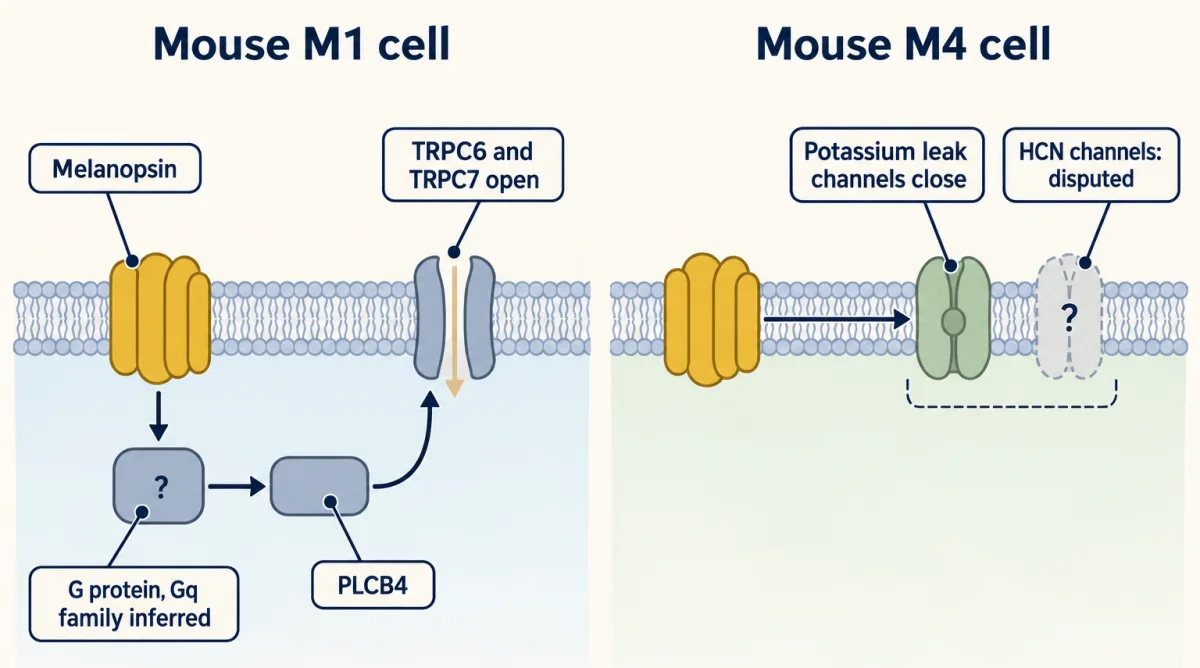
Now the polarity, which is opposite to a rod’s. Light closes cation channels in a rod, through the cGMP cascade in senses and cGMP, so less positive charge enters and the rod hyperpolarizes. Light opens channels in an M1 cell and depolarizes it.
Melanopsin is an opsin, but it is closer to the visual opsins of insects and many other invertebrates than to rod and cone opsins. In mouse M1 cells the pathway is melanopsin, a G protein, probably of the Gq family (inferred; Xue and colleagues did not identify the G-alpha), phospholipase C beta 4 (PLCB4) and the opening of TRPC6 and TRPC7 channels, the signalling pattern covered in G proteins and PLC, IP3 and DAG. A fly photoreceptor works much the same way, with Gq, PLC and TRP channels. In Xue and colleagues (2011), loss of Plcb4 essentially eliminated the intrinsic light response of M1 cells, and M1 cells lacking both TRPC6 and TRPC7 kept almost none of it under the conditions tested.
Predict. TRPC channels are nonselective cation channels, and their current reverses near 0 mV. The cell sits near rest, where potassium dominates. Recall the balance-point rule from the action potential: when the voltage is steady and no current is injected, it sits at the average of the reversal potentials, weighted by each conductance. Which way does opening TRPC channels move the voltage?
Check your prediction
Opening channels gives more weight to their reversal potential, so the balance point moves up from near EK toward 0 mV, and the cell depolarizes. With illustrative values in a passive model (not ipRGC measurements), a potassium conductance of 1 and a cation conductance of 0.2, with EK at −90 mV, give a balance point of (1 × −90 + 0.2 × 0) / 1.2 = −75 mV. Raise the cation conductance to 1 and it moves to (1 × −90 + 1 × 0) / 2 = −45 mV.
That pathway does not describe every subtype. In M4 cells, studies report that melanopsin closes potassium leak channels as well as opening TRPC channels; closing potassium channels in a cell sitting above EK also depolarizes it, by the same rule. Whether HCN channels contribute is disputed (Going deeper).
Remove the pigment, or remove the cell
Read the transcript of “Remove the Pigment, or Remove the Cell”
Film transcript: Remove the Pigment, or Remove the Cell
To learn what a part is for, take it away. But which part should you take away? The one idea gives three choices. Remove the pigment. Remove the rods and cones. Or remove the cell. First, the readouts. For the clock, keep a mouse on a light-dark cycle and watch its running. A mouse entrains if its running keeps a fixed timing relative to the cycle. A mouse whose clock is not entrained free-runs. It drifts on its own internal period. Separately, a brief light pulse at night tests how far the rhythm phase-shifts. Masking is a third readout. Bright light at night often suppresses a mouse’s running right away, independently of any clock shift. The same light can also shift the clock.
For the pupil, film the constriction over seconds to minutes of steady light. Row one is wild type. It entrains, shifts fully, and its pupil constricts fast and holds. Row two has no melanopsin. It still entrains, but its phase shifts to brief pulses are smaller. Its pupil constricts, but in steady bright light the constriction fades over minutes. Row three has no rod or cone signalling. It entrains. Its pupil is slower and less sensitive, but it holds. In bright light, it reaches its normal maximum. Row four is the triple mutant. It lacks melanopsin, rod transducin and a cone channel subunit. It does not entrain. It free-runs, shows no masking and has no significant pupil reflex.
Read down the table. Either input alone is enough to entrain and to constrict. Lose both, and nothing is left. Predict. Keep the rods and cones working, and ablate ipRGCs. If rods and cones had their own route, what would you see?
A strong separate route would let these mice keep much of their clock and pupil responses, like the melanopsin knockouts. Güler and colleagues found something else. The deficits were larger than with melanopsin loss alone, similar to those of the triple mutants. Pattern vision, which runs through other ganglion cells, remained. So in mice, these cells are the main route to the clock and the pupil. An ablation removes both inputs, so it cannot say which input supplied what was lost. Predict. Here are made-up phase shifts. Do the two single losses add up to the loss of both?
They do not. Together the two single losses are smaller than the loss when both inputs go. One possible reading is that the output saturates. A pupil cannot get smaller than its minimum. A phase shift has a ceiling too. Near a ceiling, the stronger input alone gets close, and adding the other gains little. A single knockout shows what the other input can do alone. It does not show each input's share. One limit. In the Güler mice, a weakened toxin gene at the melanopsin locus kills the cells gradually and incompletely, and more are lost with age. Losing cells during development can let targets rewire. An ablation timed in adults is better. Remove the pigment, and the relay carries on.
Remove the cell, and both inputs go.
From the film
- In mice, either input alone is enough to entrain the clock and to constrict the pupil, and losing both leaves nothing.
- Removing the ipRGCs gave larger deficits than removing melanopsin alone, close to the triple mutants.
- A single knockout shows what the other input can do alone, and it does not show each input's share.
The 2002 papers did not show what the pigment, or the cells, are needed for. For that, you take something away and watch what fails, and the one idea gives three things to remove: the pigment, the rods and cones, or the cell itself.
First, the readouts. For the clock, a mouse is kept on a light-dark cycle to see whether it entrains, keeping a fixed timing relative to the cycle, or free-runs, drifting on its own internal period. Separately, a brief light pulse at night tests how far the rhythm phase-shifts. A third readout is masking: bright light at night commonly suppresses a nocturnal mouse’s running acutely, independently of any clock shift. The same light may also shift the clock, so the two are measured separately, and both depend on the stimulus and the time of night. Because masking can make running look locked to the cycle, releasing the mouse into constant darkness shows where the clock itself was set. For the pupil, constriction is filmed over seconds to minutes of steady light.
Here are four kinds of mouse in which the cells are intact (Panda and colleagues, 2002; Hattar and colleagues, 2003; pupil phenotypes reviewed by Do, 2019):
| Mouse | Rod and cone relay | Melanopsin | Clock | Pupil in steady bright light |
|---|---|---|---|---|
| Wild type | yes | yes | entrains; full phase shifts | fast, full and held |
| No melanopsin | yes | no | entrains; smaller phase shifts to brief pulses | constricts, but the constriction fades over minutes |
| No rod or cone signalling | no | yes | entrains | slower and less sensitive, but sustained, and reaches its normal maximum in bright light |
| Triple mutant: no melanopsin (Opn4), no rod transducin (Gnat1), no cone channel subunit CNGA3 (Cnga3) | no | no | does not entrain; free-runs; no masking | no significant pupil reflex |
Read down the table with the two inputs in mind. Either input alone is enough for the clock to entrain and for the pupil to constrict. Each still leaves its own mark. The rod and cone signals make constriction quick and sensitive, especially in dim light and at the start of a light, so without them the pupil is slow and needs brighter light. Melanopsin holds constriction steady over minutes in bright light, so without it the constriction fades, and the pupil reflex is only partly impaired. The melanopsin knockout’s phase shifts to brief pulses are reduced, by an amount that differs between studies and irradiances, so do not memorize one size (Going deeper). Lose both inputs, and nothing is left.
Predict. Now keep rods and cones working, but kill the ipRGCs themselves. If rods and cones had a strong route of their own to the clock and the pupil, what would you see? If the ipRGCs are the principal route, what would you see?
Check your prediction
With a strong separate route, the ablated mice would keep much of their clock and pupil responses, as the melanopsin knockouts do. If the ipRGCs are the principal route, ablation would cost more than losing melanopsin alone and approach the triple mutants, even though rods and cones still work. Güler and colleagues (2008) found the second: pupil-reflex and entrainment deficits larger than those of melanopsin knockouts and similar to those of the triple mutants. Pattern vision, which runs through other ganglion cells, remained. So in mice the ipRGCs are the main route by which rod, cone and melanopsin signals reach the clock and the pupil.
Three details qualify this ablation. An attenuated toxin gene placed at the melanopsin locus kills the cells progressively and incompletely, with more lost as the mice age, and some pupil response remained, so a weak bypass cannot be excluded. The most strongly affected mice carried the toxin gene in both copies of the melanopsin gene, so they had no intact melanopsin gene; that is why melanopsin knockouts are the right comparison. And losing cells during development can let targets rewire, so when a manipulation can be timed, an adult ablation is better (Going deeper).
Two lessons come out of this, and both apply to every later experiment.
Removing the cell and removing the pigment answer different questions. An ablation shows that the cells are needed, but because it removes both inputs it cannot say whether the melanopsin response or the rod and cone relay supplied what was lost.
Single-loss deficits need not add up. Take some made-up phase shifts (they return in the first MCAT question): 2.0 h in wild type, 0.6 h without melanopsin, 1.8 h without rods and cones, and none after ablation.
Predict. If each input supplied a fixed share of the wild-type shift, the two inputs working alone would add up to the wild-type shift. Do they?
Check your prediction
No. Each input working alone gives 0.6 h and 1.8 h, which sum to 2.4 h, more than the wild type’s 2.0 h. Seen as losses, losing melanopsin costs 2.0 − 0.6 = 1.4 h and losing rods and cones costs 2.0 − 1.8 = 0.2 h. Together that is 1.6 h, but losing both costs the full 2.0 h.
One possible reading is that the pooled output saturates. A pupil cannot get smaller than its minimum, and a phase shift has a ceiling. Then the stronger input alone (here melanopsin, 1.8 of 2.0 h) comes near the ceiling, and adding the other gains little. These numbers do not say where the saturation happens, in the cell or downstream, and other explanations are possible. The real knockout studies show complementary inputs (mice without rods and cones still phase-shift to light, mice without melanopsin shift less, and mice without both do not entrain), but they used different readouts, so they do not test this saturation model. A single knockout shows what the other input can do alone, not each input’s share.
Separate cells for the clock and the pupil
Read the transcript of “Separate Cells for the Clock and the Pupil”
Film transcript: Separate Cells for the Clock and the Pupil
Do the same cells set the clock and narrow the pupil? Or do separate cells do each job? To find out, you need a way to mark one set of cells. Here the marker is Brn3b. Brn3b is a transcription factor. Most ganglion cells make it, but a subset of M1 cells does not. In 2011, Chen, Badea and Hattar traced where each set goes.
Brn3b-negative M1 cells reached the SCN, the clock. Brn3b-positive cells reached the olivary pretectal nucleus, which drives the pupil. Predict. Kill only the Brn3b-positive ipRGCs, leaving other ganglion cells intact. What happens to the pupil? And to the clock?
The pupil light reflex was severely impaired. But circadian photoentrainment was spared. The Brn3b-negative cells that reach the SCN were still there. So the two behaviours depend on partly different cells. The outputs can be pulled apart. Notice what the marker defines here. Brn three B marks an output set, not one morphological type. Those outputs come from a varied population. Mouse ipRGCs are named M1 to M6. They differ in shape, in where their dendrites sit, and in where their axons go. M1 cells are the classic clock and pupil cells. Their dendrites lie in the outermost stratum of that layer. That stratum is conventionally called OFF. The name describes where the dendrites sit, not the cell's own response.
M2 dendrites lie in the inner band. M3 cells have dendrites in both bands. M4 is the mouse sustained ON alpha cell, and it is intrinsically photosensitive. M5 and M6 send axons to the thalamic relay for image-forming vision. The letters name mouse cells. They are not six matching human types. A marker can separate the clock route from the pupil route. It cannot, by itself, name the subtype behind each job.
From the film
- The clock and the pupil depend on partly different ipRGCs.
- Removing only the Brn3b-positive cells severely impaired the pupil reflex but spared photoentrainment.
- The names M1 to M6 are for mouse cells, and they are not six matching human types.
Do the same ipRGCs set the clock and narrow the pupil, or do separate cells do each job? To ask, you need a way to mark one set of cells. Brn3b is a transcription factor made by most ganglion cells but absent from a subset of M1 cells. Chen, Badea and Hattar (2011) found that Brn3b-negative M1 cells innervated the suprachiasmatic nucleus (SCN) and that Brn3b-positive ipRGCs, a mixed set of more than one type, innervated the olivary pretectal nucleus.
Predict. Kill only the Brn3b-positive ipRGCs, leaving other ganglion cells intact. What happens to the pupil reflex, and what happens to photoentrainment?
Check your prediction
Genetic ablation of the Brn3b-positive cells severely impaired the pupil light reflex and spared circadian photoentrainment, because the Brn3b-negative cells that reach the SCN remained. The two behaviours depend on partly different cells. Because Brn3b marks a mixed output set, the result separates two behaviours without assigning either to one subtype.
Those outputs come from a varied population. Mouse ipRGCs are named M1 to M6, and they differ in shape, in where their dendrites sit in the inner plexiform layer (the layer where bipolar cells contact ganglion cells) and in where their axons go. ipRGC dendrites gather in two bands of that layer, an outer one and an inner one. The letters describe mouse cells, not six homologous human types.
| Subtype | What to know |
|---|---|
| M1 | The classic clock and pupil cells, mainly non-image-forming. Dendrites in the outer band, the outermost stratum, conventionally called OFF; the name gives position, not the sign of the response (Going deeper). |
| M2 | Dendrites in the inner band. |
| M3 | Dendrites in both bands. |
| M4 | The mouse sustained ON alpha ganglion cell (the ON alpha cell of retinal cell types), intrinsically photosensitive. |
| M5 | Compact, highly branched arbors; axons reach the dorsal lateral geniculate nucleus (dLGN), the thalamic relay for image-forming vision. |
| M6 | Small-field and bistratified; also project to the dLGN. |
The M5 and M6 projections to the dLGN are one reason to ask whether ipRGCs also help with seeing patterns, a smaller question taken up near the end. Subtype counts depend on method (Going deeper).
Beyond the clock: light, mood and learning in mice
Read the transcript of “Beyond the Clock: Mood, Learning, Seeing and People”
Film transcript: Beyond the Clock: Mood, Learning, Seeing and People
Light sets the clock and the pupil. Can it also change mood and learning, apart from sleep? LeGates and colleagues tested this in mice, with a T7 cycle. That is three and a half hours of light, then three and a half hours of dark, again and again. T7 mice showed depression-like behaviour. Long-term potentiation in hippocampal slices was impaired. Mice lacking ipRGCs did not show these changes. Two controls matter. Earlier work found total sleep preserved under T7. And the clock stayed rhythmic, although its circadian period lengthened. As with any ablation, this implicates the cells, not the pigment. And a mouse assay is not a diagnosis. Predict. Do the mood and learning effects run through the same output, or through different ones?
Fernandez and colleagues found two routes. Cells projecting mainly to the SCN were sufficient for the learning deficits. A separate path, to the perihabenular region next to the habenula, carried the mood effects. In the tested paradigms, silencing this region prevented the mood-like change. Activating it was enough to produce one. Whether a matching circuit exists in humans was not tested. A smaller story asks whether these cells help with seeing patterns. Some of them send axons to the thalamic visual relay. Mice without rod and cone signalling could tell a striped grating from grey. But only very coarse stripes. Mice lacking melanopsin showed deficits in contrast sensitivity.
Still, this is a secondary role. What about people? Zaidi and colleagues studied two blind adults without functional rods or cones. In one woman, the pupil was most sensitive near 480 nanometres, after correcting for the lens. She also detected a light above chance in a forced-choice test. In one man, light of 460 nanometres suppressed melatonin. Two results, from two different people. Together they are an existence proof, not a population result. Evening screens are a separate question. In one study, people read a bright device for about four hours, on five evenings. Melatonin was lower than in a dim-light test, and its evening rise came later than after a printed book.
But the study did not separate spectrum from brightness. The link to these cells is an inference. The same picture helps across these mouse results. Each cell pools two light signals, and different cells reach different targets. It does not explain the image-forming subtypes, and it cannot test necessity in people.
From the film
- In mice, ipRGCs contribute to mood-like and learning effects of abnormal light cycles, through separate outputs.
- Whether a matching circuit exists in humans was not tested.
- Two blind adults show that human light responses can run without rods and cones, but that is an existence proof and not a population result.
ipRGC signals reach brain areas beyond the clock and the pupil. Mouse experiments suggest that light acts on ipRGCs to affect mood and learning, apart from sleep. LeGates and colleagues (2012) imposed a T7 cycle, 3.5 hours of light followed by 3.5 hours of dark, so that 24 / 7 is about 3.4 cycles per day, against a control 24-hour cycle. T7 mice showed depression-like behaviour and impaired long-term potentiation in hippocampal slices (synaptic plasticity), and these changes were absent in animals lacking ipRGCs.
Two controls matter. The authors cite earlier work showing that total sleep and sleep architecture are preserved under T7, and they found the clock still rhythmic, with intact PER2 rhythms in the suprachiasmatic nucleus (SCN) and liver, although its circadian period lengthened. As with any ablation, the result implicates the cells, not the pigment, and a mouse assay of depression-like behaviour is not a diagnosis (see emotion and stress).
- Cycle length T = L + D
- 7 h
- Cycles per day, 24 / T
- 3.43
- LeGates 2012 T7 cycle?
- yes
In this sketch: square-wave light starting with the light block, no intensity or wavelength, mouse results for the T7 cycle only, set against a control 24-hour cycle whose light-dark split the lesson does not give (drawn unsplit).
It does not show: that circadian changes are absent, which input (melanopsin response or rod and cone relay) mattered, that a mouse depression-like assay is a diagnosis, or anything about human light schedules.
T is the length of one light-dark cycle, so cycles per day is 24 / T: 3.5 + 3.5 = 7 h gives 24 / 7 = 3.43, about 3.4 cycles per day. Only that schedule has a result in the lesson, and it is a result in mice.
Fernandez and colleagues (2018) then separated the effects by route in mice, using genetic ablation to leave only Brn3b-negative ipRGCs. These SCN-projecting cells were sufficient for the learning deficits under T7, although minor non-SCN innervation remained in this model and its contribution was not excluded. An SCN-independent path from ipRGCs to the perihabenular (PHb) region, next to the habenula, carried the affective effects. In the tested paradigms, silencing PHb prevented the mood-like change, and activating it was enough to produce one. Learning and mood therefore appear to run through different outputs in mice. Whether a homologous circuit exists in humans was not tested.
What people show: two blind participants and an evening screen
Human evidence comes from smaller studies. Zaidi and colleagues (2007) studied two profoundly blind adults without functional rods or cones. In one woman, the pupil’s action spectrum peaked at 480 nm after correction for absorption by the lens, and she detected a 481-nm stimulus above chance in a forced-choice test. In one man, 460-nm light suppressed melatonin. The two results come from two different people and must not be merged. Together they are an existence proof that human inner-retinal photoreception can drive a pupil response and melatonin suppression, not a population result, and they do not show restored image vision.
For screens, the best-known human evidence is a single protocol. Chang and colleagues (2015) compared reading a light-emitting device at maximum brightness with reading a printed book, for about four hours on each of five consecutive evenings. After the fifth evening of device reading, melatonin was suppressed compared with a dim-light assessment of the same time window, and melatonin onset came later than after book reading. The study did not manipulate ipRGCs and did not separate the device light’s spectrum from its brightness, so its link to these cells is an inference, and it is one protocol, not a dose-response curve for phones, filters or brief use.
A smaller extension: coarse patterns and contrast
A smaller line of work asks whether ipRGCs also help with seeing patterns. Ecker and colleagues (2010) used a Cre-based melanopsin reporter and found that the labelled cells heavily innervated the dLGN. Mice lacking rod and cone signalling (Gnat1 and Cnga3 knockouts) discriminated a striped grating from a grey field of the same average luminance. Schmidt and colleagues (2014) found behavioural contrast-sensitivity deficits in mice lacking melanopsin. Some later work, on M4 cells and on human vision, has followed up the contrast result, but this remains a secondary role next to the clock and the pupil. Discriminating a grating is very coarse vision, not normal acuity, and these results come from mice.
Design problem
Mice lacking melanopsin still entrain and keep a partial pupil reflex, while mice whose ipRGCs are killed lose much more of both, close to mice that lack melanopsin and rod and cone signalling together. Interpret these data: what does each manipulation remove, and what do the cells and the pigment each contribute to the clock and the pupil? Say how you would design the next experiments, and how far a human study can go.
Saved in this browser only.
One way to do it
Start with what each manipulation removes. The melanopsin knockout removes one input and leaves the relay, so the cells still carry light to the clock and the pupil; the ablation removes both inputs, which is why its deficits resemble the triple mutant’s. So the cells are the main route, and melanopsin adds to it rather than being required. Because single-loss deficits need not add, do not read the knockout’s deficit as melanopsin’s share.
Next, match the readout to the input. Measure phase shifts to brief light pulses as well as entrainment, since the knockout entrains but shifts less. For the pupil, measure constriction over minutes in steady bright light, where melanopsin holds the response, and quick responses in dim light, where rods and cones dominate.
Then refine the manipulation. Use an adult, timed ablation rather than a germline one, so that developmental rewiring cannot explain the result, and check how complete it is. Use an output-restricted ablation, such as removing the Brn3b-positive cells, to separate the pupil route from the clock route, remembering that the marker defines an output set and not a subtype.
Finally, state what the human evidence reaches. Pupil action spectra, the post-illumination pupil response and melatonin suppression in blind participants without functional rods or cones can show that inner-retinal photoreception drives these responses in people. They cannot remove the cells, so they cannot test necessity the way a mouse ablation does.
Going deeper
Four discoveries, four dates
A common retelling says melanopsin cells were discovered in 2000. The papers separate four steps.
In 1998, Provencio and colleagues identified melanopsin in cultured, light-sensitive dermal melanophores of the frog Xenopus laevis. That was a pigment first found in a skin cell, with mRNA also reported in frog retina, brain and iris, and it was not a mammalian ganglion-cell result.
In 1999, the two companion Science papers used different rodless, coneless mice: Freedman and colleagues measured phase shifts in rdta/cl mice, and Lucas and colleagues measured pineal melatonin suppression in rd/rd cl mice. They showed that the mouse eye holds a photoreceptor other than rods and cones, without saying which cells.
In 2000, Provencio and colleagues reported the human melanopsin gene, cloned from human retina, and used in situ hybridization, which marks where a messenger RNA is present, on retina from adult macaque and from adult and 10-day-old mice. Melanopsin transcripts lay in cells of the inner retina, in the ganglion and amacrine cell layers. It located a gene’s expression and recorded no light response; a transcript alone does not make a cell light-sensitive.
In 2002 the rat action spectrum was derived from intensity-response curves at several wavelengths; 484 nm is the value later reviews cite for this paper, and it is a rat estimate, not a human one. In the companion paper by Hattar, Liao, Takao, Berson and Yau, all 18 of the rat cells they recorded as intrinsically photosensitive were melanopsin-immunopositive, and in a mouse in which a reporter gene replaced one copy of the melanopsin gene, labelled axons reached the suprachiasmatic nucleus (SCN).
How large is the knockout’s phase-shift deficit?
Panda and colleagues (2002) described the melanopsin knockout’s phase shifts as severely reduced. Ruby and colleagues (2002) reported a more modest reduction, and the size depends on irradiance and wavelength. Rods alone can drive entrainment over a wide range of irradiances (Altimus and colleagues, 2010; Lall and colleagues, 2010).
An ablation timed in adults
Hatori and colleagues (2008) used an inducible design: a diphtheria-toxin receptor made in melanopsin cells, so that the cells are killed only when the toxin is given, which can be done in adults. That design avoids developmental rewiring as an explanation.
Subtype counts and how each was assigned
A 2023 count describes six morphologically characterized subtypes, and the counts depend on the data set and method. Ecker and colleagues (2010) identified at least five subtypes, which is not six. A 2019 anatomical study cautioned that it remains unclear whether M3 is a bona fide type.
M1 dendrites sit in the OFF stratum, yet M1 cells receive ON bipolar input. Some ON bipolar cell axons make ribbon synapses in passing as they cross the OFF layer, and M1 dendrites are among their targets (Dumitrescu and colleagues, 2009; Hoshi and colleagues, 2009).
M4 was recorded as intrinsically photosensitive by Ecker and colleagues in 2010, linked to ON alpha cells by Estevez and colleagues in 2012, and shown to be an intrinsically photosensitive ON alpha cell by Schmidt and colleagues in 2014.
In M5 cells, Stabio and colleagues recorded UV-excitatory, green-inhibitory colour opponency and traced M5 axons to the dLGN. That is a circuit result, and it does not show that melanopsin alone distinguishes colours. For M6, retrograde tracing from the dLGN confirmed a projection, which the authors took to suggest a role in pattern vision. Quattrochi and colleagues did not test M6-specific behaviour.
Channels beyond M1: an open disagreement
The M1 double-knockout result has exact conditions. M1 cells lacking both TRPC6 and TRPC7 had no detectable response at 23 degrees C, but kept a response of 1% or less of the wild-type response to strong flashes at 35 degrees C. Even for M1, the temperature and the flash strength belong to the claim.
Whether HCN channels contribute in M2 and M4 cells is an open disagreement between laboratories (see ion channels as receptors). Sonoda and colleagues (2018) studied M4 cells in isolated retina from mice aged one to three months and reported that melanopsin raised excitability by closing potassium leak channels. The two accounts have been in tension since 2018, when Jiang and colleagues in the Yau laboratory proposed a cyclic-nucleotide and HCN mechanism for M4 cells, and two 2023 papers then disagreed about HCN channels, which open when the membrane hyperpolarizes and open more readily when cAMP binds them. Li and colleagues, extending earlier work from their laboratory on M1, M2 and M4 cells, reported that PLCB4-TRPC6/7 and cAMP-HCN components coexist in individual cells of all six subtypes, at least for bright flashes, so the two papers conflict directly over M2 and M4. Contreras and colleagues recorded M4 cells in retina from mice aged 30 to 90 days at 30 to 32 degrees C and found that the photocurrent persisted when HCN currents were eliminated, and their data supported potassium-channel closure together with TRPC channel opening; they also concluded that M2 phototransduction requires TRPC channels together with T-type calcium channels. Both used 30 to 32 degrees C; the difference may reflect flash strength, blocker incubation time or other recording conditions.
Worked problem: reading a photocurrent
Contreras and colleagues recorded from a representative mouse M4 cell in isolated retina, at 30 to 32 degrees C, with synaptic transmission blocked. The holding current at a fixed voltage was +31.28 pA in the dark and -27.48 pA in light.
- Change on light. Light minus dark is (-27.48) - (+31.28) = -58.76 pA. This is an inward shift under that recording convention, and it matches the figure legend.
- What the subtraction establishes. It gives the size of a current change in one cell, not a group average. An inward shift can mean more inward current (cation channels opening) or less outward current (potassium channels closing), and the subtraction cannot tell them apart, so a current trace alone cannot settle which channels carry the M4 response. Here the voltage was held fixed, so the recording shows a current, not a depolarization. In an unclamped cell either change would depolarize. In the passive model of the Predict box, besides the cation case shown there, halving the potassium conductance (cation conductance kept at 0.2) moves the balance point from −75 to about −64 mV.
- Use the controls. After HCN blockade that was verified, the M4 light response was not reduced in the short-incubation condition, and in four cells the authors found no significant light-evoked change in HCN tail-current amplitude. That argues against HCN opening being necessary for the M4 response in this preparation. A non-significant difference is not proof of equality, and the result does not settle the competing study.
The mouse mood and learning experiments in detail
LeGates and colleagues (2012) used male wild-type mice aged 4 to 8 months and ipRGC-ablated mice aged 6 to 8 months.
Fernandez and colleagues (2018) made their model by Cre-driven genetic ablation (Opn4-Cre with Brn3b-zDTA), and they concluded that the learning effect did not depend on the SCN’s pacemaker function.
The human studies in detail
In Zaidi and colleagues (2007), the woman was 87 years old. Her pupil action spectrum was built from irradiance-response curves at eight wavelengths. The man was 56 years old, and he had 57% melatonin suppression, measured as the area under the curve, during a 6.5-hour 460-nm exposure.
Chang and colleagues (2015) studied 12 healthy young adults, mean age 24.92 years, in an inpatient crossover design. Eleven contributed melatonin data. After the fifth evening of device reading, mean suppression was 55.12% relative to the same 4-hour window in a dim-light constant-posture assessment 24 hours later.
Pattern vision: the numbers
The rod- and cone-deficient mice in Ecker and colleagues (2010) were aged 4 to 16 months. They managed 0.16 cycles per degree against 0.55 in wild type.
Your notes
Select any text in the lesson to highlight it or add a note to it. Your highlights and notes are saved in this browser and collected in your notebook.
This browser is not letting the site save anything (a private window or strict privacy settings), so highlights and notes will be lost when you leave the page.
How we measure it
In situ hybridization
A labelled nucleic-acid probe binds a messenger RNA in a tissue section, showing which cells transcribe the gene. For melanopsin it located the transcripts in inner retina, but transcript location alone says nothing about whether those cells respond to light.
Patch clamp with synaptic blockade
Whole-cell recording from a labelled ganglion cell in isolated retina, with drugs applied that stop transmission from the photoreceptors. A light response that survives a verified block points to an intrinsic response; Berson and colleagues also recorded from a cell body removed from the retina, a stronger test. It shows where the response is made, not which protein makes it.
Cre-based reporters and genetic ablation
A Cre driver line expresses a recombinase in cells that use a chosen gene, so that a reporter labels them or a gene is deleted. With a permanent reporter, the label marks cells that expressed the driver at any time, including during development (an inducible driver narrows this to the induction window), and an ablation shows necessity only for the assay tested.
Human pupillometry and psychophysics
The pupil's constriction is measured at different wavelengths and irradiances to build an action spectrum, and a forced-choice test asks whether a person can detect a light. In blind participants these separate an inner-retinal contribution from rod and cone signalling.
Practice questions on this lesson (3) → MCAT-style, original, each with an explanation
Where this lesson fits
Before thisReview first
- Parallel Channels Out of the EyeThis course · Lesson 18
You need the ganglion cell types and the mouse ON alpha cell from there to place M4 and the other ipRGC subtypes.
- Five Ways to Turn a Stimulus into a CurrentThis course · Lesson 1
It sets out the ways a stimulus becomes a current, which this lesson applies to melanopsin's depolarizing pathway.
After thisStudy next
- Calcium Feedback Sets Photoreceptor Gain, and When Photoreceptors DieThis course · Lesson 21
It takes the question of how photoreceptors set their gain and survive further, with rods and cones that this lesson showed can be lost.
- Combining Senses and Predicting ThemThis course · Lesson 29
It builds on the idea of pooling several signals into one output, here across senses weighted by reliability.
Elsewhere on the siteFilms, simulations and method films in other courses
- Pressure and the ClockFilm · Neuroscience
It shows how light resets a circadian clock, which is the job ipRGCs do for the suprachiasmatic nucleus.
- Turn the year dialSimulation · Life Science
Environmental cues timing a daily rhythm is the entrainment that ipRGC light signals drive.
- One Cell CountsFilm · Neuroscience
It shows rods and cones turned down by light, contrasting with the ipRGC depolarizing response.
Suggested by an AI model from the lessons themselves, then checked. How we use AI
Finish line
Test yourself
8 questions
Practice and MCAT-style, with explained answers.
Next · Lesson 21
Calcium Feedback Sets Photoreceptor Gain, and When Photoreceptors Die
Delete the GCAPs, proteins that turn a rod's gain down, and its response to a single photon becomes about five times larger. Yet cones lacking the GCAPs and recoverin lose sensitivity more steeply than normal cones as a steady background brightens. How can removing gain control make a cell adapt worse in the light?
Continue →