Sensory Systems · Unit 1: Transduction and sensory cell types
Five Ways to Turn a Stimulus into a Current
Rewritten on : new text, flashcards and film. Your notes and highlights stay, but the text around them may have moved.
In turtle hair cells, transduction channels can activate with a time constant as short as about 50 microseconds. A rod's response to one photon takes of order a tenth of a second or more to peak, yet the rod can report that single photon. Why would a sensory cell accept being slow, and what exactly does a quoted speed measure?
Pause when the reading reaches a film, and offer to play it. Saved on this device.
Watch the film now, or keep listening
Builds on:The Action Potential and ConductionSensing the World: Vision, Smell, Nitric Oxide and cGMPIon Channels as ReceptorsWhy these?
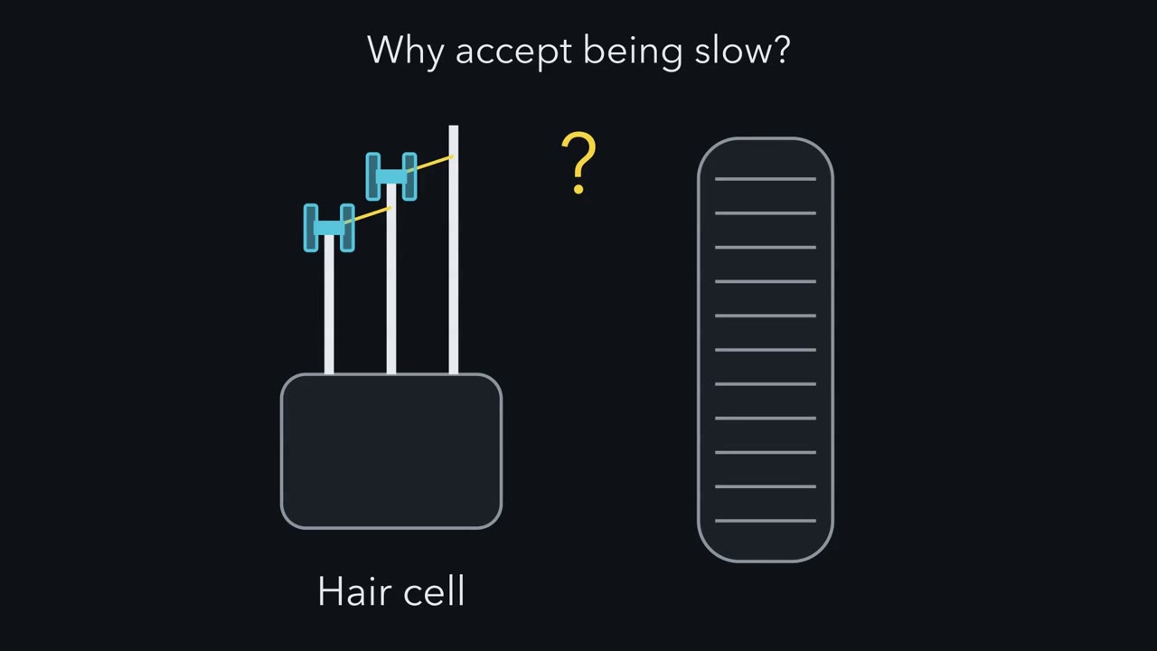 ▶ Watch Nothing in Between, or Enzymes in Between · 2:41
Read the transcript of “Nothing in Between, or Enzymes in Between”
Film transcript: Nothing in Between, or Enzymes in Between
Why would a sensory cell accept being slow? A hair cell and a rod hold the answer. Every sensory cell turns a stimulus into a current. So the first question is what sits between the stimulus and the pore. In a hair cell, no enzyme. Bending the bundle pulls on tip links, and the pull opens transduction channels directly. In turtle hair cells, these channels can activate with a time constant as short as about 50 microseconds. In a rod, there is a ladder of enzymes. Light activates rhodopsin, then transducin, then PDE6. PDE6 breaks down cyclic GMP. Channels close, less positive charge enters, and the rod hyperpolarizes. A mouse rod's response to a single photon takes of order a tenth of a second or more to peak.
On one time axis, the two sit more than three orders of magnitude apart. But one is a channel time constant, and the other a whole cell's time to peak. Read the gap as scale. Predict. What does the rod get in return for being slow?
Gain. Enzymes keep working while active. In mouse rods, one absorbed photon removes about 2,000 cyclic GMP molecules. That is one reason a rod can signal a single photon. Low dark noise and a reproducible response are needed too. A direct channel has no enzyme cascade before the current. Force acts on the channel complex itself. The rod's price is time. Molecules must meet, and enzymes must turn over, before any channel changes. That is the one idea of this lesson. What sits between stimulus and pore sets what a sensory cell can do. The second question is how the signal leaves the cell. An olfactory neuron carries it on its own axon. A hair cell or a type II taste cell hands it to a separate afferent fibre instead.
Together, the two questions make a table of four boxes, and five examples fill it. Rods and cones do not fit neatly. They synapse inside the retina and send no axon to the brain. So ask two questions of every sensory cell. What sits between stimulus and pore? And how does the signal leave?
From the film
- Every sensory cell turns a stimulus into a current, and what sits between the stimulus and the pore sets what the cell can do.
- Where no enzyme sits in between, as in a hair cell, the current can start very fast; in turtle hair cells channels can activate with a time constant as short as about 50 microseconds.
- Where enzymes sit in between, as in a rod, one stimulus event can change thousands of molecules, usually at a cost in time.
- Ask two questions of every sensory cell: what sits between stimulus and pore, and how does the signal leave the cell?
In turtle hair cells, transduction channels can activate with a time constant as short as about 50 microseconds. A rod’s response to a single photon takes of order a tenth of a second or more to reach its peak, yet the rod can report that one photon. Why would a sensory cell accept being slow?
Every sensory cell turns a stimulus into a change in ion-channel current. What differs is the machinery in between, and that machinery sets what the cell can do. Sensory coding already follows a stimulus through the receptor potential to a firing rate, ion channels as receptors treats gating as a receptor problem, and senses and cGMP works through the rod and olfactory cascades. This lesson starts where they stop and sets five examples side by side.
The five are not five separate designs but answers to two questions. The lesson has three parts: the two questions, a skill for reading what a quoted number actually measures, and a checklist for naming the transducer.
Two questions for any sensory cell
First: what sits between the stimulus and the pore (the channel’s ion-conducting path)? In the examples here, either no enzyme, so that the stimulus acts on the channel complex itself, through a mechanical link or by binding, or an enzyme cascade, so that a chain of reactions must run before any channel changes.
Second: how does the signal leave the cell? A primary sensory neuron detects the stimulus and carries the signal into the nervous system itself: an olfactory neuron sends an axon from the epithelium to the bulb, and a free nerve ending belongs to a neuron. A secondary receptor cell detects the stimulus and passes on a signal to a separate afferent, a nerve fibre that carries it into the brain or spinal cord, as hair cells do with graded receptor potentials in the mature state and type II taste cells do through CALHM1/3. This is cell architecture, not transduction mechanism, so the two questions are independent.
Here are the five examples, with the evidence each rests on.
- Hair cell: force on the pore. Deflection of the bundle pulls on tip links and opens transduction channels in the bundle (hearing and balance covers the gating). TMC1 is the leading candidate for the pore, and the evidence is the subject of the last section. The follow-up is the hair-cell transduction channel.
- Insect olfactory neuron: an odorant on the pore. Insect odorant receptors are ligand-gated channels. A tuning odorant receptor pairs with the co-receptor Orco around a central pore, and structures of purified, heterologous complexes with and without ligand indicated that binding to the tuning subunit could open the pore (Zhao and colleagues 2024).
- Heat-sensing nerve ending: heat on the pore. In a free nerve ending, transient receptor potential vanilloid 1 (TRPV1) senses heat in the same protein that forms the pore (touch and pain covers what TRPV1 is needed for).
- Rod and vertebrate olfactory neuron: enzymes in between. In a rod, rhodopsin activates transducin, which recruits PDE6; falling cGMP closes cyclic-nucleotide-gated channels, so less positive charge enters and the rod hyperpolarizes. In a vertebrate olfactory neuron, the receptor acts through Golf and adenylyl cyclase III to raise ciliary cAMP, which opens the same family of channels and recruits a chloride current, so odour depolarizes the cell. The sign is opposite; the logic of enzymes between receptor and pore is the same.
- Type II taste cell: enzymes in, a channel out. A mouse type II taste cell detects sweet, bitter or umami through G-protein-coupled receptors (GPCRs), and releases ATP through the CALHM1/3 channel instead of vesicles. Its transduction is a GPCR cascade; its output sets it apart.
Put together, the two questions sort them:
| Primary: carries the signal on its own axon | Secondary: hands the signal to an afferent | |
|---|---|---|
| No enzyme between stimulus and pore | heat-sensing free nerve ending (TRPV1); insect olfactory neuron (odorant receptor with Orco) | hair cell (force-gated channel) |
| Enzymes sit in between | vertebrate olfactory neuron (GPCR cascade) | type II taste cell (GPCR cascade, output through CALHM1/3) |
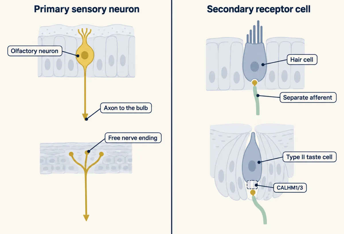
Two cases need a note. Rods and cones are sensory neurons with an enzyme cascade, but textbooks differ on their column, because they synapse inside the retina and send no axon into the brain. OTOP1, the proton channel of type III taste cells, belongs in the top row and the secondary column, with a twist: the stimulus, a proton, is also the ion that flows.
The one idea to carry through this lesson. What sits between the stimulus and the pore sets what a sensory cell can do. Where no enzyme sits in between (force on a hair-cell channel, an odorant on an insect receptor, heat on TRPV1), the current can start as fast as the channel changes shape, and the stimulus acts on the channel complex without an intervening messenger-producing enzyme cascade. Where enzymes sit in between (rhodopsin, transducin and PDE6 in a rod), molecules must meet and enzymes must turn over before any channel changes. Those turnovers let one stimulus event change thousands of molecules, and they are also a likely source of delay.
The idea compares classes of design in the examples here, at the level of the transducing cell’s own current. It is a tendency, not a timing law: each encounter, turnover and channel transition has its own rate. It does not rank cells within a class: TRPV1 is directly gated, yet the 3.5 ms time constant reported for purified rat TRPV1 after a rapid heat jump (Cao and colleagues 2013) is far slower than the turtle hair-cell value of about 50 μs. The two come from different preparations and protocols, so direct gating alone does not set a channel’s speed. It does not promise high gain at every step of a cascade, and it says nothing about behavioural latency.
The two designs side by side
| No enzyme in between (hair cell) | Enzymes in between (rod) | |
|---|---|---|
| Branching before the current | no enzyme cascade multiplies messenger molecules: bundle force changes the opening probability of mechanically linked channel complexes, and one deflection can affect many channels | one absorbed photon leads to about 2000 cGMP molecules hydrolysed (mouse rods; Arshavsky and Burns 2014) |
| How fast the current rises | channels activate with a time constant as short as about 50 μs (turtle hair cells; Ricci and colleagues 2005) | a single-photon response takes of order 100 ms or more to peak (mouse rods near body temperature) |
Both currents are then carried by many ions through each open channel, so the first row compares branching, not electrical gain. On one time axis the two speeds sit more than three orders of magnitude apart. But they are not the same kind of number. One is a channel time constant (the time a single-exponential rise takes to reach about 63% of its final size); the other is a whole cell’s time to peak. Read the gap as a picture of scale, not as a measured cost of the cascade.
Forty microseconds: what a fast number measures (Corey and Hudspeth 1979)
Read the transcript of “Forty Microseconds: What a Fast Number Measures”
Film transcript: Forty Microseconds: What a Fast Number Measures
Could an enzyme step fit inside 40 microseconds? First, you need to know what that number measures. In 1979, Corey and Hudspeth moved the hair bundles of a bullfrog sacculus, in a dish. They recorded the transepithelial microphonic. That is the summed electrical response across the whole sheet of cells. The epithelium's own electrical time constant slows that signal. So the recorded delay is longer than the true one. After correcting for it, the delay was 40 microseconds, at 22 degrees Celsius. Predict. Suppose an enzyme had to make a chemical messenger between the bundle and the channel. Could the response begin that fast?
Only with great difficulty. Over a gap of nanometres, free diffusion can be fast. But that alone does not say how soon the messenger finds and binds a channel. Making it is tight too. In a simple constant-rate model, one enzyme at 4,000 turnovers a second makes, on average, only 0.16 molecule. That is about a 15 percent chance of making even one. Many enzymes could make more. To test a messenger mechanism, we would need its enzyme numbers, activation and binding rates, geometry, and the messenger level that changes the current. This is a model check, not a result of the 1979 experiment. Now, what the number is not. It is not the opening time of one channel.
And it is not a mouse number. Channel speed was measured directly later. Ricci and colleagues found time constants as short as about 50 microseconds, in turtle hair cells. So in turtle hair cells, channels do open on this time scale. The 1979 number simply measured something else. Four kinds of number are often merged. A tissue response latency is the delay of a whole population of cells. A channel activation time constant is how fast current rises in a patch or cell. A molecular gain counts output events per input event. And a behavioural latency runs from stimulus to movement, through conduction, spinal circuits and muscle. Speed can be compatible with a candidate channel.
But speed alone does not identify it. So before you compare two speeds, ask whether they are the same kind of number.
From the film
- The 40-microsecond delay is a tissue response latency, the corrected summed response of the bullfrog sacculus at 22 degrees C, not the opening time of one channel.
- Channel activation was measured directly later, with time constants as short as about 50 microseconds in turtle hair cells.
- A tissue latency, a channel time constant, a molecular gain and a behavioural latency are different kinds of number, so check what kind before comparing two speeds.
Corey and Hudspeth stimulated the hair bundles of an in vitro bullfrog sacculus and recorded the transepithelial microphonic, the summed electrical response across the sheet of cells. The electrical time constant of the epithelium slows such a signal, so the recorded delay is longer than the true one. After correcting for that time constant, the delay was 40 microseconds at 22 degrees C. That short delay, with its modest temperature dependence, limited the possible models for transduction, which is its real value.
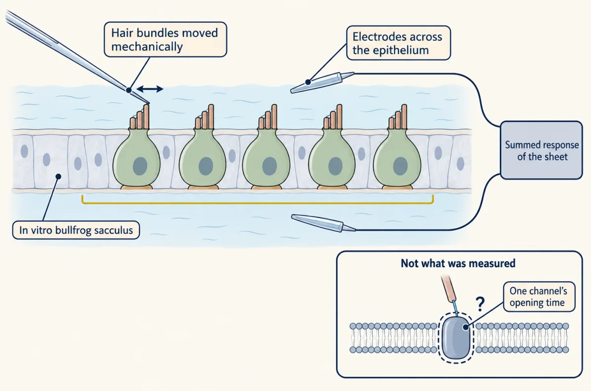
Predict. Suppose an enzyme had to make a chemical messenger between the bundle and the channel. Could the response begin within 40 microseconds?
Check your prediction
Only with great difficulty. A diffusing chemical messenger is usually judged too slow to fit, but that reading is later interpretation, not a claim of the 1979 abstract. A rough check of our own (arithmetic in Going deeper) shows how tight the budget is. Free diffusion can be fast over a gap of nanometres: a small molecule’s root-mean-square spread in 40 μs is about 270 nm, though that does not say how soon it finds and binds a channel. Production is tight too: in a constant-rate model, one already-active enzyme turning over 4000 times a second, fast for a signalling enzyme, makes on average only 0.16 molecule in 40 μs, about a 15% chance of even one. Parallel enzymes could make more. These numbers constrain a messenger mechanism but do not set the enzyme number or location it would need, or rule it out; testing one needs its activation, production, diffusion and binding kinetics, and the messenger level a detectable current needs.
Now what the number is not. It is not the opening time of one channel, and it is not a mouse number. Channel kinetics were measured directly later: Ricci and colleagues (2005) found that the transducer current of turtle auditory hair cells activated with a time constant as short as about 50 microseconds, and at least 10 times faster in rat outer hair cells. So hair-cell channels do open on this time scale; the 1979 number simply measured something else.
Four quantities that are often merged
| Quantity | What it is | Example in this lesson | What it leaves out |
|---|---|---|---|
| Tissue response latency | the delay between a stimulus and an electrical response of a population of cells | 40 μs, corrected transepithelial microphonic, bullfrog sacculus, 22 degrees C | the opening time of any one channel |
| Channel activation time constant | the time scale of the current rise in a patch or cell after a step | about 3.5 ms for purified rat TRPV1 in liposomes after a rapid heat jump; about 50 μs for turtle hair-cell channels | everything downstream of the current, such as spikes or transmitter release |
| Molecular gain | output events per input event, such as effective downstream complexes per activated receptor | about 12 to 14 effective transducin-PDE complexes per activated rhodopsin in mouse rods | time |
| Behavioural latency | the time from stimulus to movement | the time from heat on the skin to a withdrawal | nothing between stimulus and movement: it includes conduction, spinal circuits and muscle, so no channel time constant stands in for it |
These are preparation-specific numbers and must not be combined into one description. A fast latency can be compatible with a candidate channel, but speed alone does not identify the channel.
Predict. Sort four numbers from this lesson: the 40 μs corrected delay of the bullfrog sacculus; Ricci’s 50 μs in turtle hair cells; about 2000 cGMP hydrolysed per photon in mouse rods; and first spikes about 25 to 40 ms after a strong odour pulse in mouse olfactory neurons. Which kind of quantity is each, if any?
Check your prediction
The 40 μs is a tissue response latency. The 50 μs is a channel activation time constant. The 2000 cGMP is a molecular gain. The first-spike delay is none of the four exactly: it is one cell’s latency to a spike, and it includes solution exchange, membrane charging and spike initiation as well as transduction. So it should not be compared directly with the 40-microsecond tissue latency, and neither hair-cell number is a stand-in for the other.
Where a cascade’s gain comes from, and what it costs
Read the transcript of “Where the Gain Sits, and What It Costs”
Film transcript: Where the Gain Sits, and What It Costs
Where exactly does a cascade get its gain? Follow one photon down the rod's ladder. Step one. Activated rhodopsin switches on transducin, which switches on PDE6. Yue and colleagues counted each active unit as one effective complex. Step two. Each effective complex breaks down cyclic GMP, many molecules each. Step three. The fall in cyclic GMP closes cyclic-nucleotide-gated channels. In mouse rods, about 2,000 cyclic GMP molecules are hydrolysed for each absorbed photon. In a separate study, Yue and colleagues estimated about 12 to 14 effective complexes per activated rhodopsin. Predict. If these two estimates can be combined, which step multiplies the signal more?
If they combine, step two. Each complex would break down roughly 150 molecules. That is an illustration, not a measured yield. But other analyses put step one several times higher. On those, the two factors could be comparable. Either way, both steps amplify. The gain belongs to particular enzymes, not to the receptor being a G protein coupled receptor. A receptor step need not amplify at all. In frog olfactory neurons, one bound receptor rarely activates even one G protein. There, the gain is thought to come at later steps. Adenylyl cyclase makes cyclic AMP, which opens channels. The calcium that enters opens a chloride current. Now the price. Every rung is an encounter between molecules, or an enzyme turning over. So the current rises only after several steps have run.
In mouse olfactory neurons, the earliest first spikes came about 25 to 40 milliseconds after a strong odour pulse. That is a latency to a spike, which includes more than transduction. It is not a channel time constant. Spontaneous receptor activity can also enter the cascade. Neurons with different odorant receptors showed different levels of spontaneous activity. Inverse agonist odorants could quiet it. A cascade that multiplies a receptor event multiplies a spontaneous one too. So enzymes between stimulus and pore can buy gain. They often pay in time, and they can amplify spontaneous activity too.
From the film
- In mouse rods, about 2000 cGMP molecules are hydrolysed per absorbed photon, so a rod cascade can multiply one event many times.
- The receptor step need not amplify: in frog olfactory neurons one bound receptor rarely activates even one G protein, and the gain is thought to come at later steps.
- The price of a cascade is time, because every rung is a molecular encounter or an enzyme turning over, and a cascade can also amplify spontaneous receptor activity.
A direct channel has no enzyme cascade before the current. A cascade has branching steps; follow one photon down the rod’s ladder to see where it multiplies the signal.
| Step | One of these | activates or acts on |
|---|---|---|
| 1 | activated rhodopsin | transducin, which then switches on PDE6. Yue and colleagues counted each active transducin-PDE6 unit as one effective complex; how it maps onto PDE6’s two catalytic subunits depends on the activation model. |
| 2 | effective complex | cGMP, hydrolysing many molecules each |
| 3 | fall in cGMP | cyclic-nucleotide-gated channels, which close |
Predict. Arshavsky and Burns (2014) estimated about 2000 cGMP molecules hydrolysed per absorbed photon in mouse rods. Yue and colleagues (2019) estimated about 12 to 14 effective complexes per activated rhodopsin, in mouse rods lacking GCAPs, the calcium-feedback proteins that speed cGMP synthesis (Gcaps-null). If the two estimates can be combined, which step multiplies the signal more?
Check your prediction
Step 2, if they combine. Step 1 multiplies by about 13. If the 2000 cGMP and the 13 complexes described the same response, each complex would hydrolyse about 2000 / 13, roughly 150 cGMP (about 140 to 170 for 12 to 14 complexes). The two studies did not measure these together, and Yue’s rods were Gcaps-null, so 150 is an illustration, not a measured yield per complex. On this combination the larger factor is at PDE6, an enzyme that turns over again and again while it stays active. But the gain at the first step in mouse rods is preparation-dependent and debated, and other analyses give several times more (Lamb 2022). With those, the two factors could be comparable, under the conditions set out in Going deeper. Either way, both steps amplify, and the gain belongs to particular enzymes, not to the receptor’s being a GPCR.
That gain is one reason a rod can signal a single photon. Gain alone does not explain it: low dark noise and a reproducible elementary response are needed too.
The receptor step need not amplify at all. In dissociated frog olfactory neurons, Bhandawat, Reisert and Yau (2005) found a low probability that one bound receptor activated even one G protein. Whatever gain the olfactory neuron has is thought to come at later steps: adenylyl cyclase III makes cAMP, cAMP opens cyclic-nucleotide-gated channels, and the calcium that enters opens a chloride current.
The price in time. Every rung of the ladder is an encounter between molecules or an enzyme turning over, so a cascade’s current rises only after several steps have run, not after one channel’s change of shape. In dissociated mouse olfactory neurons, first spikes appeared as early as about 25 to 40 ms after a high-dose odour pulse (Ghatpande and Reisert 2011), while low-dose responses often began after the pulse had ended. That is a latency to a spike, not a channel time constant. Its roughly 600- to 1000-fold gap from the 40-microsecond tissue latency is consistent with a slower cascade, but these numbers cannot say how much of it the cascade causes.
Noise. Spontaneous receptor activity can enter the cascade too. Mouse olfactory neurons expressing different odorant receptors showed different levels of basal current fluctuation and spontaneous firing, and inverse-agonist odorants could suppress it (Reisert 2010). A cascade that multiplies a receptor event also multiplies a spontaneous receptor event.
So the trade-off is real but not a law with a fixed price in milliseconds: the price depends on how fast each rung runs. Whether that costs signal-to-noise ratio depends on where noise enters; no study in hand compares the five examples by signal-to-noise ratio under matched stimulus statistics, bandwidth and conditions, and that comparison is still open.
Detect, then send: CALHM1/3 (2018)
The second question, how the signal leaves the cell, has its cleanest test in a taste cell. Type II taste cells lack classical synaptic structures, including synaptic vesicles. In mouse isolated taste preparations, G-protein-coupled receptor (GPCR) stimulation recruits PLCβ2 and IP3-dependent calcium release, and the calcium rise activates TRPM5. So detection is a GPCR cascade. How is the message passed on?
Predict. Ma and colleagues (2018) deleted Calhm3 in mice. If CALHM1/3 is the output channel and not part of detection, what happens to the cells’ excitability, and to taste-evoked ATP release?
Check your prediction
Excitability should survive and ATP release should fail, and both held: deleting Calhm3 abolished taste-evoked ATP release and removed the nonselective current, without impairing excitability. The failure sits at the output step, after the receptor cascade has done its work.
A second argument for CALHM1/3 as the output channel comes from kinetics. In cultured mouse N2a cells, CALHM1 alone activated with a voltage time constant above 500 ms, and CALHM3 coexpression brought it to about 10 ms. Fast gating matters for a short depolarization. In a single-exponential teaching model, a channel that starts closed reaches a fraction a(t) = 1 − exp(−t/τ) of its maximal activation.
Predict. Suppose a taste cell depolarizes for 3 ms, a round number chosen for the exercise. Roughly what fraction of maximal activation does a channel reach with τ = 10 ms, and with τ = 500 ms?
Check your prediction
About 26% when the time constant is 10 ms, and about 0.6% when it is 500 ms, so the faster channel activates about 43 times as large a fraction of its channels in the 3 ms (steps in Going deeper). The measured slow time constant was above 500 ms, so in this model the slow fraction would be smaller still and the ratio larger. These are teaching assumptions: a single exponential and a square pulse are simplifications, real CALHM currents have more than one kinetic component, and the model gives an activation fraction, not the ATP released or the intensity of taste.
| Step | your τ | τ = 500 ms |
|---|---|---|
| t / τ | ||
| a = 1 − e−t/τ | ||
| fraction of maximum |
In this sketch: one exponential, a square pulse, no inactivation. The values of about 10 ms and above 500 ms are voltage activation time constants for CALHM1 with CALHM3 and CALHM1 alone in cultured mouse N2a cells; any other τ here is made up. Real CALHM currents have more than one kinetic component. Pulse durations here are made up.
A fraction of maximal activation is not the ATP released and not the intensity of a taste.
A receptor cell and its afferent may also share transduction, as Merkel cells and their touch afferents appear to (evidence in Going deeper).
The checklist for calling something the transducer
Calling a protein the transducer asks for five kinds of evidence:
- Place and time. The protein must be in the right place at the right time.
- Loss. Removing it, together with any redundant partner, must abolish or clearly alter the native current.
- Permeation. Mutating it should change permeation in a way that is hard to explain indirectly, such as a fast change after chemical modification of a single substituted residue.
- Sufficiency. Placing it, or the complex it needs, in an insensitive cell or a bilayer should confer the sensitivity; a failure may only mean a missing partner.
- Kinetics. Its kinetics must be compatible with the native response.
Applied to three proteins (criteria 1 and 5 are not tabulated; a dash means this lesson does not cover that evidence, not that none exists):
| Criterion | TMC1 (hair cell) | Piezo2 (Merkel cell, a touch-sensing skin cell) | OTOP1 (type III taste cell) |
|---|---|---|---|
| 2. Loss | mouse hair cells lacking Tmc1 and Tmc2 lose the normal, tip-link-gated current | Merkel cells lacked mechanically activated currents without Piezo2 (Woo and colleagues 2014) | in isolated type III taste cells, deleting Otop1 eliminated the proton current |
| 3. Permeation | in rescued null cells, cysteine-substituted TMC1 plus chemical modification changed permeation rapidly and irreversibly (Pan and colleagues 2018) | — | — |
| 4. Sufficiency | shown for non-mammalian orthologs: truncated turtle TMC1 and budgerigar TMC2, purified and reconstituted, responded to pressure in proteoliposomes (Jia and colleagues, 2020 issue); mammalian TMC1/2 in heterologous cells only after a trafficking intervention or an engineered tag (Fu and colleagues 2025; Chen and colleagues 2025) | Piezo2 confers mechanosensitivity on cells that lack it | OTOP1 expressed in HEK293 cells gave currents with similar properties to the native one (Teng and colleagues 2019), and sweet taste cells engineered to express OTOP1 also responded to acid (Zhang and colleagues 2019) |
| Behaviour | — | — | acid-evoked taste-nerve responses strongly reduced, yet knockout mice still avoided acids in the two-bottle assays tested |
Three readings of the table:
- TMC1. The pore case rests on criteria 2 and 3 in native cells. The sufficiency results come from non-mammalian orthologs or modified mammalian proteins, not an unmodified native complex. For the anomalous current in young null cells and the heterologous reports, see TMC1 evidence in detail.
- Piezo2. Its status in each cell type is a separate question (Piezo channels and the physics of force).
- OTOP1. Needed for the current in those cells, yet the behaviour can use other routes: necessity for a current in a cell is not necessity for the behaviour.
Heat is a reminder of the same point. Mice lacking transient receptor potential vanilloid 1 (TRPV1), TRPM3 and transient receptor potential ankyrin 1 (TRPA1) together lost the tested acute heat-withdrawal response (Vandewauw and colleagues 2018). A channel may be sufficient for heat sensitivity in liposomes and still be one of several in the animal.
Design problem
Pan and colleagues could not test mammalian TMC1 in the heterologous cells then available, because it did not reach the membrane. They worked in hair cells from mice lacking Tmc1 and Tmc2. Design experiments that separate a pore-lining residue from a trafficking or assembly partner, and say what would still be missing.
Saved in this browser only.
One way to do it
Loss of current alone cannot settle it, because removing an accessory protein would also remove the current. So first rescue: deliver wild-type TMC1 to the null cells and show that the current returns. Then deliver variants in which single residues are replaced by cysteine, and record the bundle-evoked current before and during exposure to a cysteine-modifying reagent. Wild-type TMC1 should show no change; a variant whose residue lies in the permeation path should show a rapid, irreversible change in a permeation property such as selectivity or conductance. A trafficking partner often changes how much current there is, but accessory proteins can also change conductance or selectivity, so the speed and residue specificity of the change carry the argument; a slow change would suggest an indirect effect. Controls: a cysteine variant with no change, and a check that the baseline current is normal. Even then, sufficiency is untested. That needs a naive system: reconstitution of purified protein in a bilayer, as Jia and colleagues did with turtle TMC1 and budgerigar TMC2 (a result for those orthologs), or expression in a heterologous cell once the protein reaches the membrane.
Going deeper
Insect odorant receptor structures, 2018 and 2024
In 2018 Butterwick and colleagues solved the cryo-EM structure of Orco from the fig wasp Apocrypta bakeri: a homotetramer around a central pore. In 2024 Zhao, Chen, Ryu and del Marmol purified complexes of a mosquito tuning receptor (Aedes aegypti OR10 or Anopheles gambiae OR28) with the wasp Orco and found one tuning subunit and three Orco subunits. Being purified, heterologous complexes, they do not show that every native insect receptor has a 1:3 arrangement.
Diffusion against turnover in 40 microseconds
Take a small messenger with an illustrative diffusion coefficient D of about 300 μm²/s. For free diffusion, its root-mean-square spread in three dimensions after a time t is √(6Dt). For t = 40 μs that is √(6 × 300 × 0.00004) μm, about 0.27 μm, or 270 nm. The time to find and bind a channel also depends on geometry and binding kinetics.
Now count product molecules. One active enzyme with a turnover of k per second makes, on average, k × t molecules in time t. In 40 μs, 100 per second gives 0.004 molecule, 1000 per second gives 0.04, and 4000 per second gives 0.16. Molecules come whole, so 0.16 is an expected value: if products appear at random (a Poisson process), the chance of at least one by 40 μs is 1 − e−0.16, about 15%.
Rod and olfactory recording details
Yue and colleagues (2019) recorded from intact mouse rods (1 to 3 months old, suction-pipette recordings at 37.5 degrees C), using two independent routes to the elementary event: rods with an inefficient rhodopsin mutant, and bleached opsin, which produced detectable dark electrical noise. The raw current amplitudes from the two kinds of rod cannot simply be divided to obtain the Gcaps-null estimate of 12 to 14 effective complexes, which comes from a fuller analysis.
The arithmetic of where the factors cross is illustration only. If 2000 cGMP per photon is the product of the two steps, they multiply equally at a first-step gain of about 45 (the square root of 2000); several times 12 to 14 reaches or passes that, and a first-step gain of 56 would leave about 36 per complex. This holds only for a fixed total with the same definition of an active unit in every estimate; a different activation model need not leave the total at 2000 or count the same unit.
Ghatpande and Reisert (2011) used dissociated mouse olfactory neurons at 37 degrees C with an odour pulse of 100 ms, and reported first spikes as short as about 25 ms for I7 neurons at 300 μM heptanal and about 40 ms for mOR-EG neurons at 100 μM eugenol. Solution exchange takes up to about 7 ms. Raising the stimulation rate from 2 to 5 Hz reduced spiking reliability in these neurons.
TRPV1: two protocols, two numbers
In purified rat transient receptor potential vanilloid 1 (TRPV1) in liposomes, Cao and colleagues (2013) reported a threshold of about 41.5 degrees C and a Q10 of 23.5 with slow heating, and a time constant of about 3.5 milliseconds with a rapid infrared jump. These came from different protocols and are not one description. A Q10 describes how steeply the response rises with temperature. It is not a molecular gain.
Photoreceptors: primary or secondary?
Photoreceptors are hard to place in the primary-secondary scheme. Rods and cones hyperpolarize in response to photons, and their graded voltage regulates graded transmitter release rather than triggering action potentials (Lankford and colleagues 2020). Not spiking does not make them non-neuronal. Lankford and colleagues group them with olfactory neurons as primary sensory neurons, despite their local synapses in the retina. Neuronal identity, spiking and position in the circuit are three separate properties.
Why have two architectures at all? A reasonable inference, not an experiment on evolution, is that a primary neuron skips a peripheral receptor-to-afferent synapse, while a secondary cell lets local transduction and the afferent’s firing be controlled separately.
Merkel cells and Piezo2: two sites
In mouse skin, Merkel cells sorted from newborn (P0 to P5) dorsal skin and cultured overnight lacked mechanically activated currents without Piezo2 (Woo and colleagues 2014). Piezo2 was then deleted in all epidermal cells (Krt14-Cre, so not Merkel-cell specific), and the touch afferent was recorded ex vivo from hairy hindpaw skin and its nerve at 7 to 17 weeks. The afferent kept a substantial fast (dynamic) response but lost part of its static firing, the sustained firing during a held touch. That fits a two-site model: the afferent can detect touch itself, and Merkel cells help sustain the response. The results were most consistent with two receptor sites; the manipulation alone does not assign all the fast firing to the afferent.
Ages of the animals in the taste experiments
- CALHM3 knockout (Ma and colleagues 2018): mice at least 52 days old.
- OTOP1 current (Teng and colleagues 2019): isolated type III taste cells from mice of 6 to 12 weeks; the two-bottle tests used knockout mice of 12 to 24 weeks.
The activation-fraction calculation
For a 3 ms pulse and a(t) = 1 − exp(−t/τ):
Step 1, fast case. For τ = 10 ms, t/τ = 3/10 = 0.3, so a = 1 − e−0.3 = 0.259, or 25.9% of the maximum.
Step 2, slow case. For τ = 500 ms, t/τ = 3/500 = 0.006, so a = 1 − e−0.006 = 0.00598, or 0.60%.
Step 3, the ratio. 25.9 divided by 0.598 is about 43.
TMC1 evidence in detail
Young null cells keep an anomalous current of reversed polarity (Kim and colleagues 2013). Mammalian TMC1/2 historically trafficked poorly to the surface of heterologous cells, which is why Pan and colleagues worked in hair cells. Three distinct sufficiency preparations followed. Jia and colleagues (2020) reported pressure responses of reconstituted non-mammalian orthologs in proteoliposomes. Fu and colleagues (published online December 2024; 2025 issue) reported mechanically gated mouse TMC1/2 mutants, and wild-type human TMC1/2 that reached the cell surface after a trafficking intervention found by CRISPRi screening. Chen and colleagues (2025) reported indentation-evoked currents from Fyn-tagged full-length mouse TMC1 or TMC2 in PIEZO1-knockout HEK293T cells. None of these is an unmodified native hair-cell complex.
Your notes
Select any text in the lesson to highlight it or add a note to it. Your highlights and notes are saved in this browser and collected in your notebook.
This browser is not letting the site save anything (a private window or strict privacy settings), so highlights and notes will be lost when you leave the page.
How we measure it
Whole-cell and patch-clamp recording, including outside-out patches
A glass pipette seals onto a cell or a small piece of its membrane and measures the current for a held voltage. It gives activation kinetics in a heterologous cell (CALHM1/3 in N2a cells) and resolves single-channel openings in a small excised patch (Orco).
Suction-pipette recording with fluctuation analysis
An intact rod or olfactory neuron is drawn into a pipette to record the whole-cell current or spikes. The variance and mean of small responses give the size of the elementary event, and fast solution exchange times a first spike from the arrival of an odour.
Cysteine substitution and chemical modification
A residue is replaced by a cysteine that a reagent can modify. If modification quickly and irreversibly alters permeation of the native current, the residue is probably near the permeation path.
Liposome reconstitution with rapid heating or pressure
A purified channel is placed in artificial membrane, away from every other cell protein, and stimulated. Responses that survive show the stimulus sensitivity is within the protein under those lipid conditions.
Practice questions on this lesson (3) → MCAT-style, original, each with an explanation
Where this lesson fits
Before thisReview first
- The Action Potential and ConductionNeuroscience · College: going further
You need how channels open and carry current to read a channel time constant and a receptor current here.
- Sensing the World: Vision, Smell, Nitric Oxide and cGMPCell Signaling · Graduate school and beyond
The rod and olfactory cascades are used here as the enzyme-in-between design, with no re-teaching.
- Ion Channels as ReceptorsCell Signaling · Graduate school and beyond
You need direct gating of a channel by a stimulus, such as heat on TRPV1, to read the no-enzyme design.
After thisStudy next
- Piezo Channels and the Physics of ForceThis course · Lesson 2
This takes the force-gated channel case further, asking how Piezo2 meets the transducer checklist.
- The Hair Cell's Transduction ChannelThis course · Lesson 22
It takes the TMC1 evidence and the fast hair-cell current further, testing what is needed to call it the pore.
- Merkel Cells: Touch Receptors That Are Not NeuronsThis course · Lesson 5
It develops the two-site model where a non-neuronal cell and its afferent share touch transduction.
Elsewhere on the siteFilms, simulations and method films in other courses
- Cascade amplifierSimulation · Cell Signaling
It shows how a chain of enzyme steps multiplies one photon's signal, the gain that a cascade buys at a cost.
- The Timer Inside the SwitchFilm · Cell Signaling
It shows how a G protein times its own signal, the delay inside the rod and olfactory cascades.
- How One Photon Becomes a Cellular SignalFilm · Cell Signaling
It shows how a photon's signal grows through a chain of steps, the rod gain ladder used here.
Suggested by an AI model from the lessons themselves, then checked. How we use AI
Finish line
Test yourself
8 questions
Practice and MCAT-style, with explained answers.
Next · Lesson 2
Piezo Channels and the Physics of Force
Mouse PIEZO2 threads 114 transmembrane helices through the membrane to build one ion channel, and its pore sits at the centre. Why would a channel need so much protein around one pore?
Continue →