Cell Signaling · Unit 6: Channels, the nucleus, and life or death
Ion Channels as Receptors
A chilli pepper and a hot stove open the same protein. How does its message reach your brain in milliseconds?
Pause when the reading reaches a film, and offer to play it. Saved on this device.
Watch the film now, or keep listening
Builds on:A Field Guide to ReceptorsCalcium: The Messenger Kept at Arm's LengthSensing the World: Vision, Smell, Nitric Oxide and cGMPWhy these?
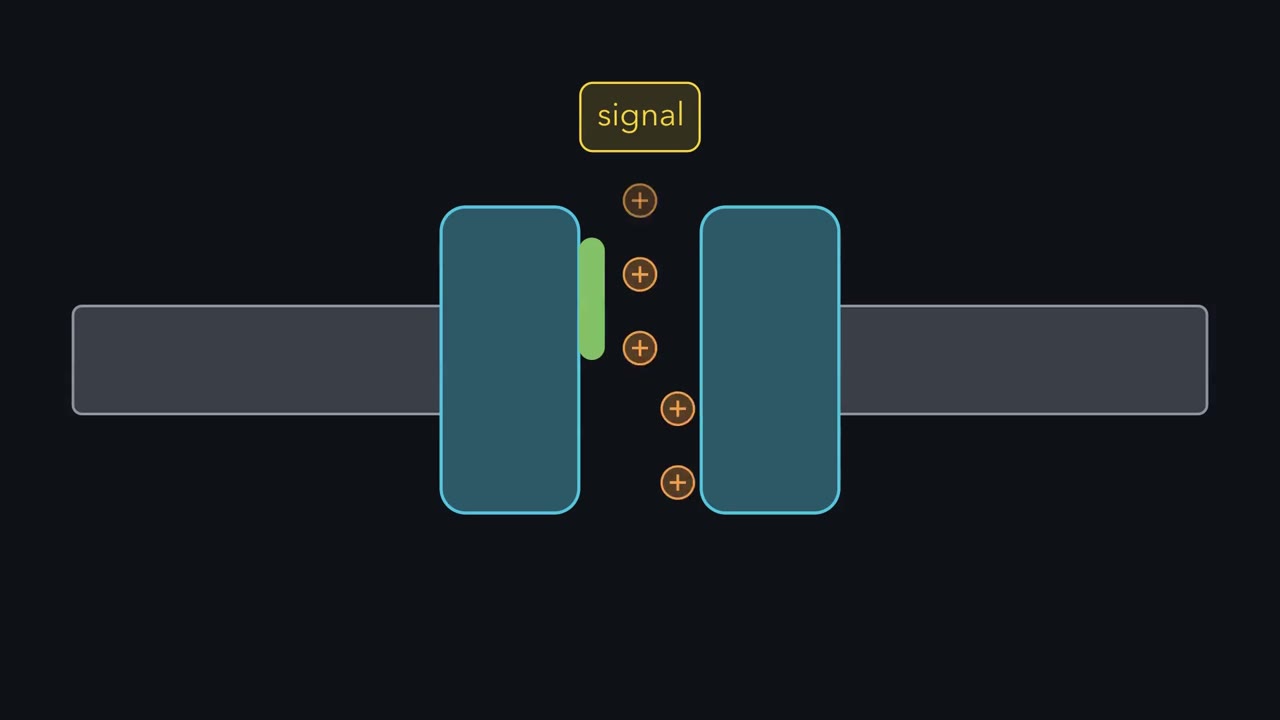 ▶ Watch The Signal Opens a Hole · 2:18
Read the transcript of “The Signal Opens a Hole”
Film transcript: The Signal Opens a Hole
How can a cell answer a signal in about a millisecond? Most receptors hand their message to something else, such as a G protein or a kinase. An ion channel hands it to nothing. The signal opens a hole, ions pour through, and the voltage across the membrane changes. One open channel lets about ten million ions cross every second, so which way do they go?
Two kinds of protein move ions across a membrane. A pump binds its ions and changes shape to carry them, uphill if it spends ATP. A channel is a pore with a gate, which a transmitter, a voltage, or heat can open. Once it is open, ions run downhill at around ten million a second, while a pump manages a few hundred. So pumps build the gradients slowly, and channels spend them fast.
Downhill has two parts: a push from the difference in concentration, and a pull from the voltage across the membrane. Take sodium. A cell keeps it mostly outside, and the inside of a resting neuron sits near minus −70 mV. For each ion, there is one voltage at which push and pull balance. That is its equilibrium potential, E, and for sodium it is about plus +60 mV.
A cell keeps potassium mostly inside. With typical concentrations, E for potassium is about minus −90 mV. An open channel drags the voltage toward the E of the one ion it passes. So the direction of flow follows from one subtraction. The driving force is the membrane voltage minus E. At minus −70 mV, sodium is 130 mV away from its E. So it rushes in, and the cell depolarises, meaning its voltage becomes less negative. Potassium is only 20 mV from its E. So it leaks out gently, and pulls the voltage back down.
Very few ions need to cross to change the voltage. So the concentrations barely shift, and nothing has to be made. The voltage is the message. So keep this rule: the signal opens a hole, and the gradient does the rest.
One open ion channel lets about ten million ions cross a membrane every second. What can a cell do with a receptor like that?
Most receptors in this course hand their message to something else, such as a G protein or a kinase. A channel hands it to nothing. The signal opens a hole, ions pour through, and the voltage across the membrane changes in about a millisecond. So what decides which way the ions go, how does a hole tell one ion from another, and what opens the gate?
The idea: the signal opens a hole and the gradient does the rest
The first point is that there are lots of ion channels and that they are in every cell type, well beyond neurons. You have met two far from any nerve already: Orai in the T cell and the inositol trisphosphate (IP3) receptor on the calcium store.
Two kinds of protein move ions across a membrane (lesson membrane-structure-function). A transporter such as the Na+/K+ pump binds its ions and changes shape to carry them, uphill if it spends ATP, at a few hundred a second. A channel is a pore with a gate, and while the gate is open ions run downhill through it at around ten million a second. Pumps build the gradients slowly and channels spend them fast.
“Downhill” has two parts, and the phrase to remember is “Electro-chemical gradient”. An ion is pushed by its difference in concentration and pulled by the voltage across the membrane, and the inside of a resting neuron sits near −70 mV (other cells differ). For each ion there is one voltage at which push and pull balance. That is its equilibrium potential, E, and the Nernst equation gives it:
E = (61.5 mV ÷ z) × log10 (concentration outside ÷ concentration inside), at 37 °C
Here z is the charge of the ion. With typical concentrations, E is about −90 mV for K+, about +60 mV for Na+ and above +120 mV for Ca2+. An open channel drags the membrane voltage toward the E of the ion it passes (or, for a channel that passes several ions, toward a value between their E’s), so the direction of flow follows from one subtraction:
driving force = membrane voltage − E
At −70 mV, sodium is 130 mV away from its E, so it rushes in and the cell depolarises, meaning that its voltage becomes less negative. Potassium is only 20 mV from its E, so it leaks out gently and pulls the voltage back down. Try the calculator: change the concentrations on the two sides and watch E move.
- Equilibrium potential EK
- —
- Concentration ratio out/in
- —
- Driving force at rest (Vm − E)
- —
Potassium is high inside and low outside, so it tends to leak out, leaving the inside negative, which is why the resting cell sits near EK.
E = (61.5 / z) · log₁₀([out]/[in]) mV at body temperature. Each ion pulls the membrane toward its own E; the resting potential is the weighted compromise, dominated by whichever ion the membrane lets through most.
Very few ions need to cross to change the voltage, so the concentrations barely shift and nothing has to be made. The voltage is the message, and when the ion is calcium the ion is a messenger too (lesson calcium-signaling).
One protein, watched opening and closing
How does anybody know what a single channel does? The current through one is a few picoamps, a few trillionths of an amp. In Göttingen, Erwin Neher and Bert Sakmann pressed the tip of a glass pipette against a frog muscle fibre and recorded the current through the small patch of membrane under it. The trace they published in 1976 sat at zero, stepped up to a fixed level and stepped back, as one acetylcholine-gated channel opened and shut.
The weak point was the seal between glass and membrane. Current that leaked around the rim reached the amplifier along with the signal and buried it in noise. By Neher’s account in his Nobel lecture, around 1980 they noticed by chance that slight suction on the pipette made the seal more than a hundred times tighter. Its resistance went from millions of ohms to billions, and single openings stood clear of the background. The method is the patch clamp.
To a good approximation a channel is either shut or open, and a stronger signal raises the share of time it spends open.
At the junction of nerve and muscle, Bernard Katz and his colleagues had found in the 1950s that acetylcholine is released in packets of roughly constant size. Each packet opens many channels at once.
How a pore passes potassium and keeps out the smaller sodium
Read the transcript of “A Pore That Pays for Potassium”
Film transcript: A Pore That Pays for Potassium
A potassium channel can pass up to about a hundred million potassium ions a second. Yet for every thousand potassium ions, fewer than one sodium ion gets through. Sodium is the smaller ion. Its radius is 0.95 Å, against 1.33 Å for potassium. So the pore cannot be a simple sieve. Think of it as a trade. In water, every ion wears a coat of water molecules, and stripping that coat costs energy. An ion will enter a narrow pore only if the pore pays the cost back. In 1998, Roderick MacKinnon's laboratory showed how, with the structure of KcsA, a potassium channel from a soil bacterium. Four identical subunits surround one pore. Near the outer end it narrows to a selectivity filter, 12 Å long, too tight for an ion to keep its water.
The filter is lined with carbonyl oxygen atoms from the protein's backbone, set in rings that surround a potassium ion much as the oxygens of water did. Potassium swaps water for protein and loses almost nothing. Sodium is too small to touch all the oxygens of a ring at once, and in the 1998 account the filter is held too stiffly to close in on it. For sodium, leaving water costs more than the filter repays, so it stays outside. Two potassium ions sit in the filter in single file, and the repulsion between them pushes one out as the next arrives. That is the answer for potassium channels; sodium and calcium channels build their filters differently.
A potassium channel can pass up to about a hundred million K+ ions a second, yet fewer than one Na+ gets through for every thousand K+. Sodium is the smaller ion, with a radius of 0.95 Å against 1.33 Å, so the pore cannot be a simple sieve.
Think of it as a trade. In water, a positive ion such as K+ or Na+ wears a coat of water molecules with their oxygen atoms turned toward it, and stripping that coat costs energy. An ion will enter a narrow pore only if the pore pays the cost back.
In 1998 Roderick MacKinnon’s laboratory showed how, with the structure of KcsA, a potassium channel from a soil bacterium. Four identical subunits surround one pore. Near the outer end it narrows to a selectivity filter 12 Å long, too tight for an ion to keep its water. The filter is lined with carbonyl oxygen atoms from the protein’s backbone, set in rings that surround a K+ ion much as the oxygens of water did. Potassium swaps water for protein and loses almost nothing. Sodium is too small to touch all the oxygens of a ring at once, and in the 1998 account the filter is held too stiffly to close in on it, so for Na+ leaving water costs more than the filter repays. Later work changed that picture. The filter flexes, and what favours K+ is the electrical character of its oxygens and the need for four sites in a row. Sodium can enter the filter and bind there, still partly wet, but it is not passed along the way potassium is.
The same structure explains the speed. K+ ions sit in the filter in single file, and the repulsion between them pushes one out as the next arrives. How many ions are in the filter at once, and whether water sits between them, is still argued. Sodium and calcium channels build their filters differently.
Three keys for one gate: a ligand, a voltage, the outside world
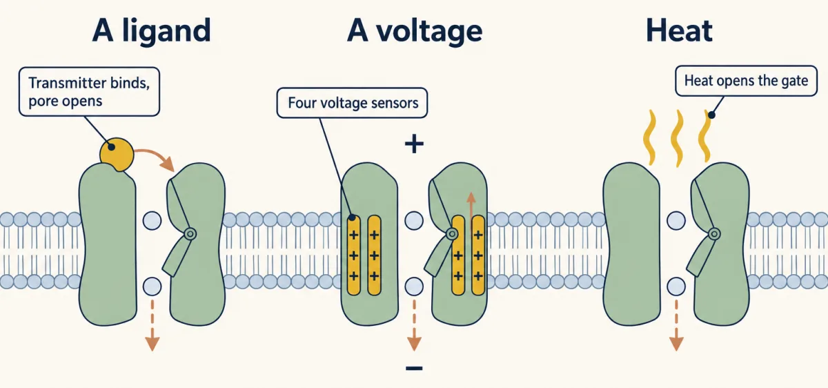
The gate is what makes a channel a receptor, and gates answer to three kinds of key.
A ligand from outside. The acetylcholine receptor of muscle is the model (lesson receptor-field-guide): transmitter binds and the pore opens. Glutamate-gated channels let Na+ in and excite a neuron, while GABA-A receptors pass Cl− and, in most mature neurons, hold its voltage down. Remember that “Both ionotropic and metabotropic can use ion channels.” An ionotropic receptor is the channel. A metabotropic receptor is a G-protein-coupled receptor that reaches a channel through a messenger inside the cell. You know four channels opened from inside: the heart’s potassium channel by Gβγ (lesson gpcrs-and-g-proteins), the rod’s channel by cGMP (lesson senses-and-cgmp), and the inositol trisphosphate (IP3) receptor and Orai (lesson calcium-signaling).
A voltage. Voltage-gated channels read the membrane itself. The sodium, potassium and calcium channels of this kind each have four voltage sensors, and in each one a helix called S4 carries a positive charge at about every third position. A voltage across a membrane is an electric field, and a field pushes on charge. Depolarise the membrane and S4 shifts outward and pulls the gate open.
This drives the action potential, the nerve impulse. A small depolarisation opens some sodium channels, Na+ enters, the membrane depolarises further and more channels open. That positive feedback (lesson signaling-circuits) carries the voltage toward the E of sodium within about a millisecond. Then a loop on the inner side of each sodium channel shuts the pore (inactivation), while slower potassium channels open and bring the voltage back. Current carried in by sodium at one spot spreads and depolarises the next stretch of membrane, so the impulse travels along the axon, the nerve cell’s long fibre, and the inactivated channels behind it keep it from turning back.
A stimulus from the world. “How does the skin detect heat??” With a channel of the TRP family, which cells use for sensing their surroundings. Before you read its name, work out what it has to be.
Design problem
Design a detector for dangerous heat in a nerve ending in the skin. It must turn a temperature above about 43 °C into a nerve impulse within milliseconds. What kind of protein do you need, which ions flow and in which direction, and how does that small local change become an impulse that travels? Then explain why a chilli pepper feels hot.
Saved in this browser only.
One way to do it
Milliseconds favour a direct route over a chain of enzymes, so make the detector itself a channel with a gate that heat opens. Let the pore pass cations of several kinds, calcium most readily. At a resting voltage near −70 mV, Na+ and Ca2+ are both far from their equilibrium potentials, so both flow in and the ending depolarises. That change is small and local and fades with distance, so put voltage-gated sodium channels in the membrane nearby. If the depolarisation reaches their threshold they open, and an action potential carries the message along the axon at full height.
One real version, and one of the skin’s detectors of painful heat, is TRPV1, cloned in David Julius’s laboratory in 1997 by looking for the protein that answers to capsaicin, the hot molecule of chilli peppers. Capsaicin binds in a pocket in the membrane-spanning part of the channel and opens the gate that heat opens. The nerve sends the same impulses either way, and the brain reads them as burning heat. Your mouth is no hotter.
A channel that needs two things at once, and what plugs a pore
The N-methyl-D-aspartate (NMDA) receptor is a glutamate-gated channel with a second lock, found at synapses, the junctions between nerve cells. At the resting voltage a magnesium ion from outside is drawn into the open pore and sticks there, so glutamate opens the gate and almost nothing flows. If the membrane is already depolarised, usually because AMPA-type glutamate receptors beside it have let Na+ in, the magnesium is driven out. The channel conducts strongly only when two things happen together: the cell before the synapse has released glutamate, and the cell after it is already active. (Glycine or D-serine must also be bound.) What enters is Ca2+ along with Na+, and calcium is a messenger (lesson calcium-signaling). From there a path runs to the nucleus and to memory, the subject of a later lesson.
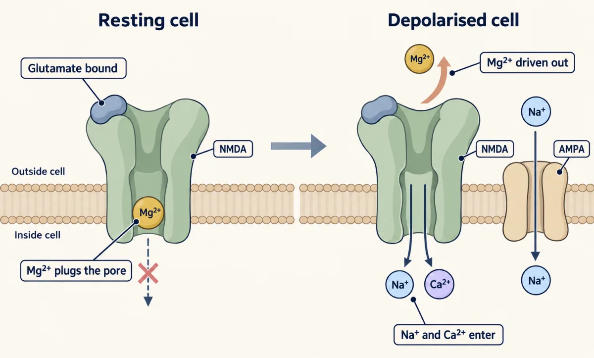
Receptors at a synapse are “Anchored in place by a bunch of different scaffolds.” At glutamate synapses the scaffold PSD-95 holds the tails of NMDA receptors with its PDZ domains (lesson protein-interaction-modules), which helps keep them opposite the site of glutamate release.
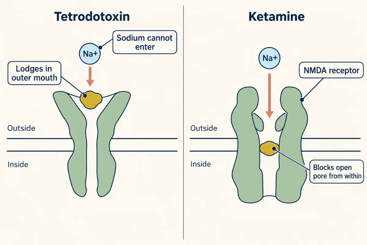
Because a channel is a hole, the simplest way to stop it is to plug it. “Pufferfish=tetrodotoxin (TTX).” Tetrodotoxin is a small molecule that lodges in the outer mouth of the voltage-gated sodium channels of nerve and skeletal muscle. Sodium cannot enter, no action potential starts, and the result is paralysis. A “UVa moment” belongs here: Edmund “Butch” Brodie of the University of Virginia studies newts that carry the toxin and the garter snakes that eat them. Resistant snakes have changed amino acids at the mouth of their sodium channel, so the toxin binds poorly.
Drugs plug pores as well. Ketamine, an anaesthetic that is also a drug of abuse, enters the open NMDA receptor channel and blocks it from within, and it also makes the channel open less often.
Channels can also be borrowed as tools: “Hacking Trp Channels”. Güler and colleagues (2012) made mice that express transient receptor potential vanilloid 1 (TRPV1) in chosen neurons, so that a dose of capsaicin switches those neurons on.
Your notes
Select any text in the lesson to highlight it or add a note to it. Your highlights and notes are saved in this browser and collected in your notebook.
This browser is not letting the site save anything (a private window or strict privacy settings), so highlights and notes will be lost when you leave the page.
How we measure it
Patch clamp
A glass pipette with a tip about a micrometre across is pressed against a cell and sealed to the membrane with slight suction. The seal has a resistance above a gigohm, so little current leaks around the rim and the picoamp current of one channel can be recorded. Variants record from the whole cell or from a patch pulled off the cell.
Intracellular microelectrode recording
A fine glass electrode is pushed into a cell to read the voltage across its membrane. Fatt and Katz used it in frog muscle at the spot where the nerve ends, and it showed the small spontaneous steps of voltage that led to the idea of transmitter packets.
Expression cloning
A library of DNA copies of a tissue's messenger RNAs is split into pools, and each pool is expressed in cells that lack the function you seek. A pool that gives the response is divided again until one clone is left. The capsaicin receptor was found this way, with a rise in calcium as the readout.
Structure determination of channels
X-ray crystallography and electron cryo-microscopy both give the positions of atoms in a channel protein. Crystallography of KcsA showed the selectivity filter. Electron cryo-microscopy, which needs no crystal, gave the structure of TRPV1.
Practice questions on this lesson (3) → MCAT-style, original, each with an explanation
Where this lesson fits
Before thisReview first
- A Field Guide to ReceptorsThis course · Lesson 4
It introduces the acetylcholine-gated muscle channel that this lesson uses as the model of a ligand-gated pore.
- Calcium: The Messenger Kept at Arm's LengthThis course · Lesson 15
Calcium as both an ion and a messenger, with Orai and the IP3 receptor, is assumed when Ca2+ enters through channels here.
- Sensing the World: Vision, Smell, Nitric Oxide and cGMPThis course · Lesson 17
The rod's cGMP-gated channel is one of the channels opened from inside the cell that this lesson names.
After thisStudy next
- Signaling to the Nucleus: CREB, AP-1 and Immediate Early GenesThis course · Lesson 32
Calcium entering through the NMDA receptor sets off a path to the nucleus and immediate early genes.
- Long-Distance Signaling in NeuronsThis course · Lesson 37
The depolarisation and impulse that channels create here must be carried along an axon to reach the cell body.
Elsewhere on the siteFilms, simulations and method films in other courses
- The length constantSimulation · Neuroscience
Resting voltage here comes from the balance of potassium and sodium gradients, which you need to predict ion direction.
- The Spike and Its PuzzlesFilm · Neuroscience
Voltage-gated sodium and potassium channels open and close in sequence to produce the spike.
- Two Pushes, One HillFilm · Cell Biology
It shows how concentration difference and membrane voltage together set the direction of ion flow.
- Why Capsaicin Feels HotFilm · Neuroscience
Capsaicin opens the heat-sensing TRPV1 channel, the same protein as the chilli and hot-stove example.
Suggested by an AI model from the lessons themselves, then checked. How we use AI
Finish line
Test yourself
8 questions
Practice and MCAT-style, with explained answers.
Next · Lesson 31
Nuclear Receptors: Receptors That Are Transcription Factors
What does a cell do with a hormone that goes straight through its membrane?
Continue →