Sensory Systems · Unit 5: Vision
Calcium Feedback Sets Photoreceptor Gain, and When Photoreceptors Die
Rewritten on : new text, flashcards and film. Your notes and highlights stay, but the text around them may have moved.
Delete the GCAPs, proteins that turn a rod's gain down, and its response to a single photon becomes about five times larger. Yet cones lacking the GCAPs and recoverin lose sensitivity more steeply than normal cones as a steady background brightens. How can removing gain control make a cell adapt worse in the light?
Pause when the reading reaches a film, and offer to play it. Saved on this device.
Watch the film now, or keep listening
Builds on:Five Ways to Turn a Stimulus into a CurrentParallel Channels Out of the EyeWhy these?
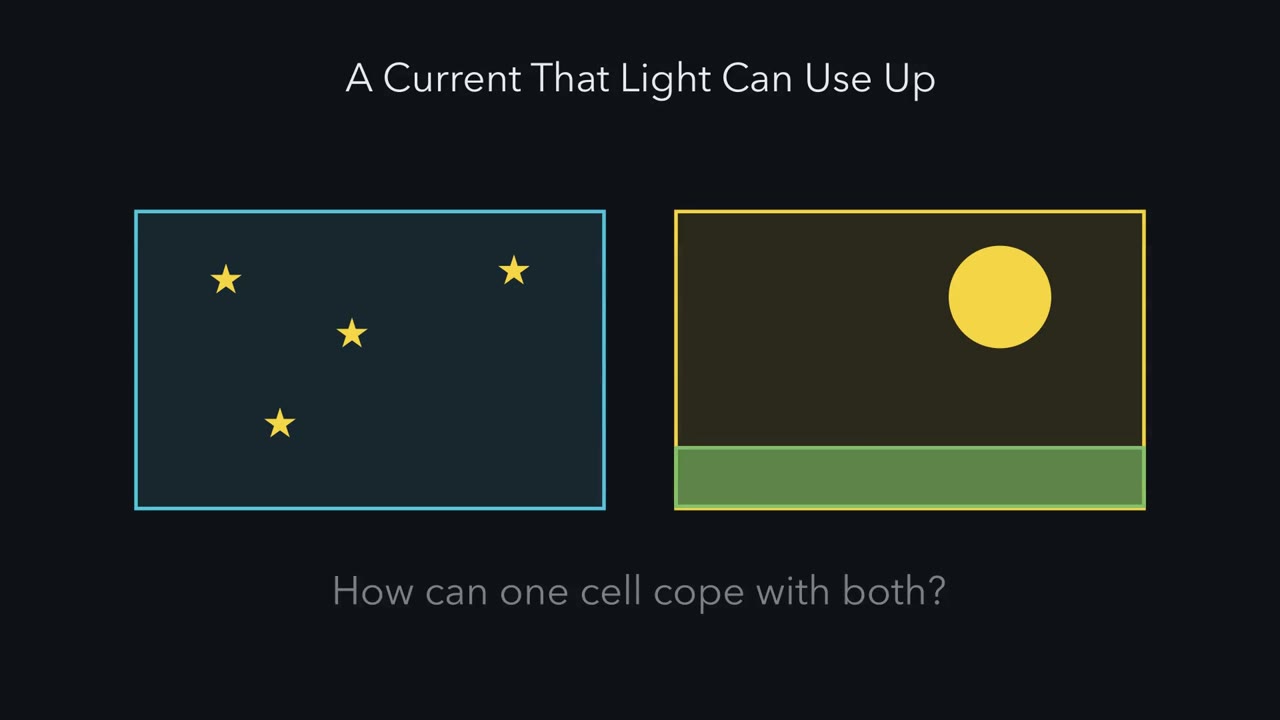 ▶ Watch A Current That Light Can Use Up · 2:52
Read the transcript of “A Current That Light Can Use Up”
Film transcript: A Current That Light Can Use Up
Your eyes work in starlight and in bright sun. How can one kind of cell cope with both? Between starlight and sunlight, the light changes a hundred-million-fold or more. Yet a dark-adapted rod goes from barely responding to saturated over about a hundredfold range of flashes. In darkness, cGMP holds channels open in the outer segment. A steady dark current flows in. Light activates PDE, which breaks down cGMP, and channels close. Each response switches off part of that current. So light spends the current. What is still flowing limits how big the next response can be. Two terms. Gain is how much current one absorbed photon switches off, and for how long.
Sensitivity is the response to a dim flash, divided by the strength of that flash. Predict. Suppose a rod's gain never changed. What would a modest steady background do to it? It would close nearly every channel. The rod would be saturated, unable to signal any further brightening. Only dimming the light would let current return. So a photoreceptor turns its own gain down as the background brightens. Calcium feedback helps. While the light stays on, it reopens some channels, keeping current in reserve. It also cuts and shortens each new response. Each photon does less. The result is called Weber behaviour. On brighter backgrounds, sensitivity falls roughly as one over the background.
Contrast is a change in light, as a fraction of the background. The payoff. On bright backgrounds, take small, brief flashes of the same contrast and the same duration. Their photon count grows with the background, while sensitivity falls just as fast. So their peak responses stay about the same. Now remove the GCAPs. In darkness, a rod's response to one photon gets larger. But on a background it keeps less current in reserve. Its sensitivity falls more steeply. This picture comes mainly from mouse rods and cones. A bright enough light can still saturate them. Pigment bleaching, light using up visual pigment, and other mechanisms extend the range in bright light.
One feedback loop does most of this work. The test is to cut it. The takeaway. Light spends a limited current, and calcium feedback keeps some in reserve.
From the film
- Light switches off part of a photoreceptor's dark current, so the current still flowing limits how big the next response can be.
- Calcium feedback turns the gain down as the background brightens, keeping current in reserve and trimming each response, mainly in mouse rods and cones.
- Over a range of bright backgrounds, sensitivity falls roughly as one over the background, so the peak response to a small, brief flash of fixed contrast and duration stays about the same.
Delete the GCAPs, proteins that turn a rod’s gain down, and its response to a single photon becomes about five times larger. Yet cones lacking the GCAPs and recoverin lose sensitivity more steeply than normal cones as a steady background brightens. How can removing gain control make a cell adapt worse in the light?
The retina lesson gave you phototransduction and the calcium loop, and senses and cGMP gave you the cascade and its shutoff. Sensory coding introduced adaptation as a property of any sense.
This lesson asks two separate questions. The first is how a photoreceptor keeps working as the background light changes enormously: the problem gain control solves, what knockout mice revealed about the calcium loop that supplies it, how to read its effect on an adaptation curve, and how rods and cones divide the range of light. The second is what happens when photoreceptors die, and what gene-therapy trials can claim.
Why a photoreceptor must turn its own gain down
In darkness, cGMP holds the outer segment’s cyclic-nucleotide-gated (CNG) channels open, and a steady inward dark current flows. Light activates phosphodiesterase (PDE), PDE breaks down cGMP, and channels close. Each light response is a piece of that current switched off. In that sense light spends the current: the current still flowing limits how big the next response can be.
Three terms carry the argument.
- Gain is how much current one absorbed photon switches off, and for how long.
- Sensitivity is the response to a dim flash divided by the strength of that flash. Gain is a property of the cell; sensitivity is how you measure it.
- Contrast is a change in light as a fraction of the background. A brief flash of the same contrast on a brighter background is a bigger flash.
That sets a problem. Between starlight and sunlight the light changes a hundred-million-fold or more. A dark-adapted rod’s responses to flashes go from barely detectable to saturated over roughly a hundredfold range of flash strength. If a rod’s gain never changed, a modest steady background would close nearly every channel. The rod would be saturated: with almost no current left to switch off, it could not signal any further brightening. Only dimming the light enough would let current return.
The one idea to carry through this lesson. A photoreceptor has a limited dark current, and light spends it. Calcium feedback turns the gain down so that a steady background switches off less of the current than it otherwise would. While the light stays on, the feedback reopens channels, keeping current in reserve, and it cuts and shortens each new response. The result is Weber behaviour: over a range of bright backgrounds sensitivity falls roughly as 1/background, while the photons in a small, brief flash of fixed contrast and duration grow in proportion to the background, so the two cancel and its peak response stays about the same. Remove the GCAPs and a rod’s single-photon response in the dark is larger, but on a background the cell keeps less current in reserve, so its sensitivity falls more steeply.
The idea covers steady backgrounds in mouse rods and cones recorded ex vivo, where most of the evidence comes from, over the range where pigment bleaching (bright light using up visual pigment faster than it is regenerated) is minor. The feedback has limits: a bright enough background can still saturate the cell, and the idea does not claim that this loop alone covers all of daylight. Other mechanisms, including bleaching itself, extend the range.
The loop: two sensors, two jobs
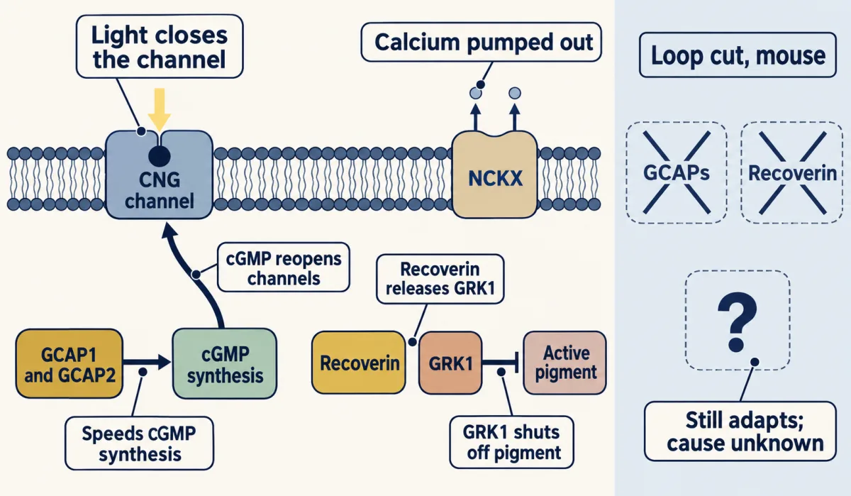
Calcium enters the outer segment through the open cyclic-nucleotide-gated (CNG) channels, and a sodium-calcium-potassium exchanger (NCKX) pumps it out all the time.
Predict. Light closes CNG channels, and NCKX keeps pumping. Which way does outer-segment calcium move? And if the cell is to win back some of the current the light has switched off, what should that calcium change do to cGMP synthesis?
Check your prediction
Calcium falls, because entry drops while extrusion continues. To win back current, falling calcium should speed cGMP synthesis, because more cGMP reopens channels. That is what the first of the two main calcium sensors does.
At low calcium, two main calcium-sensing proteins change state.
- GCAP1 and GCAP2, the guanylyl cyclase activating proteins, speed cGMP synthesis; the GCAPs can change the cyclase’s activity up to about 20-fold. This cGMP route is the strongest of the calcium feedbacks.
- Recoverin lets go of GRK1, the kinase that switches off activated visual pigment, so the pigment is shut off sooner.
A third feedback acts on the CNG channel itself; in mouse rods it is small.
Each arrow opposes the light, and that does two jobs. During a steady background the feedback reopens channels, giving back current that would otherwise stay switched off: the reserve, which keeps the cell out of saturation over a wider range of backgrounds. During any new flash it pushes against the change, so each absorbed photon closes less current, for less time: the lower gain, part of what you measure as lower sensitivity. Reviews of the field credit this feedback with most of the shift in sensitivity that lets rods and cones work over a wide range of backgrounds.
Cut the loop
The test of a loop is to cut it. Mendez and colleagues (2001) studied mouse rods lacking both GCAP1 and GCAP2, in which the calcium feedback on cGMP synthesis is gone.
Predict. (a) In darkness, is the response of these rods to a single photon larger or smaller than normal? (b) On a steady background, does their sensitivity fall at a brighter or a dimmer background than normal?
Check your prediction
(a) Larger. Without the GCAPs, cGMP is restored more slowly during the flash, so each photon closes more current for longer; pigment and PDE shutoff still work. Single-photon responses in rods lacking both GCAPs were about five times larger than in controls, and responses recovered far more slowly.
(b) Dimmer. Without the GCAPs, the cell can do less to win back current during a background, so it runs short of current sooner, although channels can still reopen through the mechanisms that remain. Adaptation to background light was severely compromised in those rods. In cones lacking the GCAPs and recoverin (Vinberg and Kefalov, 2018), sensitivity fell more steeply than in controls as the background brightened. Their fitted half-sensitivity background was about a quarter of the control value, though that shift was not statistically significant.
| Response to one photon in darkness | Sensitivity on a background | |
|---|---|---|
| Normal | smaller and brief: the feedback curbs it | falls roughly as 1/background over a range of bright backgrounds (Weber); some current held in reserve |
| Loop cut | about five times larger and slow (rods lacking GCAPs) | adapts poorly (rods lacking GCAPs); falls more steeply (cones also lacking recoverin); less current held in reserve |
So removing the gain-lowering proteins makes a rod’s response larger in the dark and the cell’s adaptation worse on a background. Both results come from the same feedback: in darkness only its trimming of each response can show, and on a background its role in holding current in reserve also comes into play.
Cones lacking the GCAPs (Sakurai and colleagues, 2011) also give larger, slower responses and adapt less well, though cyclase feedback matters less in cones than in rods. Deleting recoverin acts at the other arrow, and its effect runs the other way: in both rods and cones it speeds recovery in darkness and on dim backgrounds, as expected if recoverin normally slows pigment shutoff.
Cutting the loop does not end adaptation altogether. When GCAP1, GCAP2 and recoverin are all deleted, mouse rods and cones still show some adaptation, so other mechanisms add to this loop. What supplies the remainder is unresolved (the experiments and their limits are in Going deeper).
Reading an adaptation curve: half-sensitivity background and slope
As the background brightens, sensitivity falls. The mouse cone study of Vinberg and Kefalov (2018) fitted this with a modified Weber-Fechner relation, where S is the flash sensitivity on a background of intensity I, and Sdark is the sensitivity in darkness:
S(I) / Sdark = 1 / (1 + (I / I0)n)
I0 is the half-sensitivity background: when I equals I0, the bracket is 1 + 1 and sensitivity is 0.5 of its dark value. The exponent n sets the slope. On a log-log plot, at backgrounds well above I0, the curve falls with slope minus n. With n = 1 and backgrounds well above I0, sensitivity is roughly proportional to 1/I. Well below I0, sensitivity stays near its dark value.
The slider snaps to 12,100 and 48,800, the two I₀ values.
- Control cones, S/Sdark
- 0.500
- Cones lacking GCAPs and recoverin (fit), S/Sdark
- 0.110
- Ratio, control ÷ deficient (flash strength needed for a fixed-size response, each against its own dark sensitivity)
- 4.55
- Slope on the log-log plot well above I₀ (control; deficient)
- −1.0; −1.5
In this sketch: the curves are computed from the lesson’s fit, S/Sdark = 1/(1 + (I/I₀)n), using central fit values with no uncertainty carried through (control I₀ = 48,800 photons µm⁻² s⁻¹, n = 1.0; cones lacking GCAPs and recoverin I₀ = 12,100, n = 1.5). Isolated mouse retina 2 to 3 months old, 505-nm light, Gnat1 knockout background (Vinberg and Kefalov, 2018). The lesson does not give the range of backgrounds that was tested, so the whole curve is labelled fit extended.
Rule to keep: I₀ is the background that halves sensitivity and n sets the slope; each sensitivity is relative to its own dark value, so the ratio is not a ratio of absolute thresholds between genotypes, and the fit does not name the residual mechanism (the low-calcium bath is not a clamp of outer-segment calcium).
Predict. Take n = 1 and backgrounds well above I0. Present the same contrast, say a brief flash 10% brighter than the background, first on one background and then on a background ten times brighter. Is the response larger, smaller or the same?
Check your prediction
About the same. Keep the flash small and its duration T fixed. A brief flash of contrast C on a background I then adds C × I × T photons per square micrometre, and its peak response is about sensitivity times flash, S × C × I × T. With S proportional to 1/I, the I cancels, so for a fixed duration the response depends on C alone. That is Weber behaviour. With n above 1, sensitivity falls faster than Weber’s rule says, the I no longer cancels, and the response to a fixed contrast shrinks as the background brightens.
Now put the two published cone fits through that arithmetic. Control cones had I0 = 48,800 photons per square micrometre per second and n = 1.0. Cones lacking GCAPs and recoverin had I0 = 12,100 and n = 1.5. The knockout’s quarter is the ratio of central values, 12,100/48,800, about 0.25; unlike that shift, the steeper slope was statistically significant. Within one genotype, (S/Sdark) × I tracks the peak response to such a flash, so it shows whether contrast responses hold up as the background brightens.
| Background (photons per µm² per s) | Control cones: (S/Sdark) × I | Deficient cones: (S/Sdark) × I |
|---|---|---|
| 48,800 | 24,400 | about 5,400 |
| 106 | 46,500 | 1,330 |
| 107 | 48,600 | 420 |
The paper tested backgrounds up to about 400,000, so the 106 and 107 rows show what the fitted equations do beyond the data, not measurements.
For control cones the product rises toward a ceiling equal to I0, 48,800, so once the background is well above I0 a small contrast flash gives about the same response. For the deficient cones it falls, by about threefold for each tenfold brightening well above I0 (1,330 to 420 from 106 to 107). A fall steeper than Weber’s rule fits a cell whose background switches off current faster than the remaining mechanisms win it back, but the slope alone does not measure how much current is left.
Worked problem: two kinds of mouse cone at one background. Using the fit values above, take a background of I = 48,800 photons per square micrometre per second.
- Control: I/I0 = 1, so S/Sdark = 1/(1 + 1) = 0.500.
- Deficient: I/I0 = 48,800/12,100 = 4.03. Raise it to the power 1.5: 4.031.5 = 8.10. So S/Sdark = 1/(1 + 8.10) = 0.110.
- Compare: the control cones keep 0.500 of their own dark sensitivity, the deficient cones 0.110. The deficient cones keep 0.500/0.110 = 4.55 times less (they have lost about 89%, against 50%).
Each sensitivity is normalized to its own genotype’s dark sensitivity, so 4.55 is not a ratio of absolute thresholds between genotypes. It does set a break-even point. The deficient cones are less sensitive in absolute terms at this background unless they started more than about 4.5 times more sensitive than controls in darkness; at 106, beyond the tested range, that break-even is about 35 times. This lesson does not give the two genotypes’ dark sensitivities, so the absolute comparison stays conditional. The inputs are central fit values with no uncertainty carried through (the paper gives them as means with standard errors), and both the ratio and the sensitivities are dimensionless.
Rods and cones divide the range, and rods in daylight
Rods are the more sensitive cells: one absorbed photon gives a rod a clear electrical response. Hecht, Shlaer and Pirenne inferred this in 1942 from human threshold flashes: because so few absorptions were spread over about 500 rods, they concluded that a single quantum is enough to excite a rod. Recordings from single toad rods later showed it directly (Baylor, Lamb and Yau, 1979). Cones are less sensitive but faster. A biochemical comparison of each phototransduction reaction in purified carp rods and cones (reviewed by Kawamura and Tachibanaki, 2022) found that the reactions that generate a response are somewhat less effective in cones, while the reactions that end and recover a response are much more effective. That combination gives cones lower sensitivity and briefer responses. Rods serve dim light; cones serve daylight, colour and sharp vision.
A standard story says rods saturate once background light is bright enough, and cones then carry vision. Earlier in this course, retinal cell types and intrinsically photosensitive ganglion cells described other routes for bright-light information; here the question is whether the rod route really closes.
Tikidji-Hamburyan, Reinhard, Storchi and colleagues (2017) tested it in cone-deficient mice, recording ganglion cells in isolated retina and neurons of the visual thalamus (the dorsal lateral geniculate nucleus) in anaesthetized mice, with intact controls as well. Rod-driven responses to moderate-contrast stimuli were present at backgrounds up to 107 dark-adapted-equivalent activated rhodopsins (R*) per rod per second in isolated retina, and up to 106.35 in the thalamus in vivo. Dark-adapted-equivalent means the rate a dark-adapted rod would see at that light; once pigment bleaches, the real rate is lower.
Predict. Contrast sensitivity of the rod-driven responses fell when a bright background came on. If rods simply saturate at a fixed light level, what should happen as the background stays on? What should happen instead if gain changes and pigment bleaching keep working while the light stays on?
Check your prediction
A fixed saturation level predicts no recovery. Instead, contrast sensitivity recovered with continued exposure, and recovery was faster at higher backgrounds. A phototransduction model suggested that changes in gain and bleaching adaptation, the loss of sensitivity as pigment is bleached until it is regenerated through the visual cycle, contribute at high backgrounds. Bleaching removes pigment, so fewer R* are made each second at the same light, which would let some current return.
So the rule needs revising: in these mice, rod saturation depends on how long the retina has been exposed, not on irradiance alone. The recordings came from ganglion cells and thalamus, not rod current, so the gain-and-bleaching account is a model’s inference. A recent review of this work (Reinhard and colleagues) proposes that rods are better thought of as tuned to low contrast than to low light. The evidence here is from mouse retina and thalamus, so it sets no daylight cutoff for human rods.
Adaptation lets rods and cones keep reporting contrast as the mean light changes, at the cost of reporting less about the absolute light level. That is the other half of the picture in intrinsically photosensitive ganglion cells, which combine their own melanopsin response with rod and cone input to act as the retina’s light meter for the clock and the pupil.
Read the transcript of “Cut the Loop”
Film transcript: Cut the Loop
What happens to a photoreceptor if you cut its calcium feedback? Calcium enters through the open channels. An exchanger pumps it out all the time. Predict. Light closes the channels, and pumping goes on. Which way does calcium move?
Calcium falls. Entry drops, while pumping continues. Two main calcium sensors answer. First, the GCAPs speed cGMP synthesis, up to about twentyfold. More cGMP reopens channels. This is the strongest of the calcium feedbacks. Second, recoverin lets go of GRK1. That kinase switches off active pigment, so pigment shuts off sooner. One feedback does two jobs. On a steady background, it reopens channels. That keeps current in reserve. During each new flash, it pushes against the change. Each photon closes less current, for less time. Now cut the loop. Mendez and colleagues studied mouse rods lacking both GCAPs. Predict. In darkness, is a single photon's response bigger or smaller?
On a background, does the rod fail sooner or later? In darkness, about five times bigger. Without the GCAPs, cGMP is restored more slowly, so responses recover far more slowly. On a background, adaptation was severely compromised. In cones lacking the GCAPs and recoverin, the fitted half-sensitivity point sat at about a quarter of normal. That shift was not statistically significant. But their sensitivity fell more steeply as the background brightened, and that difference was significant. So removing the feedback makes rod responses larger in the dark, and adaptation worse on a background. In darkness, only the trimming of each response can show.
On a background, the reserve comes into play too. Now the arithmetic. Take a small, brief test flash of fixed duration. Its extra intensity is contrast times background, and its photon count is that times the duration. Its peak response is about sensitivity times that photon count. Multiply sensitivity by background. For normal cones, the product climbs to a ceiling. On bright backgrounds, contrast responses hold.
For the knockout-cone fit, well above the half-sensitivity background, the product falls about threefold for each tenfold brightening. A fall steeper than Weber's rule fits a cell running short of current. But the slope alone cannot measure the current. Some adaptation survives the cut. What supplies it is still unresolved. Rods in bright light raise the same question. On a very bright background, mouse rod-driven responses dip at first. Then they recover with continued exposure. A model suggests that gain changes and pigment bleaching help. So in these mice, rods do not simply switch off in bright light. The takeaway. The feedback trims each response and keeps current in reserve.
Cut it, and the cell runs short sooner.
From the film
- Without the GCAPs, a mouse rod's single-photon response in darkness is about five times larger, and on a background its adaptation is severely compromised.
- The calcium feedback trims each response and keeps current in reserve, so cutting it leaves the cell short of current sooner.
- Some adaptation survives deleting the GCAPs and recoverin, and what supplies it is still unresolved.
A second question: when photoreceptors die
Read the transcript of “Rods First, Then Cones”
Film transcript: Rods First, Then Cones
A new question. What happens when the photoreceptors themselves die? In typical retinitis pigmentosa, the faulty gene is often not expressed in cones. Predict. If the faulty gene is not expressed in cones, should the cones be spared?
No. They die too, typically after most of the rods around them have gone. Patients typically lose night vision first, then peripheral vision. Central vision goes later, when cones fail. So cones depend in some way on rods. And cones carry daylight, colour and sharp central vision. Why cones die is not settled. One candidate is lost support from rods. Rods make a rod-derived cone viability factor, called RdCVF. In chicken cone cultures and a mouse model, RdCVF binds basigin-1. Basigin one associates with a glucose transporter, and cone glucose entry rises. Cone starvation and oxidative damage are other candidates. Can vision be rescued? Gene therapy has begun to improve vision in specific genetic forms.
One trial added a working RPE65 gene. It was randomized and open-label, with masked grading of a mobility test. At one year, treated participants did better than controls, by 1.6 light levels. A later study delivered editing machinery for CEP290 inside the eye. It had no randomized or sham control, only the untreated fellow eye. It recorded no dose-limiting toxic effects during follow-up, and signs of possible benefit. The fellow eye shares the person and the testing days. But practice, expectation and fluctuation cannot be fully separated from treatment. The takeaway. The design limits the claim. The randomized trial supports a benefit on its mobility test.
A small single arm supports observed safety and a preliminary signal.
From the film
- In typical retinitis pigmentosa, cones die too, usually after most nearby rods have gone, although the faulty gene is often not expressed in cones.
- Why cones die is not settled; lost rod support through RdCVF is one candidate, shown in chicken cone cultures and a mouse model, not in people.
- A randomized trial supports a benefit on its mobility test, while a small single-arm study supports observed safety and a preliminary signal only.
In typical retinitis pigmentosa (RP), a heterogeneous group of progressive dystrophies, the disease gene in many cases is not expressed in cones.
Predict. If the faulty gene is not expressed in cones, would you expect the cones to be spared?
Check your prediction
No. They die too, typically after most of the rods around them have gone (Xue and Cepko, 2024). Patients typically lose night vision first, often in adolescence, then peripheral visual field; central vision is lost later, when cones fail (Verbakel and colleagues, 2018). The order suggests that cones depend in some way on rods. Because cones carry daylight, colour and sharp central vision, keeping cones alive is a major goal of research and therapy.
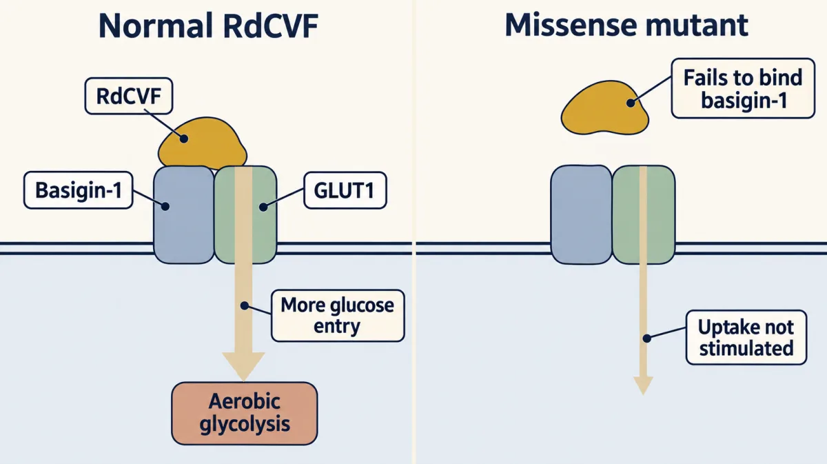
Why cones die is not settled. One candidate is lost support from rods. Léveillard and colleagues (Nature Genetics 36:755-759, 2004) screened for factors that keep cones alive and found RdCVF, the rod-derived cone viability factor, described as a truncated, thioredoxin-like protein. Aït-Ali and colleagues (Cell 161:817-832, 2015), using embryonic chicken cone cultures and the rd1 mouse, found that RdCVF binds basigin-1 on the photoreceptor surface, that basigin-1 associates with GLUT1, and that the pathway increases cone glucose entry and aerobic glycolysis. A missense mutant failed to bind basigin-1, stimulate glucose uptake or prevent secondary cone death in the model.
This is experimental work, not a human trial, and it is one proposed cause among several: Punzo and colleagues (2009) found signs that cones starve in mouse models of retinitis pigmentosa, and oxidative damage is another candidate. A gene therapy that delivers both RdCVF and its long, antioxidant form is being developed to preserve cones whatever the causative mutation; in animal models, including pigs carrying a rhodopsin mutation, it helped maintain cone structure and function. Compare the metabolic coupling in glia and metabolic partners and the wider picture in neurodegeneration and cell death.
Rescuing vision: what two gene-therapy trials can claim
Leber congenital amaurosis (LCA) typically causes profound visual impairment beginning in the first months of life, and it covers defects in phototransduction, the retinoid cycle (the pigment epithelium’s recycling of retinoids) and ciliary transport. Biallelic RPE65 disease is one retinoid-cycle form. Russell and colleagues (Lancet, 2017) ran a randomized, controlled, open-label (participants knew their group) phase 3 trial of bilateral subretinal AAV2-RPE65 gene augmentation in people with biallelic RPE65 disease (age 3 years and over) who had enough viable retina; outcome grading was masked. The outcome was a multi-luminance mobility test (MLMT), a navigation course scored by the dimmest light level at which the person passes it. At one year the between-group difference in mean bilateral change was 1.6 light levels (95% CI 0.72 to 2.41), and 13 of 20 treated participants passed at 1 lux. The US FDA approved the product, voretigene neparvovec, on 19 December 2017 according to its records, the first approved gene therapy for an inherited retinal disease. It is a genotype-specific proof that adding a working gene can improve vision. A 2026 meta-analysis of post-approval studies found improvements in full-field light sensitivity consistent with the trials, and also chorioretinal atrophy, a complication not seen in the trials, whose clinical importance is not yet clear.
Some retinal diseases are not suited to adding a gene copy, and genome editing inside the eye is being tested instead. Pierce and colleagues (New England Journal of Medicine 390:1972-1984, 2024) delivered CRISPR editing machinery for CEP290 IVS26 to the worse eye of 14 participants (see CRISPR and functional genomics for the tool, and sequencing and genomes for finding such variants). The study was open-label and single-arm, with no sham control, and recorded no dose-limiting toxic effects. Retinal implants and optogenetic vision restoration are for sensory prostheses.
Design problem
Take the RPE65 trial as described above. In the CEP290 editing study, a single-ascending-dose study with counts pooled across three dose levels, 6 of 14 participants improved on cone-mediated sensitivity by full-field stimulus testing and 11 of 14 improved on at least one of four outcomes. Interpret these data: which claims does each design support?
Saved in this browser only.
One way to do it
| Study | Design feature | What it controls | What it lets you claim |
|---|---|---|---|
| RPE65 augmentation | randomized control group; masked grading; open-label | practice and repeated testing (the control group goes through them too); grader bias | treated participants did better on mobility than controls at one year, by 1.6 light levels |
| CEP290 editing | single arm; untreated fellow eye; no sham | via the fellow eye: the person, testing days, some shared practice and change over time; not the procedure, eye-specific fluctuation or expectation | observed safety (no dose-limiting toxic effects recorded during follow-up) and a preliminary signal of possible benefit |
Start with what randomization buys: a control group that goes through the same practice and repeated testing, with masked grading limiting grader bias. The trial was open-label, so participants’ own expectations were not equalized. The mobility claim in the table is limited to participants with biallelic RPE65 disease and viable retina, to the MLMT as an endpoint, and to the follow-up of the trial. The trial’s secondary outcomes, such as full-field light sensitivity, need their own reading.
Now take what a single-arm study cannot control. Participants may improve with practice on the task, outcomes fluctuate naturally, and people who know they were treated may try harder or report more. The untreated fellow eye helps but is not a sham: it does not have the same procedure, the two eyes may differ in disease stage, and expectation can differ between a treated and an untreated eye. Without a randomized or sham comparison these effects cannot be fully separated from treatment, so the CEP290 numbers support observed safety and a preliminary signal of possible benefit, no more.
Next, separate the endpoints. Mobility, retinal sensitivity, acuity and patient-reported outcomes are different measurements. That 11 of 14 improved on at least one of four outcomes is a weaker statement than that 11 improved on all four, and a count of “at least one” rises with the number of outcomes. The 6 of 14 on cone-mediated sensitivity is a specific, more informative figure.
Neither study gives a prognosis, a claim of general benefit, or any advice about treatment for an individual.
Going deeper
The third feedback: the channel itself
A third calcium feedback acts on the cyclic-nucleotide-gated (CNG) channel. Calcium-bound calmodulin, and in fish cones a protein called CNG-modulin, lower the channel’s sensitivity to cGMP, so lower calcium in the light helps channels reopen.
GCAP1 in human disease
In people, dominant mutations in GCAP1 cause cone and cone-rod dystrophies; most keep the cyclase active when calcium is high, so they distort the feedback rather than remove it. That makes them a different experiment from the knockout mice in the main line, which remove the feedback.
When the sensors are deleted, some adaptation remains
Vinberg and colleagues (2015) showed the remainder first in mouse rods, recording ex vivo from isolated retina at 37 degrees Celsius with a transretinal electroretinogram (ERG), in which downstream synaptic responses are blocked so the voltage across the retina reports the photoreceptors. They compared wild-type rods with rods lacking the GCAPs, rods lacking recoverin, and rods lacking all three proteins. Calcium-dependent light adaptation persisted in the rods lacking both sensors.
Vinberg and Kefalov (2018) repeated the logic in cones. The mice also lacked rod transducin (a Gnat1 knockout), so rod phototransduction was gone and the recorded signal came from cones. All mice were 2 to 3 months old, and the flashes for the fits in the worked problem were 505-nm light. They compared control cones with cones lacking GCAP1, GCAP2 and recoverin, and they repeated the measurement with extracellular calcium lowered from 1.2 mM to an estimated 30 nM. The knockout cones still adapted to background light. The authors interpreted this as a sign of a calcium-independent component, which they did not find in mouse rods, and also reported a further calcium-dependent mechanism in the same cones. The same study reported dim-flash time to peak of 56 ms in control cones and 142 ms in the deficient cones (means, standard errors in the paper), so the deficient cones were also slower, consistent with weaker feedback.
Hold that conclusion at its real strength. The low-calcium bath is a change in the solution around the retina, not a direct clamp of outer-segment calcium, and the inference rests partly on fitting a model. The simplest calcium-independent explanation is that a steady background keeps phosphodiesterase (PDE) active, so cGMP turns over faster: its recovery after each flash is quicker and each flash changes it relatively less, a desensitization with no calcium involved. The authors modelled this effect and argued it does not account for all of the residual adaptation. No molecule has been identified for what remains. A direct test would hold outer-segment calcium fixed while the background series is run in cells lacking each candidate in turn. Calcium clamps of this kind have been used in amphibian photoreceptors, but the authors note they have not yet worked in mouse rods or cones.
A review by Vinberg and colleagues (2018) adds context. GCAP2 is also expressed in mouse cones and can drive cGMP synthesis when GCAP1 is missing, though GCAP1 dominates in normal cones. And, surprisingly given that cones adapt over a wider range than rods, calcium control of the cyclase matters less in cones, both in darkness and during light adaptation.
Rods in bright light: the recordings
The cone-deficient mice in the Tikidji-Hamburyan study were Cnga3 knockouts. Ganglion cells were recorded with a multi-electrode array in isolated retinas of mice 4.5 to 6 weeks old, ERGs were measured at 8 weeks, and thalamic recordings were made in mice about 6 to 8 weeks old. Two limits follow from the design. Photoisomerization rates cannot be turned into lux without assumptions this study does not supply. And whether human rods contribute to conscious vision in bright light would need a human test with rod-isolating conditions.
One photon: what 1942 showed and what 2016 added
A rod responding to one photon is not the same as a person seeing one. Two experiments show the gap.
Hecht, Shlaer and Pirenne (Journal of General Physiology 25:819-840, 1942) measured the least blue-green light that dark-adapted human observers could detect. Threshold flashes held 54 to 148 photons at the cornea. After correcting for losses in the eye, they estimated an upper limit of 5 to 14 photons absorbed by rods, and their frequency-of-seeing analysis gave 5 to 8 critical events at threshold. The arithmetic gives a feel for the losses: 5/54 and 14/148 are both near 0.09 to 0.10, so at most about one corneal photon in ten ended up absorbed by a rod, on their upper-limit estimates. Hecht did not show that a person detects one delivered photon. The inference was statistical, about a handful of absorptions.
Tinsley, Molodtsov, Prevedel and colleagues (Nature Communications 7:12172, 2016) delivered photons one at a time. Alipasha Vaziri conceived and supervised the study. Three trained men in their twenties, dark-adapted, chose which of two time intervals contained a heralded light event. Among 30,767 trials, 2,420 were post-selected as single-photon events. The pooled probability of a correct choice was 0.516 plus or minus 0.010, against 0.5 for chance, with P = 0.0545. A high-confidence subset gave 0.60 plus or minus 0.03. Combining the single-photon and attenuated-light data with Fisher’s method gave P = 0.014. A rough check: if the pooled figure rests on those 2,420 events, the binomial standard error is the square root of 0.25/2,420, about 0.010, and 0.516 is about 1.6 standard errors above chance. The authors also interpreted a dependence of detection on recently seen photons as a possible priming process lasting seconds, and that is an interpretation.
So the evidence for single-photon perception is real but analysis-dependent: it comes from three men, it depends on how trials were selected and analysed, and it does not show reliable awareness of every photon that reaches the cornea. It also separates steps that are easy to merge: a photon delivered, a rod absorbing it, the observer’s confidence, and the forced choice.
RdCVF: one gene, two forms
Léveillard’s screen was expression cloning: mouse retinal cDNAs tested on cone-enriched cultures from embryonic chicken retina. The short RdCVF it found is enzymatically inactive. The same gene, Nxnl1, also encodes a long form, RdCVFL, a thioredoxin-fold protein that helps protect photoreceptors against oxidative stress; it is the long form in the gene therapy above.
Trial details
In the retinoid cycle that RPE65 disease breaks, RPE65 in the pigment epithelium converts all-trans retinyl ester to 11-cis retinol.
Your notes
Select any text in the lesson to highlight it or add a note to it. Your highlights and notes are saved in this browser and collected in your notebook.
This browser is not letting the site save anything (a private window or strict privacy settings), so highlights and notes will be lost when you leave the page.
How we measure it
Ex vivo transretinal electroretinogram
An isolated retina is held in a chamber, downstream synaptic responses are blocked pharmacologically, and the voltage across the retina then reports the photoreceptor component of the light response. Genotype and bath controls separate cone from rod responses and test feedback components.
Multi-electrode array and visual-thalamic recording
An array under an isolated retina records spikes from many ganglion cells during controlled contrast stimuli, and electrodes in visual thalamus do the same in a living mouse. Together they test whether a retinal result survives in the intact visual system.
Heralded single-photon psychophysics
A source makes photons in pairs, and detecting one photon heralds its partner, so trials likely to hold a single photon can be identified. Optical losses mean many heralded photons never reach the eye (about 20% did in the 2016 study). The observer's forced choice between two intervals compared with chance. Performance depends on how trials are selected and analysed.
Expression cloning
A library of cDNAs is expressed and tested for a biological effect, here keeping cone-enriched cultures alive. The active clone is then sequenced, which identifies the factor without knowing its identity beforehand.
Practice questions on this lesson (3) → MCAT-style, original, each with an explanation
Where this lesson fits
Before thisReview first
- Five Ways to Turn a Stimulus into a CurrentThis course · Lesson 1
It sets out how a stimulus becomes a current, which this lesson assumes when light switches off part of the dark current.
- Parallel Channels Out of the EyeThis course · Lesson 18
It describes the rod and cone outputs whose gain and operating range are compared here.
After thisStudy next
- Restoring Senses: Implants, Prostheses and Optogenetic VisionThis course · Lesson 30
It continues from the gene-therapy and trial claims here to implants and optogenetic vision restoration.
Elsewhere on the siteFilms, simulations and method films in other courses
- Centre and surroundSimulation · Neuroscience
It shows how a rod's output falls as light closes its channels, the dark current this lesson's feedback protects.
- A Fall Can Carry a SignalFilm · Cell Signaling
It shows how light lowers cGMP and closes channels in rods, the dark current this feedback regulates.
- The Mark and the ReaderFilm · Cell Signaling
It shows how rhodopsin kinase and arrestin shut off a receptor, the step recoverin controls through GRK1.
Suggested by an AI model from the lessons themselves, then checked. How we use AI
Finish line
Test yourself
9 questions
Practice and MCAT-style, with explained answers.
Next · Lesson 22
The Hair Cell's Transduction Channel
A gene is required for hearing, and its protein sits at the right place. What else must be shown before it counts as the channel?
Continue →← Lesson 20 · Melanopsin Cells: Light for the Clock and the Pupil