Sensory Systems · Unit 5: Vision
Computing Motion in the Retina
Rewritten on : new text, flashcards and film. Your notes and highlights stay, but the text around them may have moved.
A cell in the eye fires to a spot moving left and stays quiet for the same spot moving right. Which neuron makes the difference?
Pause when the reading reaches a film, and offer to play it. Saved on this device.
Watch the film now, or keep listening
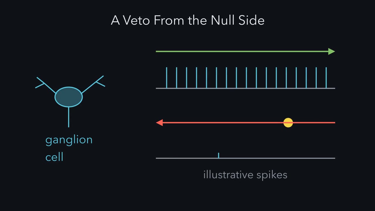 ▶ Watch A Veto From the Null Side · 2:50
Read the transcript of “A Veto From the Null Side”
Film transcript: A Veto From the Null Side
A cell in a rabbit's eye fires hard for a spot moving right. Moving left, it barely fires. How does it know? The direction that drives it is its preferred direction. The opposite way is its null direction. Barlow and Hill reported such cells in rabbit retina in 1963. Two years later, Barlow and Levick asked how it works. They flashed two neighbouring spots in the cell's field, spot A and spot B, one after the other. A then B is a small jump in the preferred direction. B then A is a jump in the null direction. If the two flashes simply added, the pair would reach the dashed line in either order. Predict. Under a veto, which order falls below the line?
Under a boost, which order rises above it? A veto leaves a deficit in the null order. A boost leaves a surplus in the preferred order. Barlow and Levick found a big deficit in the null order. The preferred order gave about the sum. So the main operation in this cell is a veto.
They also inferred that the veto is local, working in small subunits across the field. Here is a simple sketch. To cancel excitation, inhibition must overlap it in time. Null motion should start the inhibition early, so it should come from where null motion starts. Call that the null side. Preferred motion reaches the excitation first. The inhibition lags behind, so the excitation gets a head start. Barlow and Levick were tentative about which cells supply it. Most of it turned out to come from starburst amacrine cells. Its dendrites radiate like the spokes of a wheel. Each dendrite is its own small direction detector. It is most active when motion runs outward, from cell body to tip.
Take a ganglion cell that prefers rightward motion. It is wired mainly to starburst branches that point left, its null direction. Leftward motion runs outward along those branches. Their inhibition is strongest, and the ganglion cell stays quiet. Rightward motion runs inward along the same branches. They release little, so the excitation wins.
So remember the sign. The strongest inhibition comes from dendrites that prefer roughly the direction the ganglion cell ignores.
From the film
- A direction-selective ganglion cell fires to its preferred direction and little to its null direction, and Barlow and Levick found a deficit for null-order flashes, so the main operation they inferred is a veto.
- Each starburst dendrite is its own small direction detector, most active when motion runs outward from cell body to tip.
- The dendrites that give a ganglion cell its strongest inhibition prefer roughly the direction that cell ignores.
A ganglion cell in a rabbit’s retina fires hard when a spot moves one way across its field and hardly at all when the same spot moves the other way. The eye has worked out the direction before the signal reaches the brain. Which neuron makes the difference?
The retina lesson built centre-surround fields from bipolar and amacrine circuitry, and sensory coding described lateral inhibition between neighbours. Neither gave a cell a sense of direction. This lesson starts where the retina does that, one synapse before the optic nerve. Motion in the cortex belongs to visual cortex. The cell census this circuit belongs to is in retinal cell types.
The questions come in order: what the mechanism must do, which cells do it, how they are wired, how the wiring forms, and where the explanation stops.
What the mechanism must do: a veto on the null direction
Start with the cell. A direction-selective ganglion cell (DSGC) fires strongly to motion in one direction, its preferred direction, and little or not at all to motion the opposite way, its null direction. Barlow and Hill reported such cells in rabbit retina in 1963. Replacing a light spot with a black spot kept the same preferred direction, so the preferred direction does not depend on whether the spot is lighter or darker than its background.
Two years later Barlow and Levick asked how the cell does it. They recorded spikes from ON-OFF direction-selective units, which respond both when light comes on and when it goes off, in the living rabbit. In one test, they flashed two neighbouring places inside one receptive field, one after the other. Call them A and B, with A on the side that a preferred-direction stimulus comes from. A then B is a small jump in the preferred direction, and B then A is a jump in the null direction. Measure each flash alone first. If the two flashes simply added, the pair would give the sum of the two single responses in either order.
Take illustrative numbers (not Barlow and Levick’s): A alone gives 4 spikes and B alone gives 4, so simple adding predicts 8 for the pair.
Predict. For suitable positions and timing, the two orders differ, because the cell is direction selective. A cell could do that by vetoing null-direction sequences or by boosting preferred-direction ones. Which order falls below 8 under a veto, and which rises above 8 under a boost?
Check your prediction
A veto shows up as a deficit in the null order: B then A gives fewer than 8, say 2. A boost shows up as a surplus in the preferred order: A then B gives more than 8, say 14. The useful number is the remainder, the paired response minus the sum of the singles: 2 − 8 = −6 for the veto and 14 − 8 = +6 for the boost. Barlow and Levick found far fewer spikes than the sum in the null order. The preferred order gave about the sum, or a little more. The main operation Barlow and Levick inferred is a veto.
They also inferred that the veto works in small subunits spread across the receptive field, not once for the whole cell. So the specification is local. In each small patch, something must detect a null-direction sequence and cancel the excitation it would cause.
Where should that something sit? Take the simplest sketch, an offset model rather than a measurement of this circuit. Inhibition cancels excitation only while the two overlap in time. For null-direction motion to recruit inhibition early enough to overlap the excitation it causes, the inhibition should come from the side that null-direction motion approaches from. Call that the cell’s null side. For a cell that prefers rightward motion, null motion runs leftward, so it comes from the right, and the right is the null side. Preferred-direction motion reaches the excitation first, so the excitation gets a head start. How much inhibition there is matters as well as when it arrives.
- Sum of the two single flashes
- 1 + 1 = 2.00
- Paired response (this order)
- 2.00
- Remainder = paired − sum (this order)
- 0.00
- Remainder, preferred order (A then B)
- 0.00
- Remainder, null order (B then A)
- 0.00
- Reading of the two orders
- no remainder
Each flash alone counts 1, so the sum is 2.
In this sketch: single flashes add linearly, one fixed delay, a delayed veto or boost that shifts the paired response by k (the unaffected order is set to exactly the sum), relative units, no real interval or speed. Barlow and Levick’s preferred order gave about the sum or a little more. Which side carries the delayed signal is a labelling choice of this sketch; the lesson says only “one side”. The bars are computed from the rules, not recordings. The sketch does not say which cells make a negative remainder (Barlow and Levick state what the mechanism must do, not which cells), and the fly and mouse circuits reach a similar algorithm by convergent computation, not homology.
Rule to keep: measure each flash alone, add them, and subtract that sum from the paired response. A positive remainder in one order is enhancement, a negative remainder in the other order is suppression.
Barlow and Levick were tentative about which cells supply the veto, so read their paper as a statement of what the mechanism must do; the rest of this lesson is the search for those cells. (Insect vision suggested a second classical scheme, the Hassenstein-Reichardt detector, which multiplies signals and boosts the preferred direction.)
The circuit in one picture
The veto turned out to come largely from the starburst amacrine cell (SAC), an interneuron that releases both GABA, which inhibits, and acetylcholine, which excites. Its dendrites radiate from the soma like the spokes of a wheel and overlap the dendrites of direction-selective ganglion cells (DSGCs). The DSGC’s excitation comes mainly from bipolar cells, and in this picture it is about the same for either direction. Hold this picture, then test it piece by piece.
The one idea to carry through this lesson. Each starburst dendrite is its own small direction detector: it is most active, and drives the most GABA release, when motion runs outward, from the cell body to its tip. A direction-selective ganglion cell is wired mainly to the starburst dendrites that point in its null direction. So null-direction motion drives mostly the dendrites that inhibit the cell, and their inhibition overlaps and vetoes the excitation. Preferred-direction motion runs inward along those same dendrites, which release little, so the excitation wins. Remember the sign: the dendrites that give a DSGC its strongest inhibition prefer roughly the direction the DSGC ignores.
Draw it for a DSGC that prefers rightward motion, so its null side is the right. Put a starburst soma to the right of the ganglion cell, with one branch pointing left so that its tip lies over the ganglion cell’s dendrites. That is the strongly connected case; weaker connections from other branches and soma positions also occur.
| For a DSGC that prefers rightward motion | Rightward motion (preferred) | Leftward motion (null) |
|---|---|---|
| Along the left-pointing starburst branches that contact it | runs inward, tip toward soma | runs outward, soma toward tip |
| Inhibition from those branches | weak | strongest |
| The inhibition, in a simple offset sketch | lags the excitation | overlaps the excitation in time |
| Result | excitation wins; the cell fires | the veto; the cell stays quiet |
Scope. The idea is best established for the ON-OFF DSGCs of mouse and rabbit, and it describes how the adult circuit computes. It bundles two ingredients, the tuning of each branch and the placement of the inhibition on the null side; both contribute, and neither should be credited with the whole effect. It does not say how a starburst dendrite gets its outward preference (still open), how the wiring develops, how much tuned excitation, acetylcholine and timing add in some cells, or how the primate circuit is built. Later sections take these up.
Each branch is its own detector (2002)
Read the transcript of “Reading the Wiring”
Film transcript: Reading the Wiring
A starburst cell has branches pointing every way. Which ones inhibit a given ganglion cell? In 2002, Euler, Detwiler and Denk imaged calcium in single starburst dendrites, in rabbit retina. A dendrite gave larger calcium signals for motion outward from the cell body than for motion inward. Predict. The cell body sits where all the branches meet. What should its voltage show?
Little preference. Motion outward along one branch runs inward along the branch opposite it. A soma that simply summed equal spokes would see them cancel. The measured somatic voltage did not show the dendrites' preference. So the branch, not the cell, is the unit. Each branch has its own preferred direction, set by where it points. One limit here. A calcium signal is not transmitter release.
In that simple sum, input taken equally from every branch carries no direction signal. So the wiring should be selective. Paired recordings in rabbit had shown it. Branches pointing in a ganglion cell's null direction inhibited it, and preferred-pointing branches did not. In 2011, Briggman, Helmstaedter and Denk read the wiring directly, in mouse retina. First they imaged calcium during moving bars, to find each ganglion cell's preferred direction. Then they cut the same tissue for electron microscopy, and traced the cells and their contacts. Predict. Take a cell that prefers rightward motion. Which way should its contacting branches point?
Most starburst input came from branches pointing roughly left, in the null direction. And the direction of the individual branch predicted contact better than the line between cell bodies. One limit. A contact shows wiring, not synaptic strength. So the picture holds. Most starburst contacts come from branches pointing roughly in the cell's null direction.
From the film
- In rabbit starburst cells, dendritic calcium was larger for outward motion, while the somatic voltage did not show that preference, so the branch is the unit.
- In mouse retina, most starburst input to a direction-selective ganglion cell came from branches pointing roughly in its null direction.
- An electron microscopy contact shows wiring, not synaptic strength.
Euler, Detwiler and Denk imaged ON starburst amacrine cells (SACs) in isolated rabbit retina, using two-photon calcium imaging, and compared the dendrites with the voltage at the soma. Calcium signals in a dendrite were larger for motion outward from the soma, called centrifugal motion, than for motion inward.
Predict. The soma sits where all the branches meet. Each branch prefers outward motion, and outward points a different way for each branch. What should the voltage at the soma show?
Check your prediction
Little or no preference, if the soma simply summed equal spokes: motion that runs outward along one branch runs inward along the branch opposite it, so the preferences cancel. Euler and colleagues found that the somatic voltage did not show the dendrites’ directional preference under the conditions tested; they did not show that summation is the reason.
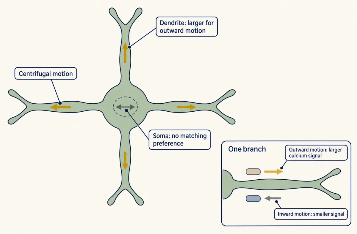
So a SAC does not have one preferred direction that its soma reports. Each branch has its own, set by where the branch points. Calcium is not release; the evidence that outward motion also gives more inhibition comes from the paired recordings of the next section. The same arithmetic shows why the wiring has to be selective: a ganglion cell that took input equally from every branch of a SAC would get no direction signal from it.
How necessary is the starburst cell? Yoshida and colleagues ablated the SAC population in adult mice with an immunotoxin-based method, then recorded ganglion-cell responses and tested the optokinetic reflex, the eye movement that follows a moving visual field. Both direction selectivity in the ganglion cells and the reflex they tested were abolished. This is necessity in that assay, and taking the whole population away removes both of its transmitters together.
Reading the wiring (2002, 2011)
Paired recordings in rabbit retina had already shown the predicted pattern in function: Fried, Münch and Werblin (2002) found that starburst amacrine cell (SAC) processes pointing in a DSGC’s null direction delivered inhibition and those pointing in its preferred direction did not, and that SAC inhibition was stronger for null-direction motion.
Briggman, Helmstaedter and Denk tested the anatomy in mouse retina. A wiring diagram alone does not say which way a ganglion cell prefers, so they read function first. In a retina from a wild-type mouse, they imaged calcium in direction-selective ganglion cells (DSGCs), using moving bars, to read each cell’s preferred direction. They then cut the same tissue with serial block-face electron microscopy, which images the face of a tissue block after each slice is removed, and reconstructed the cells and their contacts. The microscopy and images lesson covers how that stack is made.
Predict. Take a DSGC that prefers rightward motion. Which way should the starburst branches that contact it point? And which should predict a contact better: the line between the two cell bodies, or the direction of the individual branch?
Check your prediction
Left, in the null direction, and the branch, because the branch is the unit. The reconstruction showed both:
| Hypothesis | Connection should depend on | What the reconstruction showed |
|---|---|---|
| The SAC soma’s position sets the wiring | the line between the two cell bodies | a weaker predictor of connection |
| Each branch’s direction sets the wiring | which way the individual branch points | the better predictor; most input came from branches pointing roughly in the null direction |
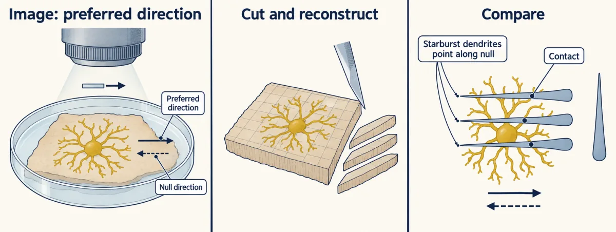
Two limits apply. Anatomy does not measure the strength of a synapse, so a contact count or area is an indirect sign of connectivity, not a conductance. And a snapshot of an adult retina says nothing about how the pattern arose. How tight must the wiring be? In a simple model (Going deeper), even fairly loose wiring around the null direction keeps a two- to threefold difference in inhibition between the two directions.
Built by strengthening, not pruning (2011)
Read the transcript of “Built by Strengthening, and Where the Idea Stops”
Film transcript: Built by Strengthening, and Where the Idea Stops
The adult wiring is lopsided. Was it built by cutting connections, or by strengthening them? Pruning would start symmetric and delete the preferred-side connections. Strengthening would keep both sides and grow the null side. Remember, the null side is the side a null-direction stimulus comes from. Wei, Hamby, Zhou and Feller recorded pairs in mouse retina. Each pair was one starburst cell and one ganglion cell. They stimulated one starburst cell at a time, and measured the inhibition the ganglion cell received. At postnatal days 4 and 7, inhibition was similar from the two sides. Predict. What should later recordings show under pruning? And what under strengthening?
Null-side inhibition grew. Preferred-side inhibition stayed about the same, and was still there in adult retina. So development strengthened one side. It did not delete the other. Later paired recordings from the same laboratory asked why. Null-side pairs had more synapses, not stronger ones. One more test. Delete the starburst cells' vesicular GABA transporter. Their GABA release drops, but their acetylcholine stays. Predict. If the veto were the whole mechanism, what would happen to direction selectivity?
Inhibition lost its direction tuning. But direction selectivity survived in a subset of ON-OFF cells. There, tuned excitation and the timing of residual inhibition supported selectivity. Direction tuning of inhibition is important, but some cells can remain selective without it.
Keep the sign. The strongest inhibition prefers roughly the direction the ganglion cell ignores. It is best established in adult mouse and rabbit. The primate circuit is less studied, and the fly uses different cells.
From the film
- In paired recordings in mouse retina, null-side inhibition grew while preferred-side inhibition stayed about constant, so development strengthened one side and did not delete the other.
- The authors concluded that null-side pairs had more inhibitory synapses, not stronger single synapses.
- When only starburst GABA release was cut in mice, inhibition lost its direction tuning but selectivity survived in a subset of ON-OFF cells, so tuned inhibition is important but not indispensable.
An asymmetric circuit could arise by pruning, deleting preferred-side connections from a symmetric start, or by strengthening, keeping both sides and making the null side stronger. Wei, Hamby, Zhou and Feller recorded pairs in mouse retina: one starburst amacrine cell (SAC) and one ON-OFF direction-selective ganglion cell (DSGC) of a labelled type whose cells all prefer the same direction. They stimulated one SAC at a time and measured the inhibitory current in the ganglion cell.
Predict. At postnatal days 4 and 7 the inhibition was similar from the two sides. What should later recordings show under pruning, and what under strengthening?
Check your prediction
| Postnatal days 4 and 7 | Later, to adult | |
|---|---|---|
| Pruning predicts | similar from both sides | preferred-side connections removed |
| Strengthening predicts | similar from both sides | null side larger; preferred side still present |
| Observed | similar from both sides | null-side unitary inhibition (from one SAC) increased; preferred-side inhibition about constant and still detectable in adult retina |
Development strengthened one side and did not delete the other.
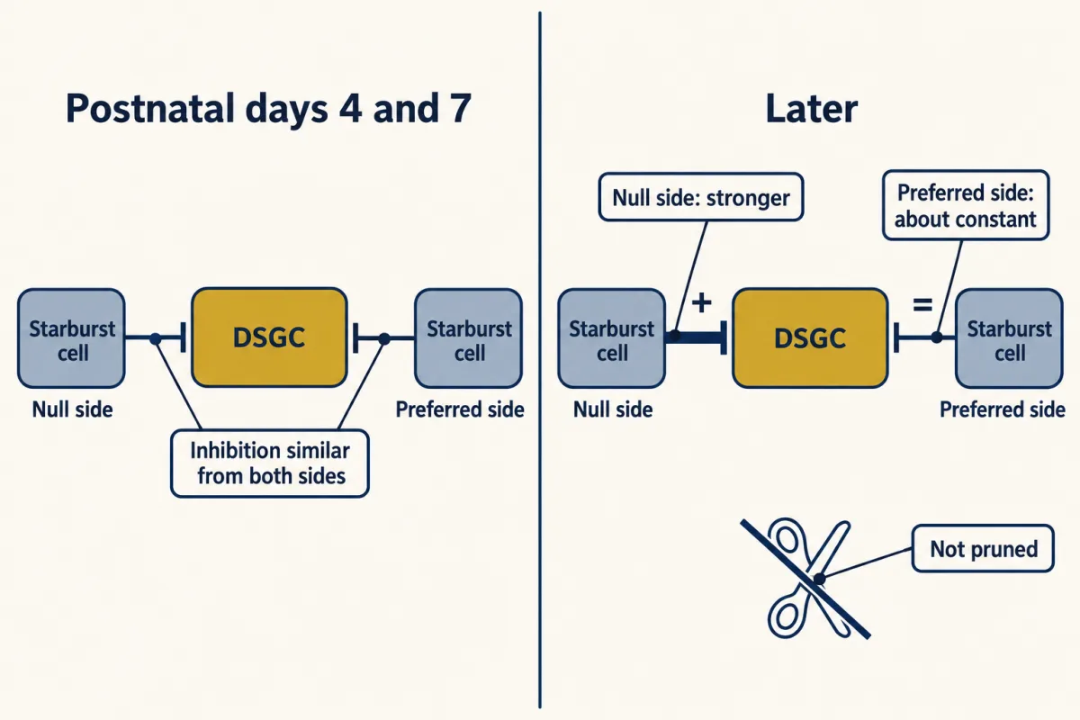
Does the strengthening need neural activity? The authors found that direction selectivity developed despite intraocular injections, during the period when the asymmetry forms, of muscimol (a GABA-A agonist used here to silence retinal activity) or gabazine (which blocks GABA-A receptors). They concluded that the null-side strengthening follows a developmental program that does not depend on neural activity, at least within that treatment window. For the activity patterns that wire the retina early on, see spontaneous activity, and for how cell types find their partners, see partners, layers and self-avoidance.
Why does the null-side connection grow? Later paired recordings from the same laboratory (Morrie and Feller, 2015) counted quantal events, single-vesicle responses to GABA released from the stimulated SAC. Larger events would mean stronger single synapses. More frequent events can mean more release sites or a higher release probability, so frequency alone does not settle it. Quantal events became more than three times more frequent for null-side pairs, with no change in their size, and the authors concluded, with further tests, that the growth comes from more inhibitory synapses per null-side pair, not from stronger single synapses.
Where the idea stops
Recall the sign from the one idea: the branches that inhibit a direction-selective ganglion cell (DSGC) most strongly prefer roughly the direction it ignores. Taking the whole starburst amacrine cell (SAC) population away abolished direction selectivity, but that removes two transmitters and every other function of the cells at once. The direct test is to remove only the GABA. In rabbit, the GABA receptor blocker picrotoxin abolished the direction selectivity of ON-OFF and ON DSGCs (Caldwell, Daw and Wyatt, 1978), though a bath-applied drug acts on every GABAergic cell.
Predict. In mouse, Pei and colleagues (2015) deleted the vesicular GABA transporter, which loads GABA into synaptic vesicles, from SACs only. That reduced their GABA release while leaving the cells and their acetylcholine. If the starburst veto were the whole mechanism, what would happen to direction selectivity? What if other mechanisms also contribute?
Check your prediction
If the veto were the only mechanism, cutting it would abolish direction selectivity in every cell; if other mechanisms can carry selectivity on their own, some would remain. Inhibition lost its direction tuning, and direction selectivity survived only in a subset of ON-OFF DSGCs, carried by tuned excitation and a timing offset: residual, untuned inhibition arriving at a different time relative to excitation, which can change how much it cancels even when its size is the same in both directions. So direction tuning of inhibition is important but not indispensable for selectivity in every tested cell. This experiment did not eliminate all SAC GABA.
Three more limits mark the edge of the idea.
How the branch gets its preference is open. One line of work points inside the dendrite: in rabbit, Hausselt and colleagues (2007) found that SAC direction selectivity persisted with GABA and glycine receptors blocked and depended on voltage-gated channels and a voltage gradient from soma to dendritic tip. Another line looks at the bipolar inputs to the starburst dendrite, and that side is under revision (Going deeper).
Excitation is not always untuned. Some DSGCs also receive direction-tuned excitation; how much is debated (Going deeper).
Other animals. In Drosophila the same job is done by T4 cells (the ON channel) and T5 cells (the OFF channel). In vivo calcium imaging of their axon terminals by Haag, Mishra and Borst found both preferred-direction enhancement and null-direction suppression in all four subtypes of each cell type, which differ in their preferred direction. The fly and the mouse reach a similar algorithm with different cells, so this is convergent computation, not homology, and no single idealised detector accounts for either circuit.
On the primate side, a textbook line says that primates lack DSGCs. That is out of date: physiological direction selectivity has been reported in macaque ON-OFF cells (Kim and colleagues, 2022) and ON cells (Wang and colleagues, 2023). The macaque ON-OFF cells form a single mosaic, one evenly tiled population, about 1.5% of peripheral ganglion cells, unlike the four ON-OFF types with different preferred directions in mouse and rabbit, so the rodent circuit should not be copied onto the primate wholesale. The 2023 human cell was identified as a candidate homolog without a human motion recording.
Worked problem (a): a direction index from a published ratio
Kim and colleagues (2022) reported, for dendritic calcium in macaque SACs (adult isolated retina, radial gratings), a mean inward-to-outward response ratio of 0.32 (SD 0.18) across 36 regions of interest on the terminal dendrites of one starburst cell. For this exercise only, normalise the outward response to 1 and define D = (Rout - Rin) / (Rout + Rin).
D = (1 - 0.32) / (1 + 0.32) = 0.68 / 1.32 = 0.515, outward-preferring
The point: a ratio of 0.32 already means a strong outward preference. This transforms a published mean ratio; it is not a published direction-selectivity index, and because the formula is non-linear it is not the mean of per-region indices, which would need each region’s own ratio. The 36 are regions of interest on one cell, not cells or animals.
Worked problem (b): when does a spike threshold sharpen the signal?
MCAT question 1 below gives a cell three times more inhibitory current for null motion than for preferred motion (120 against 40 pA). Assume the same inhibitory reversal potential and adequate voltage clamp at one holding voltage away from that reversal: the driving force is then the same, so the threefold current ratio is a threefold conductance ratio, which is what the rule below uses. How can a 3 to 1 difference in inhibition become a silent null response, and when does it not? Use the balance-point rule from the action potential and ipRGCs: when the voltage is steady and no current is injected, it sits at the average of the reversal potentials, weighted by each conductance.
Take made-up values in a passive, steady-state model (not DSGC measurements; it ignores timing). Reversal potentials: excitation EE = 0 mV, inhibition EI = −70 mV, leak EL = −60 mV. Conductances: leak gL = 1 and excitation gE = 2 in both directions; inhibition gI = 1 for preferred motion and 3 for null motion. For the nonnegative quantities used here (conductances, depolarizations from the leak potential, firing rates), compare preferred (P) and null (N) values with the index (P − N) / (P + N), defined when P + N > 0; it is positive when P > N. Do not apply it to raw membrane potentials, which are negative.
- Voltage. Preferred: (2 × 0 + 1 × −70 + 1 × −60) / (2 + 1 + 1) = −130 / 4 = −32.5 mV. Null: (2 × 0 + 3 × −70 + 1 × −60) / (2 + 3 + 1) = −270 / 6 = −45 mV.
- Indices before spiking. The inhibitory conductances give (1 − 3) / (1 + 3) = −0.50, negative because inhibition is larger for null motion; compare its size, 0.50, with the others. The depolarizations from the leak potential, 27.5 and 15 mV, give (27.5 − 15) / (27.5 + 15) = 12.5 / 42.5 = 0.29. The voltage is less selective than the inhibition: because the voltage is a weighted average, tripling one conductance does not triple its effect.
- Spikes. Let the firing rate rise in proportion to how far the voltage sits above a threshold, and be zero below it (arbitrary units).
| Threshold | Preferred rate | Null rate | Preferred : null | Spike index |
|---|---|---|---|---|
| −55 mV | 22.5 | 10 | 2.25 | 0.38 |
| −50 mV | 17.5 | 5 | 3.5 | 0.56 |
| −45 mV | 12.5 | 0 | all or none | 1.00 |
- What it shows. A threshold can turn a 3 to 1 difference in inhibition into a silent null response. In this model that happens, with the preferred response still positive, only for thresholds from −45 mV up to just below −32.5 mV. Below −45 mV both directions fire; at or above −32.5 mV neither does, and the index is undefined. With a low threshold, the spike output (0.38 at −55 mV) is less selective than the inhibition (0.50). Spike selectivity depends on where the threshold sits; it is not a general law that spiking sharpens it.
Design problem
Null-side starburst inhibition is stronger, SACs release both acetylcholine and GABA, and removing them abolished direction selectivity in the assay of Yoshida and colleagues. Does asymmetric GABAergic inhibition cause direction selectivity? Say what experiments would settle it.
Saved in this browser only.
One way to do it
Start by separating necessity from sufficiency. The ablation shows that the cells are needed, not that GABA is.
Test the transmitter directly. Remove or reduce GABA release from SACs while leaving the cells and their cholinergic output in place, and compare the result with acute, reversible silencing of the same cells. If the ratio falls but does not reach one, other mechanisms contribute. The SAC-specific deletion of Pei and colleagues, above, is the closest test so far. A genetic deletion can act through development, so the next point still matters.
Separate development from the adult requirement. A manipulation begun before about postnatal day 7 could change the wiring, and a manipulation in the adult changes signalling. The two answer different questions.
Add a control for tuned excitation. Where bipolar-bouton tuning depends partly on wide-field amacrine GABA (Going deeper), a global GABA block would remove that tuning too, while a SAC-specific removal may not. Because other work finds untuned glutamate release, measure glutamate input directly rather than assume either result.
State the species scope: rabbit, mouse and macaque DSGCs; the human cell is a candidate homolog only.
Going deeper
The Barlow and Levick preparation
Barlow and Levick recorded from unopened eyes of decerebrate rabbits (higher brain regions surgically disconnected) or lightly anaesthetized ones.
Veto, boost, and what the two-flash test measures
There are two simple ways to build a direction detector from two inputs. The Barlow-Levick scheme suppresses, with null-side inhibition vetoing the excitation as in the offset sketch above. The Hassenstein-Reichardt scheme enhances: a delayed signal from one side is multiplied with the signal from the other, so the response grows when motion runs the preferred way.
Computed from stimulus-evoked responses, baseline subtracted in matched response windows, the remainder measures the size of each nonlinearity: positive is net enhancement, negative net suppression. It does not, on its own, assign a cell to one scheme or identify a cellular mechanism. An opponent Hassenstein-Reichardt detector, which subtracts two mirror-image half-detectors, gives a surplus in one order and, in a cell with a linear term, a deficit in the other, so a deficit alone does not rule out multiplication.
The two transmitters of the starburst cell
That starburst amacrine cells (SACs) release both acetylcholine and GABA onto direction-selective ganglion cells (DSGCs) was shown in rabbit by Lee and colleagues in 2010, and the role of the cholinergic input was tested by Sethuramanujam and colleagues in 2016.
How selective must the wiring be?
A simple model makes the soma Predict box quantitative. Let each branch’s release follow 1 + k cos(motion direction − branch direction), and choose k = 0.515 so that inward release is 0.32 of outward release. That k is borrowed for illustration from the macaque calcium ratio of worked problem (a); it is not a measured release ratio for mouse or rabbit. Then let equally strong synapses, summing linearly, come from branches spread evenly within the stated angle of the DSGC’s null direction.
| Spread of contacting branches around the null direction | Null : preferred inhibition | Size of inhibition index |
|---|---|---|
| ±0° | 3.1 | 0.52 |
| ±45° | 2.7 | 0.46 |
| ±90° | 2.0 | 0.33 |
| ±180° (every branch equally; no wiring selectivity) | 1.0 | 0.00 |
In this linear, otherwise symmetric model the last row carries no direction signal, as for a summing soma, while loose wiring still gives about the 3 to 1 of the made-up currents in MCAT question 1. These are model values, not measurements.
Input kinetics onto the starburst dendrite
The EyeWire project (Kim and colleagues, Nature, 2014) reconstructed 79 OFF SACs in a mouse retinal electron microscopy volume. Contact area, an indirect measure, suggested that type BC2 bipolar cells contact proximal parts of a SAC dendrite and type BC3a cells more distal parts. Their model proposed that this arrangement, with different timing in the two inputs, could favour outward motion; that is a model prediction.
Measurements followed. Fransen and Borghuis recorded excitation in ON and OFF SACs in whole-mount retina from mice aged 1.5 to 3 months, in vitro. Distal excitation was faster and more transient than proximal excitation in both types. Srivastava and colleagues imaged glutamate on SAC dendrites directly in mouse retina in vitro and found relatively sustained proximal input. Deconvolution suggested about three times larger steady release proximally, and their constrained model gave its strongest space-time contribution below about 500 micrometres per second. They measured similar proximal and distal latencies and found the contribution of sustained and transient kinetics at low speeds. This input mechanism is a conditional contribution under revision.
Worked problem: a delay-line speed, and why it misleads
For this exercise only, take the gradients in time to peak of excitation that Fransen and Borghuis reported in mouse SACs, about 26 ms per 100 micrometres in OFF SACs and 8 ms per 100 micrometres in ON SACs, and treat them as pure delays. A pure delay line makes the signals coincide when the stimulus crosses 100 micrometres in the delay time.
OFF: 100 / 26 = 3.8 micrometres per ms, about 3,800 micrometres per second. ON: 100 / 8 = 12.5 micrometres per ms, about 12,500 micrometres per second.
At about 31 micrometres of mouse retina per degree of visual angle (a commonly used value for mouse), these are roughly 120 and 400 degrees per second, against about 16 degrees per second for the below-500 micrometres per second regime of the Srivastava model (2022). Treated as pure delays, the gradients would match only far faster stimuli. The mismatch comes from the pure-delay assumption, which ignores the difference in response shape (sustained proximal, transient distal), and is not a finding of either paper. Real latency differences depend on the radial positions and the preparation, so do not restate these values as a universal delay.
Tuned excitation: the debate
Matsumoto and colleagues found that single bipolar cells have output boutons with different preferred directions in mouse retina, and that this depended on SAC cholinergic excitation and on wide-field amacrine inhibition. The DSGCs they recorded received both direction-tuned and untuned glutamatergic input. Park and colleagues (2014) imaged glutamate arriving on ON-OFF DSGC dendrites in mouse and found no direction tuning, and they argued that apparent tuned excitation in voltage clamp comes from poor voltage control during null-direction inhibition. Either way, an upstream cell type does not make a clean feed-forward origin.
Downstream: what the cortex inherits
Hillier and colleagues reported in mice that disrupting retinal direction selectivity abolished the over-representation of posterior-motion-preferring layer 2/3 cells in primary visual cortex (V1), while other cortical motion computations persisted.
Your notes
Select any text in the lesson to highlight it or add a note to it. Your highlights and notes are saved in this browser and collected in your notebook.
This browser is not letting the site save anything (a private window or strict privacy settings), so highlights and notes will be lost when you leave the page.
How we measure it
Two-photon calcium imaging
A pulsed infrared laser excites a calcium indicator in a small volume of living tissue, so you can read calcium changes from single dendrites. Calcium rises with activity but is not a direct measure of transmitter release.
Serial block-face electron microscopy
An automated blade removes a thin slice from a block of stained tissue after each image of the block face, which gives a three-dimensional stack at synaptic resolution. Cells are then traced through the stack, and contacts between them are counted.
Paired whole-cell voltage clamp
You record from two connected cells at once, stimulate one and measure the synaptic current in the other. This gives the strength of one connection, which a population recording cannot give.
Targeted cell ablation
A genetic strategy removes one cell population from an adult animal so you can test whether it is needed for a response or a behaviour. It shows necessity in that assay only.
Practice questions on this lesson (3) → MCAT-style, original, each with an explanation
Where this lesson fits
After thisStudy next
- Melanopsin Cells: Light for the Clock and the PupilThis course · Lesson 20
It uses the same balance-point rule for conductances, applied to another ganglion cell type that reads light differently.
- Combining Senses and Predicting ThemThis course · Lesson 29
It takes motion signals further, asking how the brain weights and predicts movement signals that disagree or arise from the self.
Elsewhere on the siteFilms, simulations and method films in other courses
- One More RopeFilm · Neuroscience
Null-direction inhibition vetoing excitation follows the same rules as inhibition shunting a cell's firing.
- Shunting inhibitionSimulation · Neuroscience
It lets you see how an inhibitory input can block firing without changing voltage, the same arithmetic as the null-side veto.
- One Cell CountsFilm · Neuroscience
It shows how retinal circuits process light before the optic nerve, the setting for the starburst veto.
Suggested by an AI model from the lessons themselves, then checked. How we use AI
Finish line
Test yourself
8 questions
Practice and MCAT-style, with explained answers.
Next · Lesson 20
Melanopsin Cells: Light for the Clock and the Pupil
A mouse with no working rods or cones still sets its body clock to the day and still narrows its pupils in bright light. Which cells are measuring the light?
Continue →