Neuroscience · Unit 2: Synapses and plasticity
Synaptic Plasticity
Rewritten on : new text, figures, flashcards and film. Your notes and highlights stay, but the text around them may have moved.
A synapse that stores an association should strengthen when its own input is active at the same moment as the cell it talks to. How could one synapse know what the whole cell is doing?
Pause when the reading reaches a film, and offer to play it. Saved on this device.
Watch the film now, or keep listening
Builds on:Synaptic Transmission and Quantal ReleaseThe Resting Membrane and Passive SignalsIntegration, Inhibition and NeuromodulationWhy these?
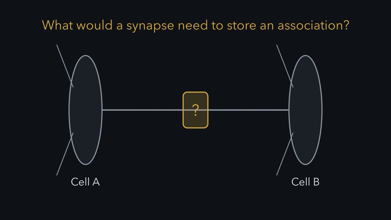 ▶ Watch A Synapse That Could Learn · 3:43
Read the transcript of “A Synapse That Could Learn”
Film transcript: A Synapse That Could Learn
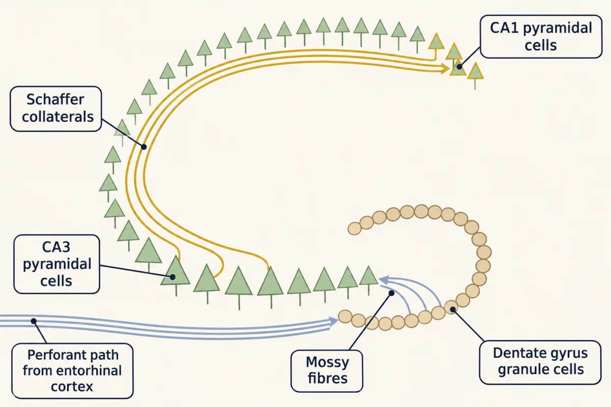
What would a synapse need, to store an association? In 1949, Donald Hebb proposed a rule. When one cell repeatedly helps fire another, the connection between them grows. Treat that as a design specification. The synapse should strengthen when two things happen together. Its own input is active. And the cell it talks to is active at the same moment. First, we need evidence that synapses change at all. Most of it comes from the hippocampus, so here is a map. Input arrives from the entorhinal cortex. Its axons, the perforant path, reach the dentate gyrus and its granule cells. The granule cells' axons, the mossy fibres, reach the pyramidal cells of CA3.
Their axons, the Schaffer collaterals, reach the pyramidal cells of CA1. Most of this story happens at that last relay. We will visit three synapses on this chain. The first is where lasting change was first measured. In 1973, Tim Bliss and Terje Lømo reported it, in anaesthetized rabbits. They stimulated the perforant path and recorded from the dentate gyrus, with an electrode outside the cells. That electrode records a field potential: an extracellular voltage made by the summed currents of many neurons. A test pulse gives two parts. A slow wave, the population EPSP, measures synaptic strength. A sharp spike riding on it shows how many cells fire, and how closely in time.
Between test pulses, they gave brief high-frequency trains, called a tetanus. One regimen was 100 pulses per second, for 3 to 4 seconds. Afterwards, the same test pulse gave a larger response, in 15 of 18 rabbits. It lasted from 30 minutes to 10 hours. This lasting rise is long-term potentiation, or LTP. How do you know the change belongs to that input? Record a second pathway to the same cells, and leave it untrained. If the preparation were drifting, that pathway would drift too. If it stays flat while the trained one rises, the change is input specific. In a made-up recording, the slope rose to 160% % of baseline. The spike rose to 250%. Is the synapse two and a half times stronger?
No. Synaptic strength is read from the slope, and it rose much less. Firing has a threshold. A modest rise in the EPSP pushes many more cells past it. So the spike grows out of proportion. To test whether the cells became more excitable, compare spike size at matched EPSP size. The record leaves three puzzles. How can a synapse detect that both cells were active together? What decides whether it goes up or down? And does memory use it?
One receptor answers the first puzzle, and offers a model for the second. At Schaffer collateral–CA1 synapses, with normal magnesium outside the cell, LTP is triggered by calcium entering through NMDA receptors. Substantial entry needs glutamate and enough local depolarization together.
From the film
- Hebb's rule asks a synapse to strengthen when its own input and the cell it talks to are active together.
- Long-term potentiation was first measured in the rabbit dentate gyrus, and a pathway that is not trained stays flat, which shows the change is input specific.
- Synaptic strength is read from the slope of the field response, and the spike can grow out of proportion to it.
A synapse that stores an association should strengthen when its own input is active at the same moment as the cell it talks to. How could one synapse know what the whole cell is doing?
In 1949 Donald Hebb proposed that when one cell repeatedly helps fire another, the connection between them grows. That is a postulate about what learning needs, not a mechanism, and it was made before anyone knew a molecule that could carry it out. This lesson follows one molecule that can, traces the experiments showing that it reads both cells, and then asks what it would take to show that memory uses it.
A synapse that could store an association
Treat the opening requirement as a design specification, with one refinement: for the receiving cell, “active” means that the spine is sufficiently depolarized, by summed synaptic input, a local dendritic event or a backpropagating spike. First we need evidence that synapses change at all, and a way to measure the change.
A map of the hippocampus
Most of the work in this lesson comes from the hippocampus, so start with a map. Picture a chain of three relays. For most of the lesson only the last one, Schaffer collateral → CA1, matters.
- Input arrives from the entorhinal cortex. Its axons, the perforant path, reach the dentate gyrus, where the main neurons are small granule cells.
- The granule cells’ axons, the mossy fibres, reach the pyramidal cells of CA3.
- The CA3 axons, the Schaffer collaterals, reach the pyramidal cells of CA1.
| Synapse | Where it appears in this lesson |
|---|---|
| Perforant path → dentate gyrus | where long-term potentiation was first measured, in rabbits |
| Schaffer collateral → CA1 | where its mechanism was worked out, in slices of rat hippocampus |
| Mossy fibre → CA3 | the exception: a potentiation that works differently |
One more synapse lies outside the chain: auditory inputs to the lateral amygdala, where the last section tests a link to memory.
The first record
Tim Bliss and Terje Lømo reported the change in 1973. They worked in anaesthetized rabbits. They stimulated the perforant path and recorded from the dentate gyrus.
Their electrode sat outside the cells, so it recorded a field potential: the voltage set up by the summed currents of many neurons at once. A test pulse gives two parts. The population EPSP (field EPSP) is the slow wave made by synaptic current flowing into many granule cells; an EPSP, an excitatory postsynaptic potential, is the depolarization one synapse’s transmitter produces. Its slope or amplitude measures synaptic strength. The population spike is a sharp deflection riding on it, made by granule cells firing together, and its size reflects how many fire and how closely in time.
Between test pulses they gave the same fibres conditioning trains. A brief high-frequency train like this is called a tetanus; one of their regimens was 100 stimuli per second for 3 to 4 seconds. Afterwards the response to the same test pulse was larger in 15 of 18 rabbits, for 30 minutes to 10 hours. They measured the population EPSP and the population spike separately, and suggested that stronger transmission and more excitable granule cells were independent contributors. This lasting rise in synaptic efficacy is long-term potentiation (LTP).
Reading the record
Worked problem, part 1. The numbers in this made-up field recording are illustrative, not data from a paper. Two pathways converge on the same cells. You measure field EPSP slope in mV/ms.
Step 1: baseline. Five test pulses on the test pathway give 0.48, 0.52, 0.50, 0.49 and 0.51 mV/ms. The mean is 2.50 / 5 = 0.50 mV/ms. Every later value is divided by this.
Step 2: after the train. Sixty minutes after a 100 Hz train the slope is 0.80 mV/ms.
percent of baseline = (value after / baseline mean) × 100
0.80 / 0.50 × 100 = 160% of baseline, which is a 60% potentiation. Do not report 160% as the potentiation.
Step 3: the control pathway. The untrained pathway was 0.51 mV/ms before and 0.50 mV/ms after: 0.50 / 0.51 = 98%. It did not move, so the rise is input specific, confined to the stimulated input, and is not a drift of the whole preparation.
Step 4: the population spike. It grew from 2.0 to 5.0 mV, 250% of baseline.
Predict. Is the synapse now 2.5 times stronger?
Check your prediction
No. Synaptic strength is read from the field EPSP, and its slope rose to 160%. Does the larger rise in the spike mean the cells also became more excitable, the component Bliss and Lømo proposed? Not necessarily. Firing has a threshold, so a modest rise in the EPSP pushes many more cells past it, and the spike grows out of proportion. These ratios alone cannot separate a stronger synapse from more excitable cells. The clean test compares spike size at matched EPSP size: if the same EPSP now gives a bigger spike, the coupling from EPSP to spike has risen, so the same drive fires more cells or fires them more in step. That can come from the cells’ own excitability, from less inhibition, or both.
Three puzzles
The record raises three puzzles.
- Coincidence. How can a synapse tell that its input was active at the same moment as the cell it talks to?
- Direction. The same synapse can also be turned down for hours. What decides up or down?
- Memory. What would show that memory actually uses these changes?
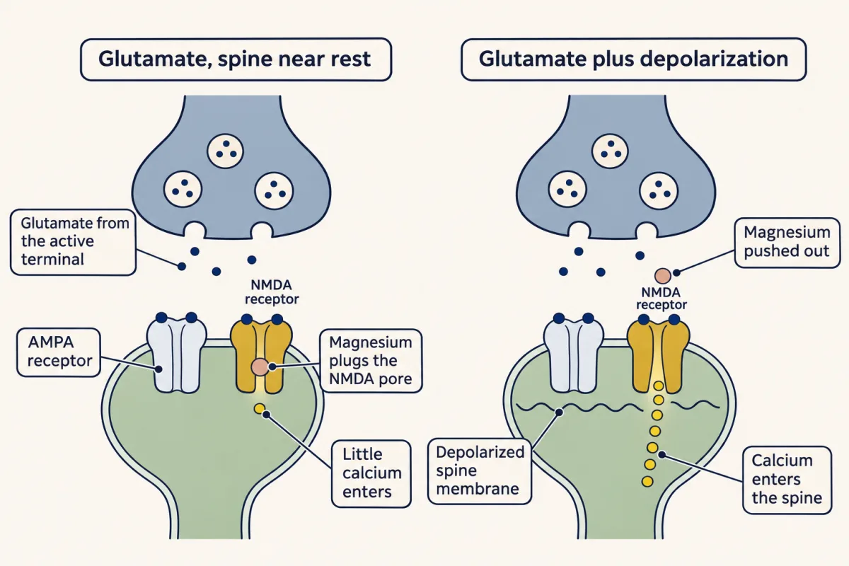
One receptor answers the first, and gives the standard model’s answer to the second. The N-methyl-D-aspartate (NMDA) receptor is a glutamate receptor. At resting potential its pore is plugged by a magnesium ion, Mg2+, and once open it lets calcium, Ca2+, into the cell. It sits on a spine, a small bud on a dendrite that holds the receiving side of one synapse.
The one idea to carry through this lesson. At an NMDA-type synapse, LTP is triggered by Ca2+ that enters the spine through NMDA receptors. At normal Mg2+ levels, substantial entry needs two signals together: glutamate from the active terminal, and depolarization of the postsynaptic membrane, which clears the Mg2+ block. In the standard model, the amount and time course of that Ca2+ also set the direction: a large, fast rise turns the synapse up (LTP), and a modest, prolonged rise turns it down (LTD). The first half, for LTP, rests on firm biophysics and experiment. The second half is a model: no single calcium threshold between LTD and LTP is established, and whether NMDA-receptor LTD needs Ca2+ flow at all is debated.
The rule’s scope: it covers NMDA-receptor-dependent LTP and LTD at Schaffer collateral-CA1 synapses and synapses like them, not every form. Harris and Cotman found in 1986 that NMDA antagonists did not block mossy-fibre LTP in guinea-pig CA3, so “LTP” names a family of changes. Oliet, Malenka and Nicoll showed in 1997, in young rats, that a second form of LTD at Schaffer collateral-CA1 synapses, triggered through metabotropic glutamate receptors instead of NMDA receptors, exists alongside the NMDA-receptor form. Short-term plasticity is a different machine, covered near the end.
The gate that needs two signals
Read the transcript of “A Gate That Needs Two Signals”
Film transcript: A Gate That Needs Two Signals
A synapse must know that its input fired, and that its own cell is active. What molecule could read both? Look at one synapse, from CA3 onto CA1. The terminal releases glutamate onto a spine, a small bud on the dendrite. Two kinds of ionotropic glutamate receptor sit there. AMPA receptors need only glutamate to open. They carry most of the ordinary EPSP. NMDA receptors also bind glutamate. They need a co-agonist too, glycine or D-serine, which is normally present. But at rest, a magnesium ion from outside sits in their pore and blocks it. Once the block clears, they pass sodium, potassium and calcium. What clears the block? Depolarization. Magnesium is positive, held in by the negative inside.
Make the inside less negative, and it is pushed out. Where does the depolarization come from? Recall the balance point from the action potential lesson. It works like a tug of war. Take an isolated compartment, with fixed ohmic conductances, and no injected or axial current. Its voltage relaxes toward a weighted average. Each current's reversal potential counts in proportion to its conductance. AMPA current reverses near 0 mV. When many receptors open at once, they pull the local balance point up from near minus −70 A spike fired by the cell and spreading back into the dendrite also depolarizes the spine. So the spine voltage reports what the postsynaptic cell is doing.
And the magnesium block reads that voltage. A spike is one source of depolarization, not the only one. Now put the signals together, as a product of three factors. First, glutamate must be bound. Second, depolarization clears the block. Third, calcium still needs an inward push. The active terminal supplies glutamate. The other two factors are set by the voltage of the postsynaptic membrane. With no glutamate, no calcium enters through these receptors, at any voltage. That makes the NMDA receptor a coincidence detector. Near rest, with glutamate bound, about one receptor in twenty is unblocked. Depolarize the spine, and about one in three is.
Glutamate arrives at a spine sitting near rest. Does any calcium get in? A little. The gate is graded, not a switch. Even at rest a few receptors are unblocked, so glutamate alone lets in a trickle. more depolarization is not always better. In this model, with normal magnesium, calcium entry through the receptor peaks near minus −20 mV, then falls. That is the third factor. Depolarization weakens the inward push on calcium. So for the same receptor, minus −30 instead of minus −70 speeds calcium entry only about four times. Where does the big difference come from? Mainly from summation. During a fast train, glutamate keeps arriving faster than the receptors close.
And the depolarization builds. The receptors stay activated while the responses summate, and the calcium in the spine climbs steeply. The takeaway: at normal magnesium levels, an NMDA receptor lets in much calcium only when glutamate and depolarization coincide.
From the film
- At normal magnesium levels, an NMDA receptor lets in much calcium only when glutamate and depolarization coincide.
- Depolarization clears the magnesium block, and the spine's voltage reports what the postsynaptic cell is doing.
- The gate is graded rather than a switch, so a little calcium enters at rest, and summation during a fast train drives the large rise.
The mechanism was worked out mostly at Schaffer collateral synapses onto CA1 pyramidal cells, in slices of rat hippocampus, where drugs reach the synapses directly. Each Schaffer terminal releases glutamate onto a spine, and two kinds of ionotropic glutamate receptor sit there.
- AMPA receptors need only glutamate to open; they have no Mg2+ block. They carry most of the ordinary excitatory postsynaptic potential (EPSP).
- N-methyl-D-aspartate (NMDA) receptors also bind glutamate, and they need a co-agonist, glycine or D-serine, which is normally present. At resting potential, a Mg2+ ion from the fluid outside sits in their pore and blocks it. Once the block clears, they pass Na+, K+ and Ca2+.
Receptor structure and gating are covered in ion channels as receptors. Keep two steps apart. Induction is what starts the lasting change (here, the NMDA receptor’s job); expression is what carries it afterwards (here, more or fewer AMPA receptors).
What clears the block is depolarization. Mg2+ is a positive ion, held in the pore by the negative inside of the cell. Make the inside less negative and Mg2+ is pushed back out.
Where does that depolarization come from? The balance-point rule from the action potential answers. In an isolated passive compartment with fixed ohmic conductances and no injected or axial current, the voltage relaxes toward the average of the currents’ reversal potentials, weighted by their conductances: a tug of war in which the rope with more open channels pulls the voltage closer to its own end. That is a simplified local balance point. A real dendrite is not isolated: current flows along it, and the voltage at a spine can differ from the voltage at the cell body. AMPA receptors pass both Na+ and K+, so their current reverses near 0 mV. When many AMPA receptors on a stretch of dendrite open at once, their conductance pulls the local balance point up from near −70 mV toward 0 mV. A spike fired by the cell and spreading back into the dendrite, a backpropagating spike, also depolarizes the spine, though less far out on the dendrite. So the voltage at the spine reports what the postsynaptic cell is doing, and the Mg2+ block reads that voltage.
Put the two signals together:
Ca2+ entry through NMDA receptors ∝ (glutamate bound) × (fraction of the block cleared by depolarization) × (inward push on Ca2+)
The active terminal supplies the first factor. The other two are set by the voltage of the postsynaptic membrane: depolarization raises the second and lowers the third. Without glutamate, no Ca2+ enters through these receptors at any voltage, and near rest the Mg2+ block keeps entry small. That makes the receptor a coincidence detector.
| Spine near rest (−70 mV) | Spine depolarized (−30 mV) | |
|---|---|---|
| No glutamate | no Ca2+ | no Ca2+ |
| Glutamate bound | a little Ca2+ (about 1 receptor in 20 unblocked) | about 4 times more (about 1 in 3 unblocked) |
Two features of the gate matter later.
- It is graded, not a switch. Even at rest a few receptors are unblocked, so glutamate alone lets in a trickle of Ca2+.
- More depolarization is not always better. Ca2+ entry is greatest at about −20 mV and falls beyond it, because depolarization also weakens the push that drives Ca2+ inward.
The table’s 4-fold gain at −30 mV is modest because about 8 times as many receptors are unblocked but each passes Ca2+ about half as hard (model constants in Going deeper). The steep difference between a tetanus and a single pulse comes mainly from summation. During a 100 Hz train, glutamate keeps arriving faster than the receptors close, so they stay activated while the EPSPs summate and the depolarization builds, and the Ca2+ in the spine climbs steeply. The enzymes downstream then respond nonlinearly to that Ca2+.
Testing the gate
Read the transcript of “Testing the Gate”
Film transcript: Testing the Gate
A rule earns trust when it predicts experiments. How would you test the two-signal gate? Work in a slice of rat hippocampus. Stimulate the Schaffer collaterals, and record from CA1. Give the input its usual fast train, 100 pulses per second. But hold one CA1 cell very negative, near minus −90 mV. Will that cell's input potentiate?
No, if the current keeps the active spines negative. Then the magnesium block stays strong, and glutamate arrives without the second signal. In 1986, Malinow and Miller found that hyperpolarizing the cell during the train prevented LTP, under their conditions. The reverse also works. Slow input, too weak to potentiate by itself, was paired with current that depolarized the cell. That pairing produced LTP. Two groups reported it in 1986. So which matters: how fast the input fires, or whether the cell is depolarized when it does?
Coincidence. High frequency is not required. A fast train is one way to make the cell supply its own depolarization. Three experiments tested the gate directly. You have just seen one. First, block the receptor. In 1983, Collingridge, Kehl and McLennan applied the NMDA antagonist APV. They used doses that abolished responses to an NMDA agonist. The baseline EPSP was unaffected. But a fast train no longer produced LTP. NMDA receptors were needed to start the change, not to carry the everyday response. Second, block the calcium. The same year, Lynch and colleagues injected the calcium buffer EGTA into CA1 cells. LTP failed at those cells. So induction needs a calcium rise inside the postsynaptic cell.
With the APV result, that points to calcium entering through NMDA receptors in active spines. One caution about scope. Not every LTP uses this gate. Mossy-fibre LTP, in guinea-pig CA3, needed no NMDA receptors. The takeaway: block the NMDA receptor, the calcium, or the depolarization, and LTP fails. Pair glutamate with enough depolarization, and it appears.
From the film
- Block the NMDA receptor, the calcium or the depolarization, and LTP fails.
- Pairing glutamate with enough depolarization produces LTP, so coincidence matters and high-frequency input is not required.
- Not every LTP uses this gate, because mossy fibre LTP in guinea pig CA3 needed no NMDA receptors.
A rule earns trust when it predicts experiments. Here is one.
Predict. Give the Schaffer input its usual 100 Hz train, but inject current into one CA1 cell to hold it hyperpolarized, near −90 mV, throughout the train. Does that cell’s input potentiate?
Check your prediction
No, provided the injected current keeps the stimulated spines sufficiently negative during the train. Then the Mg2+ block stays strong (at −90 mV only about 1 receptor in 75 is unblocked), and glutamate arrives without the second signal, so the NMDA-mediated Ca2+ signal stays below what induction needs. Malinow and Miller found in 1986 that postsynaptic hyperpolarization during the train prevented LTP under their conditions. Holding the cell body near −90 mV does not by itself guarantee the voltage at every spine: strong synaptic or dendritic depolarization can escape it. The reverse also works. Low-frequency input, too weak to potentiate by itself, produced LTP when each pulse was paired with depolarizing current injected into the cell (Kelso, Ganong and Brown, 1986; Wigström and colleagues, 1986). So high frequency is not required; what the gate needs is glutamate and depolarization together. A fast train is one way to make the cell supply its own depolarization, and frequency still shapes how much Ca2+ builds up.
Three experiments tested the gate directly.
- Block the receptor. In 1983, Collingridge, Kehl and McLennan applied the N-methyl-D-aspartate (NMDA) antagonist APV in rat slices, at doses that abolished responses to an NMDA agonist. The baseline field EPSP was unaffected, but a high-frequency train no longer produced long-term potentiation (LTP). NMDA receptors were needed to induce the change, not to carry the everyday response.
- Block the Ca2+. The same year, Lynch and colleagues injected the Ca2+ buffer EGTA into CA1 cells, and LTP failed at those cells. So induction needs a Ca2+ rise inside the postsynaptic cell. With the APV result, that points to Ca2+ entering through NMDA receptors in active spines.
- Withhold the depolarization. That was the prediction above.
Worked problem, part 2. Repeat the made-up experiment with APV in the bath. Baseline is 0.50 mV/ms. Sixty minutes after the same train, the slope is 0.51 mV/ms: 0.51 / 0.50 = 102%, within the drift of the control pathway. Induction under this protocol was NMDA-receptor dependent.
What the rule explains: specificity, cooperativity, associativity
Read the transcript of “What the Rule Explains”
Film transcript: What the Rule Explains
If calcium entry needs glutamate and depolarization together, what happens when several inputs reach one cell? Picture one CA1 cell receiving separate input pathways. You can stimulate each one on its own. Three properties were measured before the gate was known. Can the gate explain them? First, an input that stays silent during the train. Does it change? No. It released no glutamate, so its NMDA receptors let in no calcium, however depolarized the cell became. This mechanism does not potentiate it. That is input specificity. You saw it in the first recording, where the untrained control pathway stayed flat. Next, a train to just a few fibres.
It can fail to induce LTP. The same train to many fibres succeeds. The balance point shows why. A few synapses barely move the local voltage, so the gate barely moves. Many synapses together pull the balance point up, close to where calcium entry is greatest. That is cooperativity. These voltages come from a single patch of membrane. Real dendrites differ, so read the direction, not the exact values. Now take two pathways, one weak and one strong. The strong one can potentiate on its own. Stimulated alone, the weak pathway fails. Its spines get glutamate, but too little depolarization to clear the block. Now stimulate the weak pathway during the strong pathway's train.
A third pathway gets no stimulation at all. Predict each pathway's slope an hour later. The strong pathway is potentiated, as before. The weak pathway is potentiated too. It supplied its own glutamate. The strong input supplied the depolarization that cleared the block.
The third pathway stays near baseline, because it released no glutamate. That is associativity, a Hebbian rule in a dish. A spike fired by the cell and spreading back into the dendrite can play the strong input's part. This is the shape an association needs. A weak input, like a cue, gains strength when active with a strong one, like an outcome. The takeaway: one gate, reading two signals, explains input specificity, cooperativity and associativity. That holds when the inputs depolarize the right spines enough.
From the film
- One gate reading two signals explains input specificity, cooperativity and associativity.
- An input that releases no glutamate is not potentiated, however depolarized the cell becomes.
- A weak input paired with a strong one can be potentiated, because the strong input supplies the depolarization that clears the block.
Picture one CA1 cell receiving several separate input pathways, each of which you can stimulate on its own. Three properties of long-term potentiation (LTP) were measured before the N-methyl-D-aspartate (NMDA) gate was known. The product rule explains all three, as long as the inputs depolarize the right spines enough.
Input specificity. An input that was silent during the train released no glutamate, so its NMDA receptors let in no Ca2+, however depolarized the cell became. It is not potentiated by this mechanism. That is the control pathway in worked problem 1.
Cooperativity (McNaughton, Douglas and Goddard, 1978, in the dentate gyrus). A train to a few fibres can fail to induce LTP, because it cannot depolarize its spines enough. The same train to many fibres succeeds. The balance point shows why. Take the isolated compartment from above, with leak reversing at −70 mV and AMPA current reversing at 0 mV. These numbers are illustrative, and they show the direction of the effect, not the voltage at any real spine. With AMPA conductance one tenth of the resting conductance, the balance point is (1 × (−70) + 0.1 × 0) / 1.1 ≈ −64 mV.
| AMPA conductance, compared with the resting conductance | Local balance point | What the gate does |
|---|---|---|
| one tenth (a few synapses) | about −64 mV | barely moves |
| equal | about −35 mV | opening |
| twice (many synapses together) | about −23 mV | near the range of greatest Ca2+ entry |
Associativity (Levy and Steward, 1979, in the dentate gyrus; Barrionuevo and Brown, 1983, at CA1 in hippocampal slices). Now take a weak pathway that cannot potentiate on its own, and a strong one that can.
Predict. The weak pathway is stimulated alone and fails. Now it is stimulated during the strong pathway’s tetanus. A third pathway gets no stimulation at all. Predict each pathway’s slope an hour later.
Check your prediction
The strong pathway is potentiated, as before. The weak pathway is potentiated too: it supplied its own glutamate, and the strong input supplied the depolarization that cleared the block at its spines (this works when that depolarization reaches them). The third pathway stays near baseline, because it released no glutamate. This is a Hebbian rule in a dish: an input that is active while other inputs strongly depolarize the cell gets stronger.
This is the shape an association needs: a weak input, like a cue, gains strength when it is active together with a strong one, like an outcome. A backpropagating spike can play the strong input’s part, which is how a synapse can read whether its own cell fired.
Up or down: what the Ca2+ does
Read the transcript of “Turning It Up or Down”
Film transcript: Turning It Up or Down
The same synapse can be turned up or turned down for hours. What decides which way it goes? Start with up. In the standard model, the large, fast calcium rise after a tetanus recruits a kinase, CaMKII. This kinase can stay active after calcium falls. So a brief calcium signal can leave a change that outlasts it. Expression includes more AMPA receptors at the synapse. In 2000, Hayashi, Malinow and colleagues saw tagged AMPA receptors driven into synapses. More AMPA receptors mean a bigger response to the same glutamate. LTP that lasts many hours often needs new protein synthesis. Now down. In 1992, Serena Dudek and Mark Bear gave the same input a long, slow train. It was 900 pulses, at 1 to 3 Hz.
The response fell and stayed down for more than an hour, only at the conditioned input. This is long-term depression, or LTD. Now repeat the slow train with the NMDA blocker APV. Does LTD develop? No. NMDA antagonists prevented it. The same receptor that gates LTP is needed for its opposite. So what decides the direction? In the standard model, how much calcium comes through, and how fast. A modest, prolonged rise preferentially recruits protein phosphatases, rather than CaMKII. The model's reason: the phosphatase pathway responds to smaller Ca2+/calmodulin signals than CaMKII does. So a small, long signal tips toward removing receptors. A large, fast one tips toward inserting them.
In 1993, Mulkey, Herron and Malenka found that phosphatase inhibitors blocked LTD induction in rat CA1. Expression of LTD includes removing AMPA receptors by endocytosis. Keep the hedge. This calcium rule is a model. No single calcium threshold between LTD and LTP is established. Whether LTD needs calcium flow through the receptor is still debated. The takeaway: in the standard model, a synapse turns up or down with how much calcium enters, and how fast.
From the film
- In the standard model, a synapse turns up or down with how much calcium enters and how fast.
- A large, fast calcium rise recruits CaMKII and adds AMPA receptors, while a modest, prolonged rise recruits phosphatases and removes them.
- This calcium rule is a model, and no single calcium threshold between LTD and LTP is established.
Turning it up. In the canonical model, the large, fast Ca2+ rise after a tetanus recruits calcium/calmodulin-dependent protein kinase II (CaMKII), a kinase that can stay active after Ca2+ falls (see the phosphorylation switch and calcium signaling). That persistence matters: a brief Ca2+ signal can leave a change that outlasts it. Expression includes more AMPA receptors at the synapse. Hayashi, Malinow and colleagues found in 2000 that long-term potentiation (LTP) drove electrophysiologically tagged AMPA receptors into synapses in rat hippocampal neurons. More AMPA receptors mean a bigger response to the same glutamate. LTP that lasts many hours often needs new protein synthesis, and its longest-lasting forms also need new gene transcription, which signaling to the nucleus takes up.
Turning it down. In 1992 Serena Dudek and Mark Bear gave the Schaffer collateral input in rat slices 900 stimuli at 1 to 3 Hz. The population excitatory postsynaptic potential (EPSP) fell and stayed down for more than an hour, only at the conditioned input. This is long-term depression (LTD), a lasting fall in synaptic efficacy.
Predict. Repeat Dudek and Bear’s 900 slow pulses with APV in the bath. Does LTD develop?
Check your prediction
No. NMDA antagonists prevented it, so the same receptor that gates LTP is needed for its opposite. In the standard model the trigger is a modest Ca2+ signal through the receptor, though whether LTD needs that ion flow is debated (see Going deeper).
What, then, decides the direction? The second half of the lesson’s rule: how much Ca2+ comes through, and how fast. In the canonical model the synapse works like a dimmer switch with one input wire. A modest, prolonged Ca2+ rise preferentially recruits protein phosphatases, which lead to receptor removal, rather than CaMKII, which leads to insertion. The model’s reason is that the phosphatase pathway (calcineurin, a phosphatase that in turn activates protein phosphatase 1) responds to smaller Ca2+/calmodulin signals than CaMKII does.
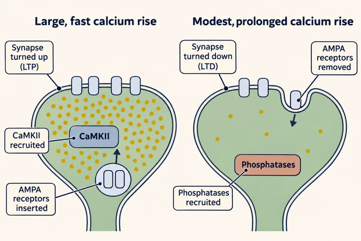
The evidence fits. Mulkey, Herron and Malenka found in 1993 that phosphatase inhibitors blocked or reduced LTD induction in rat CA1, whether applied in the bath (okadaic acid, calyculin A) or loaded into the postsynaptic cell (microcystin LR). Expression includes removal of AMPA receptors by endocytosis (see endocytosis).
Similar rules shape cortex during development, when vision rewires the visual cortex; ocular dominance and critical-period control cover that.
A different machine: short-term plasticity
Read the transcript of “Fast Changes, and the Road to Memory”
Film transcript: Fast Changes, and the Road to Memory
Some changes last milliseconds, not hours. Do they follow the same rule? No. The facilitation and depression shown here are a different machine. They need no postsynaptic coincidence, and they work mainly in the presynaptic terminal. Short-term plasticity also includes other mechanisms. Give two pulses close together. Divide the second response by the first. That is the paired-pulse ratio. Above one is facilitation. Below one is depression. The classic explanation for facilitation is residual calcium, left in the terminal from the first spike. In 2016, Jackman, Regehr and colleagues deleted synaptotagmin 7 in mice. Facilitation was lost at the synapses tested, though residual calcium was unchanged.
So at those synapses, synaptotagmin 7 is a sensor that reads the leftover calcium. Depression has a simple candidate. The first spike uses up vesicles. In a simple depletion model, with no refilling and no facilitation, the first pulse releases 40% % of the pool. Will the second response be larger or smaller?
Smaller. 60% % of the pool is left, so the second response is 60% % of the first. But a ratio like that does not prove depletion. Other processes can give the same number. Now back to Hebb. Does memory actually use LTP? A correlation, learning alongside potentiation, is not proof. In 1986, Morris and colleagues infused an NMDA blocker into rats. Place learning was impaired, a visual task was spared, and hippocampal LTP was suppressed. But the drug reached many synapses. It showed the receptors were needed for both, not that the learning was LTP. In 2014, Nabavi, Malinow and colleagues used light on auditory inputs to the rat lateral amygdala, as a cue.
After light was paired with shock, the light suppressed lever pressing. Optical LTD of those inputs removed the response. Optical LTP afterwards restored it. Is LTP at these inputs enough, on its own, to create the memory?
No. In rats never given the pairing, LTP at these inputs produced no response. Something set up by the pairing had to be there. That holds for this pathway, in rats, in this task. The takeaway: fast changes are mostly a separate, presynaptic machine. And linking LTP to memory takes tests of necessity and sufficiency, not correlation alone.
From the film
- Facilitation and depression are mostly a separate, presynaptic machine that needs no postsynaptic coincidence.
- At the synapses tested, synaptotagmin 7 is a sensor that reads leftover calcium to produce facilitation.
- Linking LTP to memory takes tests of necessity and sufficiency, not correlation alone.
Now a different machine, with a different rule. The facilitation and depression examples here need no coincidence of pre- and postsynaptic activity, mostly last milliseconds to seconds, and happen mainly in the presynaptic terminal, the one that releases transmitter (see synaptic transmission). Short-term plasticity also includes other mechanisms.
Most synapses change from one spike to the next. Give two pulses a few to a few hundred milliseconds apart and divide the second response by the first: the paired-pulse ratio. Above 1 is facilitation; below 1 is depression. Paired-pulse changes fade within about a second or a few seconds, though depression can take seconds to tens of seconds to recover and post-tetanic potentiation after a train lasts minutes.
Facilitation can raise release for hundreds of milliseconds after one spike. The classic explanation is residual Ca2+: calcium left in the terminal from the first spike adds to the second. In 2016 Jackman, Regehr and colleagues found that deleting synaptotagmin 7, a Ca2+-binding protein of the terminal, eliminated paired-pulse facilitation at the mouse synapses they tested, although the residual Ca2+ signal was unchanged. Wild-type synaptotagmin 7 restored facilitation and a mutant with a calcium-insensitive C2A domain did not, so at those synapses synaptotagmin 7 is a Ca2+ sensor that reads the leftover calcium.
Depression. Depression has a simple candidate: the first spike uses up vesicles.
Worked problem, part 3. Use the simplest depletion model, with these assumptions: no refilling between pulses, no facilitation, a fixed release fraction F and a fixed current per vesicle i. Let F = 0.40, a releasable pool S = 100 vesicles and i = 10 pA. These are teaching values.
EPSC1 = F × S × i
Predict. Will the second response be larger or smaller than the first, and by what ratio?
Check your prediction
Smaller. EPSC1 = 0.40 × 100 × 10 pA = 400 pA. The first pulse releases 40% of the pool, leaving 100 × (1 − 0.40) = 60 vesicles, so EPSC2 = 0.40 × 60 × 10 pA = 240 pA. The ratio is 240 / 400 = 0.60, which equals 1 − F: a 40% depression.
Now the limit. A measured ratio of 0.60 does not prove depletion, since other processes can give the same number, and a simple depletion model cannot explain short-term depression at many synapses.
- EPSC1 = F × S × i
- —
- Pool left after pulse 1
- —
- EPSC2 = F × pool × i
- —
- Ratio EPSC2 / EPSC1
- —
—
In this sketch: the simplest depletion model, no refilling between pulses, no facilitation, a fixed release fraction F and a fixed current per vesicle (i = 10 pA); pulses 3 onward simply apply the same rule again, the lesson works only a pair; the F and S ranges are teaching ranges and all values are teaching values, vesicle numbers are averages, and the dots are rounded to whole vesicles.
Rule to keep: in this model the ratio EPSC2 / EPSC1 equals 1 − F whatever the pool size, so a measured 0.60 (for F = 0.40) is what 40% depletion would give but does not prove depletion.
How the postsynaptic cell adds these weights is the subject of synaptic integration.
From synapse to memory: what the evidence licenses
Return to Hebb. NMDA-receptor long-term potentiation (LTP), input specific and associative, fits his rule. But fitting is not proof.
Three kinds of evidence differ in strength.
- A correlation: learning goes with potentiation at a pathway.
- A necessity test: blocking or reversing the change impairs the memory. Morris and colleagues found in 1986 that the N-methyl-D-aspartate (NMDA) antagonist AP5 infused into the ventricles of rats impaired place learning, spared visual-discrimination learning and suppressed hippocampal LTP in vivo. The drug reached many synapses, so this shows the receptors were needed for both, not that the learning was LTP.
- A sufficiency test: inducing the change produces the memory’s effect. The design problem below is an attempt at both necessity and sufficiency in one pathway.
The timing rule is also not settled. In 2017 Bittner and colleagues found that in mouse CA1, inputs arriving seconds before or after a dendritic plateau potential could be potentiated, enough to form a place field, the spot in an environment where the cell fires: a rule that works over seconds, not the milliseconds of classic Hebbian pairing. How these lessons map onto memory is the business of memory systems, plasticity, networks and consolidation and emotion and stress.
Design problem
Is potentiation at auditory inputs to the lateral amygdala needed for a conditioned fear response? Nabavi, Malinow and colleagues (2014) expressed a light-gated channel in those inputs in rats and used light as the cue. After optical cue-shock pairing, the light suppressed lever pressing. Optical long-term depression (LTD) of the inputs abolished the response at testing. Optical LTP given afterwards restored it. LTP in rats never given the pairing produced no response. What do these results show, and what do they not show?
Saved in this browser only.
One way to do it
Name the scope first: rats, auditory inputs to the lateral amygdala driven by light, and a conditioned suppression of lever pressing.
LTD after conditioning removed the response. That shows the potentiated state of these inputs was necessary for expressing this response at the time of testing.
LTP after LTD brought the response back. The association was not simply lost; the pathway could be reactivated, which argues that LTD turned the response down rather than erasing everything learned.
LTP in rats never paired produced nothing. So LTP at these inputs is not sufficient to create the association on its own; something set up by the pairing had to be there.
What the data do not show: that the whole memory lives at one set of synapses, that LTD destroyed information (the restoration argues against it), or that the same holds for other fear tasks or for people. (In a separate experiment, the paper also showed that optical LTD right after tone-shock pairing reduced the tone-evoked response, in rats with the opposite amygdala ablated.) A good next control is to test whether a different set of inputs, given the same LTD, leaves the response intact.
Going deeper
The first observation, and the variable signs of LTP
Lømo first saw the effect in 1966, in the same rabbit preparation, in Per Andersen’s laboratory in Oslo, so 1973 is the date of the full report, not of the first observation. The 1973 rabbits were under urethane anaesthesia. Potentiation did not always look the same: the population excitatory postsynaptic potential (EPSP), the spike size and the spike latency (the delay from stimulus to spike) all improved together in only 29% of experiments, and a shorter spike latency was the commonest sign.
Who first described the Mg2+ block
In 1984 two groups reported the voltage-dependent Mg2+ block of N-methyl-D-aspartate (NMDA) receptors: Nowak, Ascher and colleagues in mouse central neurons, and Mayer, Westbrook and Guthrie in mouse spinal cord neurons.
The co-agonist
The NMDA receptor’s co-agonist binds its GluN1 subunit. In rat CA1, D-serine becomes the main co-agonist at synaptic receptors as the synapses mature, taking over from glycine.
The numbers behind the gate
The fractions in the gate table come from the standard Mg2+-block function of Jahr and Stevens (1990) at 1 mM external Mg2+, combined with a Goldman-Hodgkin-Katz Ca2+ flux at 2 mM external Ca2+, 100 nM internal Ca2+ and 35 °C.
| Spine voltage | Fraction of NMDA receptors unblocked | Ca2+ entry, relative to glutamate at −70 mV |
|---|---|---|
| −70 mV | 0.044 (about 1 in 20) | 1.0 |
| −50 mV | 0.14 | 2.3 |
| −30 mV | 0.36 (about 1 in 3) | 3.8 |
| −20 mV | 0.51 | 4.2 (near the peak, −18.6 mV) |
| 0 mV | 0.78 | 3.3 |
| +40 mV | 0.98 | 0.65 (15% of peak) |
The peak moves to about −16 mV at 1.3 mM Mg2+ and −11 mV at 2 mM, so the rise-then-fall shape does not depend on the exact Mg2+ level.
Receptors cycle all the time
When Lüscher and colleagues blocked endocytosis in CA1 pyramidal cells in 1999, AMPA receptor EPSCs (excitatory postsynaptic currents, the current counterpart of an EPSP) gradually grew. The receptors are inserted and removed all the time.
Does NMDA-receptor LTD need ion flow?
The standard model treats NMDA-receptor long-term depression (LTD) as driven by Ca2+ flowing through the receptor. One line of work reported LTD that needs glutamate binding to NMDA receptors but not ion flow (Nabavi and colleagues, 2013, PNAS). Another reported that ion flow is required (Babiec and colleagues, 2014, Journal of Neuroscience). Treat the question as open.
Your notes
Select any text in the lesson to highlight it or add a note to it. Your highlights and notes are saved in this browser and collected in your notebook.
This browser is not letting the site save anything (a private window or strict privacy settings), so highlights and notes will be lost when you leave the page.
How we measure it
Extracellular field recording
An electrode placed among many neurons records the voltage set up by their summed currents. In the hippocampus a stimulus to a fibre bundle gives a slow field EPSP, which measures synaptic strength, and, if enough cells fire, a sharp population spike riding on it, which reflects how many fire and how closely in time. Reading them separately hints at a stronger synapse or a more excitable cell; comparing spike size at matched field EPSP size is the clean test.
Receptor-antagonist pharmacology
Apply a selective blocker, show that it works by abolishing the response to its agonist, and check whether it changes the baseline response. A drug that leaves the baseline alone but prevents a lasting change shows that the receptor is needed for induction under that protocol.
Paired-pulse recording
Two stimuli are given a few to a few hundred milliseconds apart, and the second response is divided by the first. A ratio above 1 is facilitation and below 1 is depression; a change in the ratio after a manipulation is often taken as a sign of a presynaptic change, though it is not proof.
Optogenetic stimulation
A light-gated ion channel expressed in a defined set of axons lets light drive them at a chosen frequency. Driving them in an LTP or LTD pattern changes those synapses on purpose, so the effect on behaviour can be tested rather than only correlated.
Practice questions on this lesson (3) → MCAT-style, original, each with an explanation
Where this lesson fits
Before thisReview first
- Synaptic Transmission and Quantal ReleaseThis course · Lesson 5
You need glutamate release and EPSPs from there to follow what a train of pulses does at the synapse.
- The Resting Membrane and Passive SignalsThis course · Lesson 3
The balance-point rule for the spine's voltage comes from how leak and synaptic conductances set the resting potential.
- Integration, Inhibition and NeuromodulationThis course · Lesson 7
Summation of EPSPs there is what depolarizes the spine enough to clear the Mg2+ block.
After thisStudy next
- Plasticity, Networks and ConsolidationThis course · Lesson 23
It takes LTP further, testing whether networks of potentiated synapses are needed and enough for stored memories.
- Memory SystemsThis course · Lesson 22
It asks which kinds of memory the hippocampus and its plastic synapses actually support.
- Emotion and StressThis course · Lesson 20
It uses plasticity at amygdala inputs to explain how a tone comes to predict a shock.
Elsewhere on the siteFilms, simulations and method films in other courses
- Quantal analysisMethod film
It shows how to tell whether a change in synaptic strength is presynaptic or postsynaptic from the sizes of quantal responses.
- The Muscle Proposes, the Nerve DisposesFilm · Developmental Neuroscience
It shows how a synapse's receptors are clustered and kept, the machinery that LTP changes by adding AMPA receptors.
Suggested by an AI model from the lessons themselves, then checked. How we use AI
Finish line
Test yourself
8 questions
Practice and MCAT-style, with explained answers.
Next · Lesson 9
Principles of Sensory Coding
A heavier weight on a frog's muscle receptor did not make bigger impulses, only more of them. How do identical spikes carry a whole world?
Continue →