Cell Biology · Unit 2: Membranes and compartments
New to this? Review it at high school level: Organelles as a System
Protein Sorting into Compartments
Rewritten on : new text, flashcards and film. Your notes and highlights stay, but the text around them may have moved.
Made in a test tube with no membranes, a secretory protein comes out longer than the one the cell secretes. Why?
Pause when the reading reaches a film, and offer to play it. Saved on this device.
Watch the film now, or keep listening
Builds on:Organelles as Distinct EnvironmentsMembrane Organization and TopologyExperiments That Distinguish MechanismsWhy these?
 ▶ Watch Where the Reader Works · 3:45
Read the transcript of “Where the Reader Works”
Film transcript: Where the Reader Works
A secretory protein made in a test tube with no membranes comes out longer than the one cells secrete. What is the extra piece, and why does the cell lose it? The extra piece is an address, a stretch of the protein's own chain called a signal sequence. A ribosome builds a chain from its amino-terminal end and hides the newest part in a tunnel. So that end, here the address, emerges first. SRP binds near the ribosome's exit and reads the address as it emerges. In the classic preprolactin experiment, translation slowed or stopped until ER membranes arrived. SRP docks on its receptor, next to Sec61, the channel into the ER. Like two hands gripping only while both hold GTP, they hand the ribosome to the channel.
The chain begins threading in, and GTP hydrolysis opens both hands. The chain enters unfolded, as it is made. Signal peptidase snips off the address: the piece the test tube kept, with no membrane. Back to the test tube. Add the membranes only after the chain is finished and released. Does the protein get inside?
In this test it stays outside, and long: a released chain gives SRP no ribosome to steer. Some ER proteins enter after release, by other routes. So here is the idea. An exposed address helps specify where a protein goes.
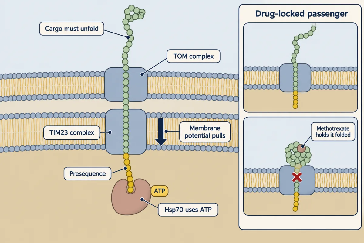
Where and how its reader works helps decide when it is read: as the chain grows, or after release. A mitochondrial address also comes out first. But its reader, TOM, the outer membrane's import gate, sits on the mitochondrion. Most of these chains are released before TOM reads them. And a ribosome sitting on the mitochondrion? Hold that question. Released chains may start to fold, though helper proteins called chaperones often keep them loose. To cross TOM and the inner membrane's TIM23 complex, a chain must be at least partly unfolded. The membrane potential, a voltage across the inner membrane, pulls on the positive address. Inside, the chaperone Hsp70 uses ATP to grip the chain.
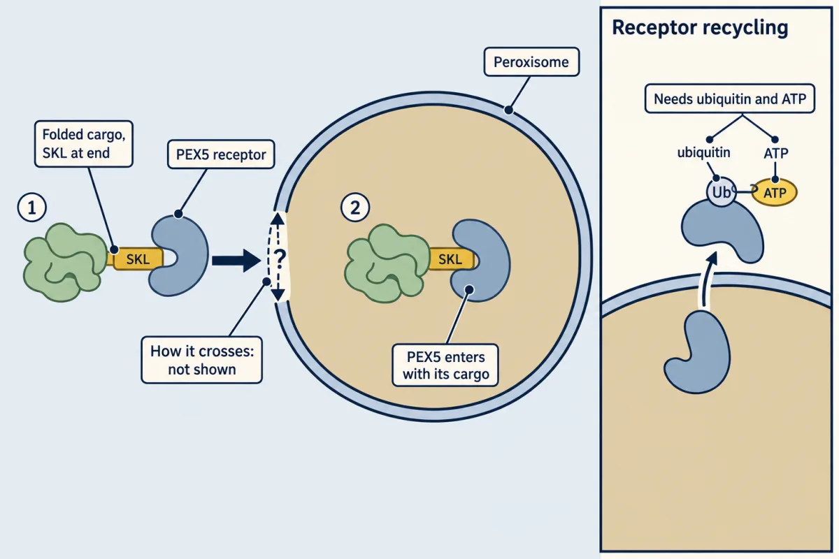
Put that address on a mouse enzyme, and the enzyme goes in too. Lock the enzyme folded with the drug methotrexate, and import stops. It had to unfold. A peroxisomal address is often the last three amino acids, such as SKL. They leave the tunnel only as the chain is released, so they are read afterwards. Its reader, PEX5, is a receptor free in the cytosol. It carries the finished protein in. Unlike mitochondria, peroxisomes can import cargo that stays folded. How it crosses is still debated. So the address points the way, and where its reader works shapes the timing. SRP meets growing chains; other readers mostly meet finished ones. Mitochondria need those unfolded; peroxisomes can take them folded.
From the film
- A protein's address, its signal sequence, is a stretch of its own chain; moved onto a suitable passenger, it can carry that passenger to the same place.
- For many secretory proteins, SRP catches the ER signal at the ribosome's exit, the chain enters while still being made, and signal peptidase cuts the signal off; some ER proteins enter after release by other routes.
- TOM sits on the mitochondrion and PEX5 works in the cytosol, and most mitochondrial and peroxisomal proteins are read after the chain is released.
- To enter the mitochondrial matrix a protein must be at least partly unfolded, as methotrexate-locked DHFR shows; peroxisomes can import cargo that stays folded.
- An exposed address helps specify where a protein goes; where and how its reader works helps decide when it is read, as the chain grows or after release.
Made in a test tube with no membranes, a secretory protein comes out longer than the one the cell secretes.
The first film answered why; here is what it leaves out. The text uses the field’s words for the film’s: the “address” is a signal sequence, reading “as the chain grows” is cotranslational, and reading “after release” is post-translational.
The address
Almost every protein in a eukaryotic cell starts on a ribosome in the cytosol, the fluid outside the organelles (a few are made inside mitochondria and chloroplasts from their own genes), yet thousands must reach the ER, mitochondria, chloroplasts, peroxisomes or the nucleus. The inside of a membrane-bound organelle is its lumen (in mitochondria and chloroplasts the innermost space is called the matrix and the stroma, respectively).
A signal sequence is a continuous stretch of amino acids that carries sorting information, like a zip code on a parcel. A protein with no signal stays in the cytosol by default, which gives the field its two tests. If moving a stretch onto a protein that normally stays in the cytosol moves that protein, the stretch is sufficient to sort it. If deleting the stretch leaves the protein in the cytosol, it is necessary.
Which way a membrane protein faces, its topology, is set at insertion (membrane organization and topology); nuclear import, through pores rather than across a membrane, has its own lesson. Here the question is how a protein reaches the right membrane at all.
Into the ER while being made
The amino terminus leaves the ribosome first (translation), and the ER signal is a hydrophobic stretch, usually near the amino terminus. Here is the experiment behind each of the first film’s four steps.
- Recognition. Signal recognition particle (SRP) is a complex of six proteins and an RNA. Evidence: in a translation extract made from wheat germ, SRP stopped the growth of preprolactin chains at about 70 residues: the 30-residue signal, just clear of the ribosome, plus roughly 40 residues still hidden in its tunnel. Adding ER vesicles released the stop. (In other extracts SRP only slows chain growth for a while; Going deeper.)
- Docking. Evidence: washing dog-pancreas ER vesicles in high salt removed their ability to bind ribosomes making prolactin, and purified SRP restored it (Walter and Blobel).
- Handover. The first film’s two “hands” are GTPases, subunits of SRP and of its receptor that bind GTP and then hydrolyse it. Evidence: in the bacterial versions of the two GTPases (3D viewer), the GTP sites face each other, so each helps the other hydrolyse GTP and the complex comes apart.
- Cleavage. Signal peptidase works on the lumenal side of the membrane. Many membrane proteins keep an uncleaved signal that stays in the membrane as an anchor.
Predict. A signal’s position sets the earliest moment it can be read. A secretory protein is only 60 residues long, with its signal at residues 1 to 20, and about 40 residues stay hidden in the ribosome. When has the whole signal cleared the ribosome, and what does that leave SRP?
Check your prediction
Only when the chain is about 60 residues long (20 + 40), that is, when it is finished. SRP has almost no time to catch it, and very short secretory proteins like this can enter after release by an SRP-independent route (Going deeper). A signal at the carboxyl end is the extreme case: it leaves the ribosome only as the chain is released, too late for SRP but not for readers that work after release.
SRP is the main reader at the ribosome, not the only one, and some ER proteins, such as tail-anchored ones, enter after release (Going deeper).
After release: mitochondria, chloroplasts, peroxisomes
The first film gave the idea and its mitochondrial and peroxisomal cases; here are the readers for all three organelles. They work away from the ribosome’s exit: TOM on the mitochondrial outer membrane, TOC on the chloroplast outer envelope, and PEX5 in the cytosol. Their cargo is usually read after release. The reader’s place does not settle the timing alone; the exceptions are in Going deeper.
The table sets the four routes side by side; they differ most in the last two rows.
| ER lumen | Mitochondrial matrix | Chloroplast stroma | Peroxisomal matrix | |
|---|---|---|---|---|
| Signal | hydrophobic stretch, usually amino-terminal | amino-terminal presequence: an amphipathic helix, positive on one face, hydrophobic on the other | amino-terminal transit peptide | carboxyl-terminal PTS1 such as serine-lysine-leucine (SKL); less often an amino-terminal PTS2 |
| Reader, and where it works | SRP, at the ribosome’s exit | TOM receptors, on the outer membrane | TOC receptors, on the outer envelope | PEX5 (PTS1) or PEX7 (PTS2), in the cytosol |
| Usually read | while the chain is made | after release | after release | after release |
| Way across | Sec61 channel | TOM, then the TIM23 complex | TOC, then TIC | PEX5 docks and inserts with cargo; in Xenopus egg extract it accompanied cargo into the lumen, but the general crossing is still debated |
| Energy, and what it pays for | GTP for targeting by SRP and its receptor; chain growth feeds the chain in; BiP, a lumenal Hsp70, helps some chains | membrane potential acts on the presequence; ATP for matrix Hsp70 | GTP at TOC; ATP in the stroma | loaded PEX5 may enter without ATP; ubiquitin and ATP recycle PEX5 for continued import |
| Cargo as it crosses | threads in unfolded as it is made | must unfold, at least partly | crosses largely unfolded | can stay folded, even as oligomers (several chains bound together) |
| Signal cut off? | often, by signal peptidase | often, in the matrix | usually, in the stroma | PTS1 usually not |
Helices are explained in protein structure; the 3D viewer shows a presequence helix lying in a groove on Tom20, one of the TOM receptors.
Mitochondria: what the methotrexate result proves. In the first film’s experiment (Eilers and Schatz, 1986), methotrexate binds the passenger, the enzyme dihydrofolate reductase (DHFR), and holds it folded. The control is what makes it decisive: the authentic precursor still entered the same mitochondria, so the organelles and their membrane potential were working and the block was in the passenger. Inner-membrane carriers with internal signals take another route (Going deeper).
Predict. You give isolated mitochondria a matrix precursor, with or without an uncoupler (a chemical that collapses the membrane potential), then add protease. Which sample gives a protected band slightly smaller than the precursor?
Check your prediction
Only without the uncoupler. Then the presequence is pulled through the TIM23 complex, is often cut off in the matrix, and the membranes shield the chain from protease. With the uncoupler, complete TIM23-mediated import and processing are blocked; the precursor may bind or partly engage the machinery, but that is not matrix entry. Protected, processed protein that disappears with an uncoupler is the standard sign of real import.
Predict. The first film left a question. Some ribosomes making mitochondrial proteins sit on the mitochondrial surface. Could their presequences be read before the chain is finished?
Check your prediction
They could: the ribosome is now within TOM’s reach. Being close makes early reading possible, not certain. Studies of ribosomes near mitochondria suggest that some mitochondrial proteins may be imported while still being made (Going deeper).
Chloroplasts follow the mitochondrial pattern, through TOC in the outer envelope and TIC in the inner. Finished precursors of the Rubisco small subunit entered purified intact chloroplasts and were processed (Chua and Schmidt, 1978). Which ATP-driven motor pulls hardest is still debated (Going deeper).
Peroxisomes: the exception in the last rows. PEX7 reads PTS2 cargo as PEX5 reads PTS1. The evidence that cargo can stay folded came in 1995: Walton, Hill and Subramani found that mammalian peroxisomes imported proteins held folded by disulfide bonds or chemical cross-links, and even gold particles coated with signal-bearing protein.
Predict. A protein ends in SKL and goes to peroxisomes. You add green fluorescent protein (GFP) to its carboxyl end, so SKL is no longer last. Where do you expect the fusion to go?
Check your prediction
Most likely it stays in the cytosol. PEX5 holds the free carboxyl end of a PTS1 deep in its groove (3D viewer), so an SKL buried inside the chain should not be read. This is an inference from the structure, not a cited experiment; a PTS2 elsewhere in the protein could still send it to peroxisomes.
Every claim so far rests on an experiment. Start by reading the protection test as numbers.
How do you know where a protein went?
Here is the protection logic as numbers.
Worked problem: reading a protection gel
Here is a made-up data set, patterned on the Blobel and Dobberstein experiments. A secretory protein’s messenger RNA is translated in reticulocyte lysate, an extract of immature rabbit red blood cells that makes protein from added messenger RNA. Some reactions include microsomes, sealed vesicles formed from ER membrane when cells are broken up. You read band intensity in arbitrary units. The precursor, still carrying its signal, runs at 26 kDa and a processed form at 23 kDa.
- Lane 1, no microsomes. 26 kDa = 100. After protease: nothing.
- Lane 2, microsomes present during translation. 26 kDa = 40, 23 kDa = 60. After protease: 23 kDa = 57, 26 kDa = 0. After protease plus detergent: nothing.
- Lane 3, microsomes added after translation. 26 kDa = 100, no 23 kDa. After protease: nothing.
Assume the added protease cannot cross an intact microsome membrane and that the bands are proportional to the amount of protein.
Predict. In lane 2, 40 units kept the full 26 kDa size. Were those chains inside the vesicles?
Check your prediction
No. After protease no 26 kDa band survives, so every unprocessed chain was outside and was digested. Those chains kept their signal because they never entered: the scissors are on the inside.
Step 1: how much was processed, and was it protected? In lane 2, 60 of 100 units are processed, so 60 percent of chains lost their signal. Of those 60, 57 survive protease:
protected fraction = 57 / 60 = 0.95, or 95 percent
So nearly all processed chains are inside sealed vesicles.
Step 2: was it the membrane? Detergent dissolves the vesicles and the 23 kDa band vanishes. The protein is not resistant to protease on its own; the membrane protected it.
Step 3: does timing matter? This is the first film’s predict, as data. In lane 3 there is no processing and no protection. In this assay, translocation needs ongoing translation.
Step 4: how big was the signal? The shift is 26 − 23 = 3 kDa. With an average residue mass of about 110 Da:
residues removed ≈ 3,000 Da / 110 Da per residue ≈ 27 residues
That fits a signal of the usual size; preprolactin’s is 30 residues.
Protease cannot cross an intact membrane, so a band that survives protease but not protease plus detergent was inside a sealed vesicle; bands are proportional to the amount of protein. This is the lesson’s made-up data set, a sketch of the logic, not a real gel.
The second film now connects that protection test to experiments that identify a sorting signal.
Read the transcript of “How Do You Know It Went In?”
Film transcript: How Do You Know It Went In?
In chapter one, a stretch of the protein's own chain worked as an address. How do you know a protein really went inside? And how do you prove which stretch sent it? Start with the classic test. Make the protein in a test tube that also holds microsomes, sealed vesicles of ER membrane. Then add a protease, an enzyme that chews up exposed protein. The added protease cannot cross an intact microsome membrane. A chain outside the vesicles is chewed up. A chain inside is shielded, so its band survives on the gel. But maybe that protein simply resists protease. So add a detergent, which dissolves membranes. If the band now vanishes, the membrane was the shield.
In 1975, Blobel and Dobberstein tested an antibody light chain, a secreted protein. Globin, which stays in the cytosol, was made the same way. Which survives the protease? The light chain came out trimmed and protected. That happened only when the vesicles were there while it was being made.
Globin was not protected. The parts came from three species: rabbit ribosomes, dog membranes, and mouse messenger RNA. They still worked together. That ruled out a special secretory ribosome and supported an address in the growing protein. So here is the idea. A sorting claim needs two kinds of test. One shows the protein got in; the other shows what sent it. To find the address, there are two tests. Move a stretch onto a protein that stays in the cytosol. If that passenger moves too, the stretch is sufficient. Or delete the stretch from its own protein. If the protein is stranded in the cytosol, the stretch is necessary.
Chapter 1's mouse enzyme was the first kind. Yeast cytochrome oxidase IV precursor's first 53 amino acids carried mouse DHFR into the matrix. Adding SKL to a bacterial enzyme's carboxyl end sent it into mammalian-cell peroxisomes. Now the other test. Deleting luciferase's carboxyl tail, no more than 12 amino acids long, left it in the cytosol. A signal in that tail is necessary. Two cautions. Working for one passenger does not prove it works for every passenger. And SKL is picky. In luciferase, only a few swaps to similar amino acids kept it working. And when a protein carries two signals, only an experiment can say where it goes. Protection shows it got in; moving and deleting show what sent it.
From the film
- The added protease cannot cross an intact microsome membrane, so a protected protein is inside; if detergent removes that protection, the membrane was the shield.
- In 1975 an antibody light chain was trimmed and protected only when ER vesicles were present while it was made; globin was not protected.
- Moving a stretch onto a passenger that stays in the cytosol tests whether it is sufficient; deleting it from its own protein tests whether it is necessary.
- A signal that works for one passenger may not work for every passenger, and when a protein carries two signals, only an experiment can say where it goes.
Each classic result is one of the two tests. Sufficient: the first 53 residues of a yeast mitochondrial precursor carried mouse dihydrofolate reductase (DHFR) into the matrix of yeast mitochondria (the film’s mouse enzyme), and SKL added to the end of a bacterial enzyme sent it into peroxisomes of mammalian cells. Necessary: deleting the short region at luciferase’s tail, at most twelve residues, left luciferase in the cytosol.
Three warnings keep predictions honest. First, sufficiency for one passenger is not universality. Second, SKL is a specific signal; only a few conservative substitutions kept it working in luciferase. Third, position matters: a signal that works at one end may fail at the other or inside the chain. When a protein carries two signals, the outcome is empirical. Sorting continues past the ER, through vesicle traffic and the secretory pathway; lipid tails that pin proteins to membranes are in lipid anchors.
Design problem
Protein Y has an amino-terminal amphipathic helix and ends in SKL. Where does it go, and how would you test it?
Saved in this browser only.
One way to do it
Predict first. The amino-terminal helix looks like a matrix presequence and SKL is a PTS1, so Y could go to mitochondria, peroxisomes or both. The presequence emerges first and, if accessible, may win, but both signals are usually read after release, so treat that only as a hypothesis.
Build four GFP fusions: full-length Y, Y without the helix, Y without SKL, and Y without both. Put GFP where it hides neither signal, for example just after the helix, so SKL stays at the free carboxyl end. Image each in cultured cells with mitochondrial and peroxisomal markers, and score colocalization. Expect the double deletion in the cytosol. If deleting the helix moves Y to peroxisomes and deleting SKL leaves it in mitochondria, each signal works and the helix dominates in the full protein.
Then test import directly. Incubate radiolabelled Y with isolated mitochondria, with and without an uncoupler, and treat with protease. Protected, processed Y that disappears with the uncoupler means import, not sticking to the surface. To show it reached the matrix, open the outer membrane by swelling and repeat the protease test. A similar protease test on isolated peroxisomes checks the SKL route.
Going deeper
How the signal idea was found
In 1971 Günter Blobel and David Sabatini proposed that proteins bound for the ER carry a signal that sends the ribosome making them to the membrane. In 1972 César Milstein’s group translated the messenger RNA for an antibody light chain in a cell-free system and got a product slightly larger than the light chain that myeloma cells secrete, which they proposed was a precursor. In 1975 Blobel and Bernhard Dobberstein tested it directly, showing that the extra piece is an address read while the chain is still on the ribosome.
SRP details from the cell-free experiments
The 70-residue stop came from Peter Walter and Blobel in 1981 and belongs to preprolactin in the wheat germ system; in reticulocyte lysate signal recognition particle (SRP) was later found to cause only a transient pause (Wolin & Walter, J Cell Biol 109:2617, 1989). In 1982 Reid Gilmore, Walter and Blobel used SRP as bait to isolate a 72,000-dalton receptor protein. Signal peptidase is a different enzyme from the SRP receptor: the SRP affinity column used to isolate the receptor separated the two activities. A 2016 structure of an active Sec61 showed the signal sitting in the channel’s lateral gate after displacing one of its helices.
Other roads into the ER
In budding yeast, rapid SRP depletion in 2018 left many proteins with cleavable signals still targeted while being made, although proteins with transmembrane domains generally needed SRP. Some proteins, especially in yeast, cross after translation through Sec61 with its partners Sec62 and Sec63; very short secretory proteins, whose signal clears the ribosome only as the chain finishes, can enter this way. Tail-anchored proteins, whose only hydrophobic segment is at the very end, are inserted after translation by other machinery (the GET, or TRC40, route).
Mitochondria: early reading, carriers and the conducting path
In human cells, nearly a fifth of mitochondrial proteins have been reported to be imported while still being made. Such studies map ribosomes near mitochondria, so they show more directly where a protein is made than when it crosses: a sizeable minority appear to be made on the mitochondrial surface, and some may be imported during synthesis. Metabolite carriers of the inner membrane have internal signals and use a separate TIM22 route. A 2023 study of the yeast TIM23 complex placed the protein-conducting path along its Tim17 subunit rather than along Tim23, as older diagrams show; the complex keeps its name.
Chloroplast motors
In the stroma, chaperones such as Hsp70 and Hsp93 have long been proposed as the ATP-driven motor; recent structures implicate another, Ycf2-FtsHi, at the inner envelope, and which does most of the work is still debated. The table says the cargo crosses largely unfolded, not that it must unfold, because some folded or partly folded passengers have been reported to cross the envelope.
The chloroplast precursor experiment and its correction
Nam-Hai Chua and Gregory Schmidt’s 1978 precursors, from pea and spinach, were 4,000 to 5,000 daltons larger than the mature protein. In that paper the precursor from the alga Chlamydomonas did not enter those higher-plant chloroplasts, but a later study (Mishkind, Wessler and Schmidt, 1985) found it imported efficiently by spinach and pea chloroplasts, so the negative result did not hold.
How PEX5 crosses
In Xenopus egg extracts in 2022, PEX5 went all the way into the lumen with its cargo, and was unfolded when pulled back out. How the membrane passes large folded cargo without leaking small solutes, and whether there is a stable pore at all, is still open.
Your notes
Select any text in the lesson to highlight it or add a note to it. Your highlights and notes are saved in this browser and collected in your notebook.
This browser is not letting the site save anything (a private window or strict privacy settings), so highlights and notes will be lost when you leave the page.
How we measure it
Cell-free translation with microsomes and protease protection
Messenger RNA is translated in a cell extract with or without ER vesicles (microsomes), and the products are treated with protease, with or without detergent. A protein that survives protease only while the membranes are intact is inside the vesicles. Adding membranes during or after translation tells you whether entry needs the chain to be growing.
Salt extraction and add-back
Membranes are washed in high salt to strip loosely bound proteins, tested for a lost activity, then given back a purified candidate factor. If a factor purified from the wash restores the activity, it is a strong candidate for what the wash removed. This is how SRP was shown to be the selective ribosome-binding activity.
Import into isolated organelles
A radiolabelled precursor made in vitro is incubated with purified mitochondria or chloroplasts, then treated with protease. Protected, processed protein has been imported. Uncouplers, ATP depletion or a drug that locks the cargo folded test what import needs.
Signal transplantation and mutagenesis
A candidate signal is fused to a passenger protein that normally stays in the cytosol, or the signal is mutated residue by residue, and the fusion's location is read by microscopy or import. Moving a passenger shows that a signal is sufficient; losing location after a deletion shows that it is necessary.
Practice questions on this lesson (3) → MCAT-style, original, each with an explanation
Where this lesson fits
Before thisReview first
- Organelles as Distinct EnvironmentsThis course · Lesson 6
It shows what each compartment is like inside, which explains why proteins need addresses to reach the right one.
- Membrane Organization and TopologyThis course · Lesson 4
You need how proteins are inserted and oriented in a membrane to follow the ER signal and anchor cases here.
- Experiments That Distinguish MechanismsThis course · Lesson 3
Protease protection and fractionation logic from there is used here to decide whether a protein went inside vesicles.
After thisStudy next
- The Nuclear Envelope and Nuclear TransportThis course · Lesson 8
Nuclear import reads a different signal and crosses through pores, so it extends the idea of sequence addresses.
- The Secretory PathwayThis course · Lesson 12
Proteins that enter the ER here continue through the Golgi, so it follows their route after translocation.
- Protein Quality Control and Organelle TurnoverThis course · Lesson 9
Proteins that fail to fold or sort correctly after entering compartments are handled by the quality control described there.
Elsewhere on the siteFilms, simulations and method films in other courses
- Centrifugation and subcellular fractionationMethod film
Microsomes used in the protection experiment are made by centrifuging broken cells into fractions.
- From Gene Copy to Protein ChainFilm · Biology
It shows how a ribosome reads mRNA to build the chain whose signal sequence is read here.
- One Cell, One Shipping LineFilm · Biology
It follows insulin from ribosome on the rough ER to secretion, the route entered here.
Suggested by an AI model from the lessons themselves, then checked. How we use AI
Finish line
Test yourself
8 questions
Practice and MCAT-style, with explained answers.
Next · Lesson 8
The Nuclear Envelope and Nuclear Transport
If one pore carries traffic both ways, what decides which way each protein goes?
Continue →