Cell Signaling · Unit 7: Space, time and distance
Endocytosis and Signaling Endosomes
When a receptor is pulled into the cell, does its signal end, or does it move somewhere new?
Pause when the reading reaches a film, and offer to play it. Saved on this device.
Watch the film now, or keep listening
Builds on:GTPase Switches: The TimerUbiquitin and Regulated Proteolysis as SignalsReceptor Tyrosine KinasesWhy these?
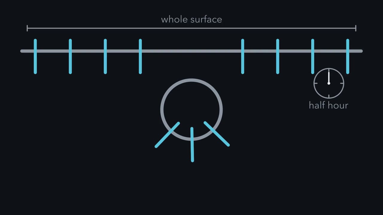 ▶ Watch Sealed Inside, a Receptor Goes Silent · 2:45
Read the transcript of “Sealed Inside, a Receptor Goes Silent”
Film transcript: Sealed Inside, a Receptor Goes Silent
A macrophage swallows the equivalent of its whole surface about every half hour, and it does not shrink. Many receptors in that membrane make the trip too. At the surface, the tail of a receptor points into the cell. So where does the receptor go once it is inside? And what does the trip do to its signal? A receptor can enter a pit through a tail sequence that an adaptor can read. The adaptor links the tail to a coat of clathrin. The pit deepens until it hangs by a thin neck. A protein called dynamin builds a collar around the neck. When it breaks down GTP, the neck is cut and the vesicle is free.
The vesicle reaches the early endosome, the first stop for almost all cargo that is taken in. On the outer membrane, the tail still points into the cytosol and can still reach its partners. So a receptor can go on signaling from here. A receptor with no other instruction spreads into thin tubes. The tubes pinch off, and that receptor mostly goes back to the surface. A receptor leaves that default route when it carries a tag. The tag is ubiquitin. For an active EGF receptor, an enzyme called Cbl adds it. A set of proteins on the endosome, called ESCRT, binds the tag. They gather the tagged receptors into a patch. ESCRT pushes the patch inward until it pinches off as a small vesicle inside the endosome. Now ask which way the tail faces.
Inside that vesicle, the tail is sealed in and cannot reach the cytosol at all. Its partners are out of reach, so the signal ends. That happens before the lysosome, a bag of digestive enzymes, destroys anything. Now break the sorting. In some lung cancers, the receptor MET loses the docking site for the enzyme that adds ubiquitin. Poorly tagged, it escapes the lysosome and keeps signaling for longer than it should. The fault is not an overactive kinase, but a receptor that misses its turn. So keep this rule: taken in, a receptor can still signal, but sealed in, it cannot.
Taking a receptor into the cell can end its signal or move it somewhere new, and where the receptor is sorted decides which. Toll-like receptor 4 is one example: at the plasma membrane it signals through the adaptor MyD88, and after it is taken in it signals through a second adaptor, TRIF, which switches on interferon genes. The traffic behind this is heavy. A macrophage takes in about 3 percent of its surface membrane every minute. It swallows the equivalent of its whole surface about every half hour, and it does not shrink. The membrane must come back as fast as it goes in, and the receptors sitting in that membrane make the trip too, some often and some rarely. So for any receptor you have met in this course, ask where it goes once it is inside the cell, and what the trip does to its signal.
What’s endocytosis used for?
Endocytosis is the pinching of a patch of plasma membrane inward to make a vesicle, a small closed bag of membrane, that carries in whatever was bound to the patch. It does three jobs.
The first is food. Cholesterol travels in the blood packed into particles of low-density lipoprotein (LDL), which are far too large for any channel or carrier. Michael Brown and Joseph Goldstein found that cells bind LDL to a surface receptor and carry the pair inside, a process that came to be called receptor-mediated endocytosis. Iron enters the same way, bound to the protein transferrin.
The second is membrane upkeep. Secretion adds membrane to the surface and endocytosis takes it back, and worn membrane proteins are pulled in for disposal.
The third is why this lesson sits in a signaling course. For a signal, endocytosis can mean two things. It can end the signal, by taking the receptor off the surface and destroying it. Or it can move the signal, by carrying an active receptor to a new platform inside the cell, where it keeps signaling or signals differently. Which one happens is decided mostly after the receptor is in.
It all starts with endocytosis: a coat, then a cut
Read the transcript of “A Coat, a Reader, and a Cut”
Film transcript: A Coat, a Reader, and a Cut
Clathrin is a protein shaped like a three-legged pinwheel. Many copies lock together into a curved cage on the inner face of the membrane. Yet clathrin touches neither the membrane nor the cargo. So what does? Adaptor proteins do. The main adaptor at the plasma membrane is AP2. It holds the lipid PIP2, a short sequence in a receptor tail, and clathrin. So a receptor enters a pit because its tail carries a sequence that an adaptor can read. Brown and Goldstein met the proof in a patient known as J.D. This patient's LDL receptors could bind LDL. But they stayed scattered over the surface instead of gathering in pits, and the cells could not take LDL in.
The fault was later traced to one tyrosine in the receptor's tail, replaced by cysteine. A pit deepens until it hangs from the surface by a thin neck. Dynamin, a large GTPase, assembles into a collar around the neck. When it hydrolyzes GTP, the neck is cut. The coat is then stripped off, and the bare vesicle can fuse with its target.
The best understood way in is the clathrin-coated pit. Clathrin is a protein shaped like a three-legged pinwheel, and many copies lock together into a curved cage on the inner face of the membrane. Clathrin touches neither the membrane nor the cargo. Adaptor proteins do that. The main adaptor at the plasma membrane, AP2, holds the lipid phosphatidylinositol 4,5-bisphosphate (PIP2) (an address from the lesson on lipid anchors), short sequences in receptor tails, and clathrin. So a receptor enters a pit when its cytoplasmic tail carries a sequence that an adaptor can read, or when it binds a protein, such as arrestin, that links it to the coat.
Brown and Goldstein met the proof in a patient known as J.D. This patient’s low-density lipoprotein (LDL) receptors could bind LDL but stayed scattered over the surface instead of gathering in pits, and the cells could not take LDL in. The fault was later traced to one tyrosine in the receptor’s tail, replaced by cysteine.
A pit deepens until it hangs from the surface by a thin neck. Dynamin, a large GTPase, assembles into a collar around the neck, and when it hydrolyzes GTP the neck is cut. The coat is then stripped off and the bare vesicle can fuse with its target.
There are other doors. Caveolae are small flask-shaped pits built around a protein called caveolin, macropinocytosis gulps large volumes of fluid, and several more routes that need no clathrin have been described and are less well mapped.
A receptor’s signaling can influence which internalization pathway it takes. How hard a receptor is signaling can change how it enters and where it ends up. The best-studied case, the epidermal growth factor (EGF) receptor, is under Going deeper.
The early endosome is a sorting station
The early endosome is the first stop for almost all endocytosed cargo. It lies near the plasma membrane, and it sorts cargo into separate patches of its own membrane that set the cargo’s fate: back to the surface, back to the Golgi, or on to the lysosome to be destroyed.
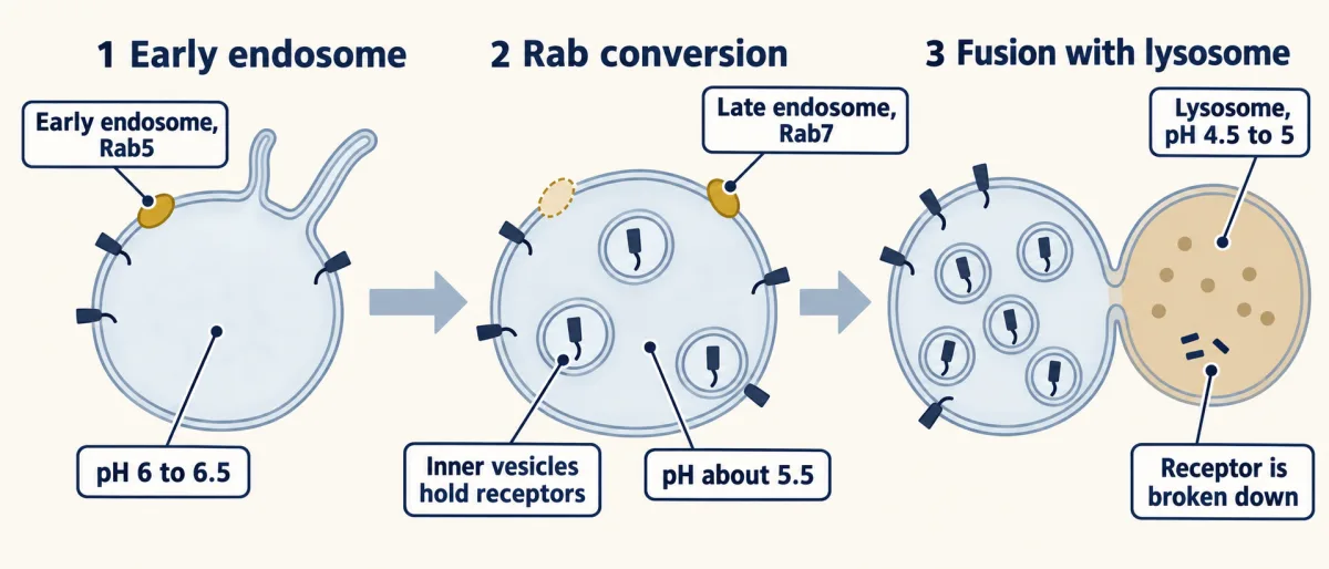
Two physical features do much of the sorting. One is acid. A proton pump in the endosome membrane holds the inside at about pH 6 to 6.5, and at that pH many ligands let go. Low-density lipoprotein (LDL) falls off its receptor, and iron falls off transferrin. The other is shape. An early endosome has a round body with thin tubes growing out of it. A tube is nearly all membrane and almost no volume, so membrane proteins spread into the tubes while dissolved contents stay in the body. The tubes pinch off and go back to the surface, so a receptor with no other instruction mostly goes back too.
There is a fast way back, taking a few minutes, straight from tubes rich in the protein Rab4. There is a slow way, taking tens of minutes, that passes through a second compartment and uses Rab4 and Rab11. An LDL receptor makes the round trip about every ten minutes, again and again.
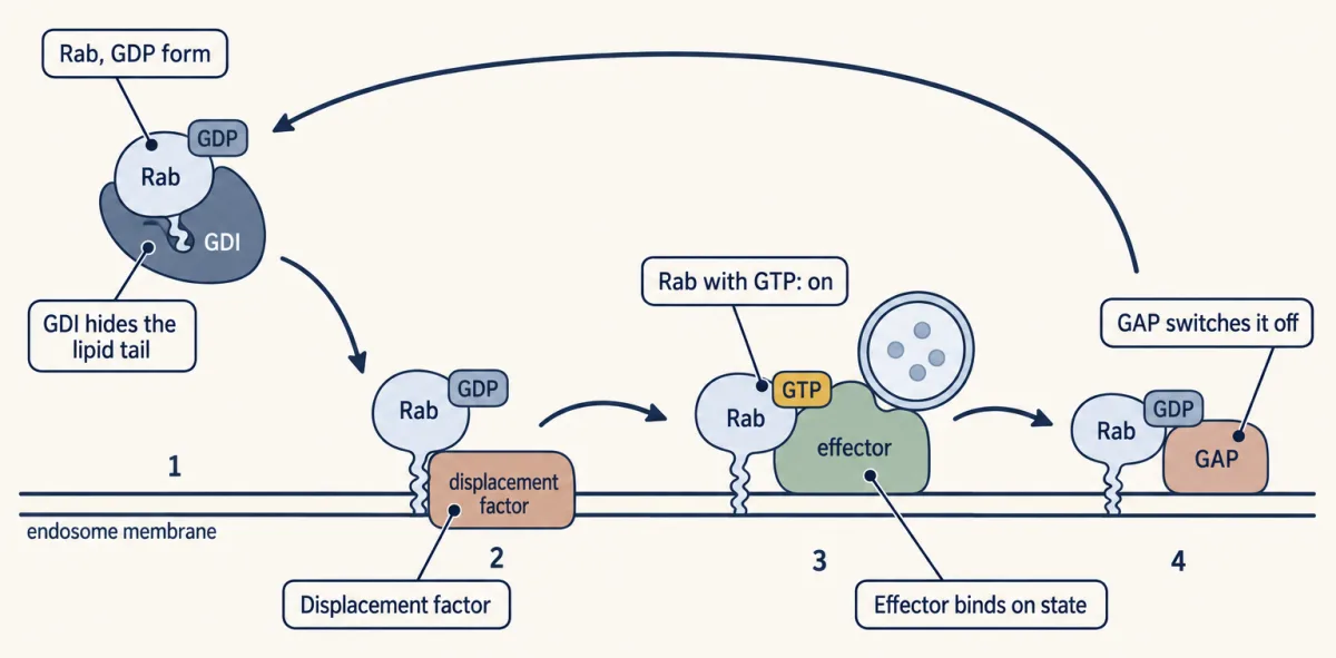
Rab proteins define and drive the maturation state. Rabs are small GTPase switches of the kind in the lesson on GTPase switches: on with GTP, off with GDP, flipped by guanine-nucleotide exchange factors (GEFs) and GAPs. Human cells have more than sixty, and most mark a particular compartment, a domain within one, or a step of traffic. Rab5 marks the early endosome. In its GTP form it sits on the membrane and recruits effectors, proteins that bind only the on state, which tether incoming vesicles and help them fuse.
A Rab’s lipid tail would stick to any membrane, so cells keep GDP-bound Rabs soluble with a protein called GDI, and later work points to a Rab’s own GEF as a main part of its address, since a Rab is switched on, and so held, on the membrane where its GEF sits. The details are under Going deeper.
Headed to the lysosome: the receptor is sealed inside
A receptor leaves the default route when it carries a tag, and the tag is one you know: ubiquitin. For an active epidermal growth factor (EGF) receptor the ligase Cbl adds it, as the lesson on receptor tyrosine kinases describes. This ubiquitin is not read by the proteasome. A set of proteins on the endosome called ESCRT binds it, gathers tagged receptors into a patch, and pushes the patch inward until it pinches off as a small vesicle inside the endosome. An endosome filled with such vesicles is a multivesicular endosome (MVE), also called a multivesicular body.
What is the purpose of MVEs? Think about which way the receptor faces. On the outer membrane of an endosome, the receptor’s tail still points into the cytosol and can still reach its partners. On an internal vesicle, the tail is sealed inside and cannot reach the cytosol at all. That step ends the signal before anything is destroyed. It also puts the whole receptor where the lysosome’s enzymes can reach it.
Meanwhile the compartment itself changes. Marino Zerial’s laboratory filmed single endosomes in living cells and saw Rab5 leave as Rab7 arrived, which they called Rab conversion. With Rab7 come new effectors, stronger acid (about pH 5.5 in late endosomes) and a growing load of internal vesicles. The late endosome then fuses with a lysosome, a bag of digestive enzymes at about pH 4.5 to 5, and the receptor is broken down.
A receptor that keeps talking, and one that escapes
Between the coated pit and the internal vesicle, a receptor spends time on the outer membrane of an endosome with its tail in the cytosol. Is it still signaling? In 1996 Sandra Schmid’s laboratory tested this by blocking endocytosis in HeLa cells with a dynamin mutant defective in GTP binding and hydrolysis. If endocytosis only ended signals, every response to epidermal growth factor (EGF) should have gone up. The cells did divide more, but some responses fell, including full activation of the MAP kinase extracellular signal-regulated kinase (ERK). On this evidence part of the EGF signal needs the receptor to be inside, although later experiments that blocked endocytosis in other ways did not all find the same fall in ERK.
Other receptors change what they say when they move. Toll-like receptor 4 signals through the adaptor MyD88 at the plasma membrane. After it is taken in, it signals through a second adaptor, TRIF, which switches on interferon genes. The Toll-like receptors that detect nucleic acids, such as TLR3, TLR7 and TLR9, work only in endosomes, where the remains of digested microbes arrive. And some G-protein-coupled receptors, taken in with arrestin as the lesson on turning signals off describes, go on activating G proteins from endosomes.
Now break the sorting. In roughly 3 percent of non-small-cell lung cancers, a splicing error removes exon 14 from the messenger RNA for MET, a receptor tyrosine kinase. The missing stretch holds tyrosine 1003, the docking site for Cbl. The receptor is poorly tagged with ubiquitin, escapes the lysosome and keeps signaling for longer than it should. The fault is not an overactive kinase. It is a receptor that misses its turn to the lysosome. Two MET kinase inhibitors, capmatinib and tepotinib, are approved for lung cancers with this change.
A neuron poses the hardest version of the keep-talking problem, because its endosomes must carry a signal the length of an axon. That is the next lesson.
Design problem
Two receptors on the same cell bind their ligands at the same moment. One must signal for about ten minutes and then be silent for hours. The other must keep signaling for hours. Using only the sorting decisions made at the early endosome, design the difference. Say what tag or sequence each receptor would need.
Saved in this browser only.
One way to do it
Give the brief receptor a docking site for a ubiquitin ligase that appears only when the receptor is active, for example a tyrosine the receptor phosphorylates on its own tail, plus lysines for the ligase to tag. Give it a ligand that stays bound at pH 6, so the receptor stays active and tagged all the way in. ESCRT then seals it inside internal vesicles and the signal stops. The silence lasts for hours because the cell has to make new receptors to replace the ones it destroyed.
Give the lasting receptor no ligase site, so it is never tagged, and a ligand that falls off at pH 6. It takes the tubes back to the surface in minutes, empty and ready to bind again, and it keeps signaling for as long as ligand is outside. To make the return more certain, add a tail sequence that recycling machinery reads. The β2-adrenergic receptor has one at its very end, a sequence that binds PDZ domains.
Cells do use this logic. The EGF receptor has two ligands with different fates. EGF tends to stay bound in the endosome and takes the receptor to the lysosome, while transforming growth factor alpha lets go in the acid and the receptor recycles.
Going deeper
How a Rab finds its membrane
How does a Rab find its membrane? Most Rabs carry one or two geranylgeranyl groups, 20-carbon lipids, on their tail, and a greasy tail would stick to any membrane it met. Suzanne Pfeffer’s laboratory worked on this with Rab9. A protein called GDI wraps the GDP-bound Rab and hides the tail, so the Rab stays dissolved in the cytosol. Her group found an activity on endosome membranes, a displacement factor, that frees the Rab from GDI, and showed that GDP is exchanged for GTP once the Rab reaches the membrane. Later work points to the Rab’s own guanine-nucleotide exchange factor (GEF) as a main part of the address: a Rab is switched on, and so held, on the membrane where its GEF sits. GDI does not pull off the GTP form, so the Rab stays put until a GAP switches it off.
A lipid of late endosomes
In late endosomes, the internal vesicles are rich in an unusual lipid, lysobisphosphatidic acid, which is scarce in early endosomes.
Dose and route for the EGF receptor
The best studied case is the epidermal growth factor (EGF) receptor. In HeLa cells, Sara Sigismund, Pier Paolo Di Fiore and their colleagues reported that at low doses of EGF the receptor enters almost entirely through coated pits and is mostly recycled. At high doses the receptor also becomes tagged with ubiquitin, and a share of it enters by a route without clathrin and is mostly destroyed. Treat that as a result in one cell line, since other laboratories find clathrin carrying most of the receptor at high doses too. The modest version is safe: how hard a receptor is signaling can change how it enters and where it ends up.
Your notes
Select any text in the lesson to highlight it or add a note to it. Your highlights and notes are saved in this browser and collected in your notebook.
This browser is not letting the site save anything (a private window or strict privacy settings), so highlights and notes will be lost when you leave the page.
How we measure it
Electron microscopy with ferritin-labeled LDL
LDL is coupled to ferritin, an iron-rich protein that shows up as a dark dot in the electron microscope, and added to cells in the cold so that it binds but is not taken in. The dots show where the receptors are. Warming the cells and fixing them at intervals shows the route in. This is how LDL receptors were seen clustered in coated pits.
Dominant-negative dynamin (K44A)
A dynamin with lysine 44 changed to alanine is defective in binding and hydrolyzing GTP. Expressed in cells, it blocks the pinching-off of coated pits. Comparing a response with and without the mutant shows which parts of a signal depend on dynamin. Because dynamin has other effects, other ways of blocking uptake are needed to show that a response needs the receptor to be taken in.
Two-color live imaging of Rab proteins
Two Rabs are fused to fluorescent proteins of different colors and made in the same cell. Filming single endosomes shows which Rab is on a compartment and when one replaces another. This is how the exchange of Rab5 for Rab7 was observed.
Cell-free reconstitution of Rab delivery
Cytosol, isolated membranes and a Rab bound to GDI are mixed in a tube to ask which parts a step needs. Pfeffer's laboratory used this to show that delivery of Rab9 to membranes comes with exchange of GDP for GTP. Because a lipid-tailed protein can stick to any membrane, controls with the wrong membrane matter.
Practice questions on this lesson (3) → MCAT-style, original, each with an explanation
Where this lesson fits
Before thisReview first
- GTPase Switches: The TimerThis course · Lesson 7
Rab proteins are on-off GTPase switches flipped by GEFs and GAPs, and this lesson uses that timer logic directly.
- Ubiquitin and Regulated Proteolysis as SignalsThis course · Lesson 8
Ubiquitin is the tag that sends a receptor into the lysosome route, and the lesson assumes you know how it is added.
- Receptor Tyrosine KinasesThis course · Lesson 18
The EGF receptor, its activation and its tagging by Cbl are the main example followed through the endosome here.
After thisStudy next
- Long-Distance Signaling in NeuronsThis course · Lesson 37
It takes the idea of signaling endosomes and asks how they carry a signal the full length of an axon.
Elsewhere on the siteFilms, simulations and method films in other courses
- A Loop and a One-Way TripFilm · Cell Biology
This film follows the LDL receptor through coated pits and endosomes, showing the same sorting between recycling and lysosome.
- Binding or entry?Simulation · Cell Biology
The simulation lets you separate binding from entry for a receptor, the same trafficking step this lesson uses.
Suggested by an AI model from the lessons themselves, then checked. How we use AI
Finish line
Test yourself
8 questions
Practice and MCAT-style, with explained answers.
Next · Lesson 37
Long-Distance Signaling in Neurons
How does a neuron that takes in its survival signal at the tip of its axon get that message to a nucleus centimeters away?
Continue →← Lesson 35 · The Shape of a Signal: Size, Duration, Frequency and Place