Cell Signaling · Unit 3: G-protein-coupled receptors and second messengers
Turning the Signal Off: Arrestins, RGS Proteins and Phosphodiesterases
Rewritten on : new text, flashcards and film. Your notes and highlights stay, but the text around them may have moved.
The hormone is still bound and the response has faded. What switched the receptor off?
Pause when the reading reaches a film, and offer to play it. Saved on this device.
Watch the film now, or keep listening
Builds on:GTPase Switches: The TimerGPCRs and Trimeric G ProteinsPhosphorylation: Kinases and PhosphatasesWhy these?
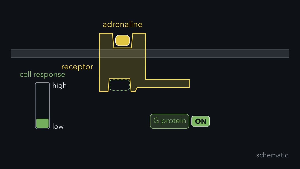 ▶ Watch The Mark and the Reader · 3:36
Read the transcript of “The Mark and the Reader”
Film transcript: The Mark and the Reader
Adrenaline still fills this receptor, but the cell's response has faded. How can an occupied receptor lose its voice? Start with the receptor while it is active. Its inner face can meet a G protein and start another round of signaling. A dedicated receptor kinase then approaches that active shape. The kinase is called a G-protein-coupled receptor kinase, or GRK. The kinase adds phosphate marks to the receptor's cytoplasmic side. But a mark is not the same as an off switch. That distinction emerged when researchers purified the kinase and rebuilt signaling from isolated parts. Rebuilding the pathway made each requirement testable. The receptor sat in a lipid vesicle beside purified G protein.
Researchers added candidate proteins one at a time. Receptor coupling became the readout, not phosphate alone. A partly purified preparation added phosphate and strongly reduced receptor coupling. A highly purified kinase added phosphate too, yet coupling fell only a little. The phosphate signal remained. Most of the functional effect vanished. The writer survived purification, but switching off did not. What missing part would make the phosphate mark matter?
A clue came from the retina. Its light receptor used a second protein, later called arrestin. Adding retinal arrestin to the rebuilt system brought back part of the lost effect. Arrestin alone could not act on an unmarked receptor. The controls narrowed the claim. An unrelated protein could not replace arrestin. Heat-damaged arrestin could not replace it either. The effect required an intact reader and a receptor carrying the kinase's mark. Now the two jobs separate cleanly. GRK writes the mark on the active receptor. Arrestin reads that mark and covers the surface needed by the G protein. The writer identifies the target. The reader produces the block.
This is one layer of a larger rule. During a GPCR response, each active layer fades when removal outruns production. Here, new receptor signaling stops because the mark and reader together prevent new G-protein activation. Notice what the mechanism does not require. The agonist need not leave first. The receptor can remain occupied while its access changes.
Desensitization therefore records regulation inside the cell, not simply disappearance of the outside signal. The receptor is not the whole pathway. Active G proteins, second messengers, and phosphorylated targets can persist after receptor coupling falls. Their removal processes decide how long the rest of the response lasts.
From the film
- A GRK mark does little until arrestin reads it.
- A GRK marks the active receptor; arrestin reads the mark and covers the surface the G protein needs.
- Controls ruled out look-alikes: an unrelated protein, heat-denatured arrestin and an unmarked receptor all failed.
- The agonist can stay bound while the receptor is silenced.
Adrenaline is still bound to its receptor, yet within minutes the response fades. What removed the signal while its source remained?
A response that fades while the stimulus continues is desensitization. The first film gave the answer at the receptor, a kinase mark read by arrestin; here is the evidence it skipped, and what happens below the receptor.
The one idea. During a G-protein-coupled receptor (GPCR) response, each active layer fades when removal outruns production.
Scope: the rule covers the active forms of a G-protein-coupled receptor response: signaling receptors, GTP-bound G proteins, cyclic nucleotides and phosphorylated targets. Desensitization works from the production side: arrestin uncoupling or a loss of inositol trisphosphate (IP3) receptors cuts the active material made per unit of stimulus, so removal wins at an unchanged stimulus. A pathway need not use every removal process.
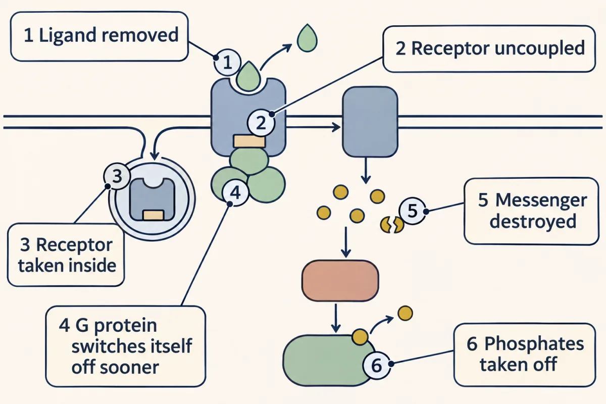
You met the “on” pathway in biochemistry: adrenaline, receptor, G protein, adenylyl cyclase, cyclic AMP, protein kinase A, phosphorylated targets. From the outside in, six removal steps can end it: the ligand leaves; the receptor loses access to its G protein; the receptor is taken inside in a vesicle; G-alpha hydrolyzes its GTP; a phosphodiesterase destroys cyclic AMP; and phosphatases reset the targets.
The experiment: the mark is necessary but not sufficient
Robert Lefkowitz’s laboratory at Duke University knew that phosphate appeared on the beta-adrenergic receptor as its response faded. Protein kinase A, activated by cyclic AMP, was the obvious suspect.
Jeffrey Benovic tested it with the kin⁻ line of S49 mouse lymphoma cells, which has no working protein kinase A. He mixed an extract with hamster lung receptor rebuilt into lipid vesicles and radioactive ATP.
Predict. Why test an extract from kin⁻ cells instead of relying only on a protein kinase A inhibitor? What would phosphorylation in that extract establish?
Check your prediction
The genetic control removes working protein kinase A instead of trusting a drug’s completeness and specificity. Phosphorylation still occurred, so another kinase was at work. Isoproterenol increased that phosphorylation, whereas alprenolol blocked the increase, showing that the kinase recognized the active receptor.
The first film showed the purified kinase marking the receptor without silencing it; the missing reader came from vision. Hermann Kühn, at Jülich in West Germany, had described an abundant retinal protein, then called the 48K protein, that works with rhodopsin kinase to shut down rhodopsin, the rod cell’s light receptor. It was later named arrestin because it arrested rhodopsin’s function. The writer, a G-protein-coupled receptor kinase (GRK), preferentially marks the active receptor.
In the lesson's test-tube experiment a kinase marks only an active receptor and arrestin reads the mark: with both, the receptor lost about 40% of its ability to switch on Gs; with one missing, little changed.
The retinal protein was a stand-in. Three years later Martin Lohse, a fellow in the same laboratory, found the non-retinal version, now called beta-arrestin. It reduced signaling by the kinase-marked receptor by more than 75% and did not act on rhodopsin. Arrestin can also bind only the marked receptor tail, leaving the core accessible to a G protein; that refinement matters when receptors signal inside the cell.
Arrestin carries the receptor into the cell
Clathrin cages a patch of surface membrane and pulls it inward as a coated pit. In 1996 Benovic’s laboratory, then at Thomas Jefferson University in Philadelphia, reported with James Keen and Vsevolod Gurevich that beta-arrestin 1 and beta-arrestin 2 bind clathrin directly and tightly. In intact cells the three came together when agonist was present.
Predict. A hybrid arrestin still binds the marked receptor but cannot bind clathrin. What happens first to G-protein coupling, and what happens to receptor uptake?
Check your prediction
It should still uncouple the receptor if it covers the core, but carry the receptor into the cell less well. In the 1996 study, hybrids lacking receptor binding or clathrin binding supported less uptake, so uptake needs both contacts: the protein that blocks the G protein’s site also pulls the receptor into a coated pit.
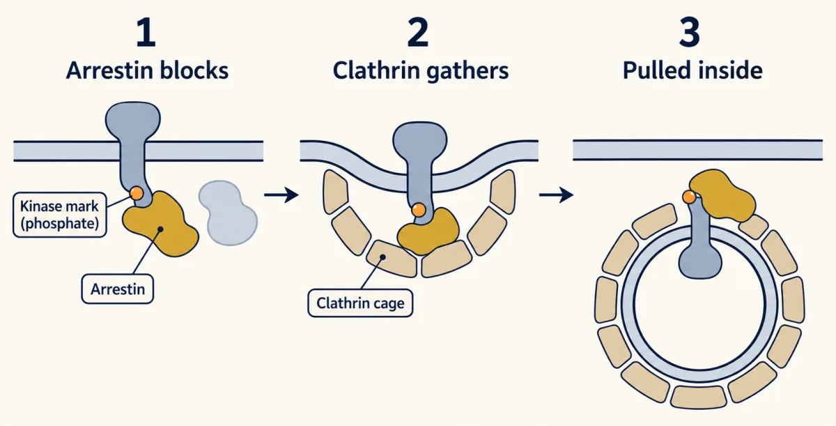
G-protein-coupled receptors (GPCRs) can be unfaithful to trimeric G proteins. Once arrestin replaces the G protein, the receptor’s new partner has contacts of its own: besides clathrin and the adaptor AP-2, it can bind Src and parts of the extracellular signal-regulated kinase (ERK) and c-Jun N-terminal kinase (JNK) kinase cascades. How much signaling this supports without a G protein remains debated.
An endosome receives incoming vesicles and was long assumed to take receptors out of service. In 2013 Roshanak Irannejad, in Marc von Zastrow’s laboratory, found active receptor and G protein at the early endosome membrane, which a later lesson takes up.
The receptor rule, then: an occupied receptor loses its G protein only when a dedicated protein acts on it, and where it sits does not tell you whether it is still signaling. Two findings would break the rule: an occupied receptor that uncouples with nothing acting on it (no mark, reader or other bound protein), or a receptor that stops signaling every time it leaves the surface, in every cell tested.
So measure the active forms themselves; the second film follows them below the receptor.
Read the transcript of “Where the Signal Runs Out”
Film transcript: Where the Signal Runs Out
The first film stopped new G-protein activation with a mark and its reader. What ends the active signal already downstream? Each layer is an active pool with an input and an exit. Receptors produce active G proteins. Active G proteins help produce second messenger.
Messenger activates a kinase, which adds phosphate marks to targets. These pools overlap in time. Stopping new receptor output does not instantly erase GTP, messenger, or target phosphate already present. Each pool keeps changing until its own input and exit settle the balance. G-alpha carries GTP while active and hydrolyzes it to GDP. An RGS protein speeds that reaction for compatible G proteins.
Faster hydrolysis shortens the lifetime of the active green pool. A phosphodiesterase drains a cyclic-nucleotide pool by opening the messenger's phosphate ring. When synthesis and breakdown match, the pool holds steady. Slower breakdown makes the same input build a higher, longer-lasting pool. Now shut messenger synthesis off, then inhibit the drain. Can protecting the remaining messenger create a new signal?
No. No new messenger is made. An empty pool stays empty. Messenger already present is protected and decays more slowly. With continuing input, inhibition raises and prolongs the pool. The last pool here is phosphorylated target protein. A kinase adds each gold phosphate badge. A protein phosphatase removes it. The phosphorylated pool fades when removal overtakes new phosphorylation. If that phosphate activates the target, its activity fades too. Changing an exit changes both amplitude and timing. A narrow exit allows an active pool to accumulate. A wide exit keeps its level lower and clears it sooner after production stops. The same bookkeeping shapes time.
Under constant input, an active pool rises toward a balance. When input stops, removal makes it decay. A faster removal process lowers the balance and clears the pool sooner. Some systems also build a slower adaptive brake while stimulation continues. A gap can let that brake reverse.
Separated pulses offer recovery intervals that one steady input never provides. Separated pulses can preserve responsiveness because each gap allows recovery. Constant input gives no such interval. Pituitary responses to gonadotropin-releasing hormone show this contrast. In gonadotrope cells, the main adaptation appears to lie downstream of the receptor. Receptor uncoupling is therefore only one timer. GTP hydrolysis, messenger breakdown, and dephosphorylation set later timers. Each downstream layer fades when removal outruns production, and the slowest surviving layer can outlast the receptor's activity.
From the film
- Each downstream layer fades when removal outruns production.
- RGS proteins speed GTP hydrolysis, phosphodiesterases destroy cyclic nucleotides, and phosphatases remove target phosphates.
- A breakdown inhibitor makes no new messenger; it slows the decay of messenger already present and, with continuing input, raises and prolongs the pool.
- Pulses leave time for adaptation to reverse; a steady GnRH signal does not.
Three downstream removal processes
Which G proteins count as “compatible” for the second film’s RGS protein? Regulators of G-protein signaling, or RGS proteins, from the lesson on GTPase switches, are selective. RGS4 and GAIP accelerated GTP hydrolysis by Gi alpha-1 at least 40-fold and acted on the other Gi-family subunits tested. They did not act on Gs, the alpha subunit in the beta-adrenergic example. A 1997 study from the same group showed that RGS4 and GAIP also act on Gq alpha, so making more of a compatible RGS protein can shorten a response.
Predict. In otherwise identical tubes, add RGS4 to active Gi alpha-1 or active Gs alpha. Which GTP-bound pool should disappear faster?
Check your prediction
The Gi alpha-1 pool. “RGS protein” names a family, not a guarantee that every member acts on every G-alpha subunit.
A phosphodiesterase turns cyclic AMP into AMP, which does not activate protein kinase A. To quantify the pool, assume synthesis is constant and breakdown is first-order, meaning proportional to the amount present. At steady state synthesis equals breakdown, so:
messenger level at steady state = rate of synthesis ÷ rate constant of breakdown
Predict. If synthesis doubles while the breakdown rate constant stays fixed, what happens to the steady messenger level?
Check your prediction
It doubles. Slowing breakdown at fixed synthesis also raises the level, and the messenger lingers.
Two drugs show the second film’s limit in practice. Caffeine inhibits phosphodiesterases in a test tube, but at the concentrations a few cups produce, most of its effect is thought to come from blocking adenosine receptors. Sildenafil, the cleaner example, inhibits phosphodiesterase 5, which breaks down cyclic GMP, a close relative of cyclic AMP. Nitric oxide starts cyclic GMP synthesis in the smooth muscle of some blood vessels, and the muscle relaxes; sildenafil makes that existing signal larger and longer.
At the last layer, a protein phosphatase removes the phosphate protein kinase A added, as in the lesson on the phosphorylation switch. At each layer, ask whether active material is being made faster or removed faster.
Constant GnRH shuts the pituitary down
Gonadotropin-releasing hormone, or GnRH, is a hypothalamic peptide that tells the pituitary to release gonadotropins, the hormones that drive the ovaries and testes. In 1978 Belchetz and colleagues studied rhesus monkeys whose hypothalamic lesions had abolished gonadotropin release. GnRH given once an hour restored secretion; a constant infusion did not. Clinics use sustained GnRH agonists to suppress gonadotropin output.
The second film’s adaptive brake, which reverses in the gaps, is a general possibility, not a location. The mammalian GnRH receptor lacks a C-terminal tail, resists agonist-dependent phosphorylation, and resists rapid desensitization. A GRK mark and arrestin therefore cannot be the whole explanation.
Predict. If adding a suitable C-terminal tail restores phosphorylation sites, what should happen to rapid agonist-dependent desensitization?
Check your prediction
It should increase. Tail-addition experiments support this, tying the missing tail to resistance against the rapid route, but they do not show that the tail explains long-term pituitary shutdown.
To locate the longer-term block, count surface receptors, measure second messenger made per dose, and bypass the receptor. A receptor-level failure lowers receptor number or messenger output; a downstream failure can leave both relatively intact while secretion falls. A bypass stimulus then places the defect relative to where the bypass enters: rescue implicates a bypassed step, and continued failure puts the block farther downstream. In gonadotrope cell lines, evidence points mainly downstream: sustained GnRH causes a fast loss of the inositol trisphosphate (IP3) receptors that release calcium, so the calcium response fades. This is the best-studied change; how much it explains in a patient receiving a GnRH agonist is less settled.
Design problem
Design a scheme to down-regulate a receptor using protein kinase C (PKC) and protein kinase A (PKA). Say which signals each kinase reports, why this differs from the kinase that marks mainly the occupied receptor, and what the cell gains by having both.
Saved in this browser only.
One way to do it
Protein kinase A, switched on by cyclic AMP, reports Gs-coupled receptor activity somewhere in the cell. Conventional and novel protein kinase C forms, switched on by diacylglycerol (conventional forms also need calcium), report phospholipase C branch activity. Neither kinase knows which receptor produced the messenger.
Give the receptor cytoplasmic serines matching both kinases’ target sequences; when either messenger rises, its kinase phosphorylates them and weakens coupling. That is negative feedback, the motif from the lesson on signaling circuits.
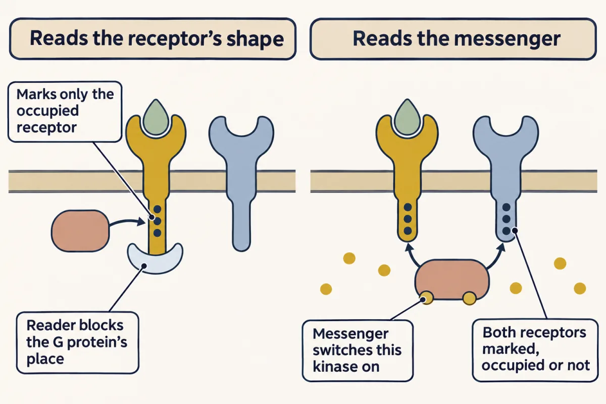
A GRK reads receptor shape, so it marks mainly agonist-occupied receptors. Kinases A and C read messengers, so they can mark any receptor carrying their sites, occupied or empty, including receptors for a different hormone. The first is homologous desensitization; the second is heterologous desensitization.
The two controls differ in reach: messenger-reading kinases act cell-wide when messenger is high, whereas a GRK and arrestin silence mainly the receptors being hit, leaving the cell able to answer a different hormone.
Going deeper
The reconstitution numbers
Benovic purified the kinase from cow brain cortex 20,000-fold. A batch purified only 350 times had cut the receptor’s ability to activate its G protein by up to 80%, and the pure enzyme cut it by only 10 to 15%. With the pure kinase alone, the receptors carried an average of about 8 phosphates each and could still switch on Gs. With kinase and arrestin together, activity fell to about 59% of the untreated receptor’s, a loss of about 40%. Arrestin added to a receptor the kinase had never marked left activity at 98.1% of the value without arrestin, across 13 measurements. Neither bovine serum albumin, an unrelated protein, nor heat-denatured arrestin could substitute for arrestin.
Where GAIP sits
GAIP is held on a membrane by palmitate, a fatty acid bonded to the protein. In 1998 Marilyn Farquhar’s laboratory reported it on clathrin-coated vesicles, some budding from the surface and some from the Golgi: the first GTPase-activating protein found on such vesicles.
The GnRH receptor has no tail
The finding that the missing C-terminal tail makes the gonadotropin-releasing hormone (GnRH) receptor resist agonist-dependent phosphorylation and rapid desensitization comes from Willars and colleagues in 1999. The receptor works mainly through Gq and phospholipase C, so cyclic AMP is not the main messenger here.
Your notes
Select any text in the lesson to highlight it or add a note to it. Your highlights and notes are saved in this browser and collected in your notebook.
This browser is not letting the site save anything (a private window or strict privacy settings), so highlights and notes will be lost when you leave the page.
How we measure it
Reconstitution in lipid vesicles
Purify each protein, then rebuild the pathway from its parts: the receptor is inserted into phospholipid vesicles, and the G protein, the kinase and any candidate protein are added one at a time. If an effect appears only when a given part is present, that part is required. Here the readout was the receptor's ability to activate Gs.
Activity-guided purification
Pass a tissue extract through a series of columns, and after each one measure how much protein is left and how much activity. Fold purification is activity per milligram compared with the starting extract. When one activity survives and another is lost as purity rises, the columns have separated two components.
A mutant cell line as a negative control
The kin⁻ line of S49 lymphoma cells has no working protein kinase A. If an event still happens in an extract of these cells, protein kinase A cannot be required for it. A genetic control of this kind tests one suspect without relying on a drug, which may block its target only partly or hit other kinases too.
Low-stringency library screening
Probe a library of cloned DNA with a labeled piece of a known gene, under conditions that tolerate mismatches. Clones that bind the probe imperfectly are candidate relatives of the known protein. Beta-arrestin was found this way, with retinal arrestin as the probe.
Practice questions on this lesson (3) → MCAT-style, original, each with an explanation
Where this lesson fits
Before thisReview first
- GTPase Switches: The TimerThis course · Lesson 7
RGS proteins here speed up the G-alpha GTP cut, which that lesson explains as the GTPase timer.
- GPCRs and Trimeric G ProteinsThis course · Lesson 12
You need the receptor, G protein and adenylyl cyclase on-pathway to see what each off switch removes.
- Phosphorylation: Kinases and PhosphatasesThis course · Lesson 6
The kinase mark on the receptor and the phosphatase reset both rest on how kinases and phosphatases act as switches.
After thisStudy next
- Endocytosis and Signaling EndosomesThis course · Lesson 36
It takes further the arrestin-clathrin trip into the cell and asks whether the receptor keeps signaling from endosomes.
- Sensing the World: Vision, Smell, Nitric Oxide and cGMPThis course · Lesson 17
It uses phosphodiesterase breakdown of cGMP and rhodopsin kinase with arrestin to shape the rod's response.
- The Shape of a Signal: Size, Duration, Frequency and PlaceThis course · Lesson 35
It builds on how pulses versus steady signals are read, as in the GnRH case.
Suggested by an AI model from the lessons themselves, then checked. How we use AI
Finish line
Test yourself
8 questions
Practice and MCAT-style, with explained answers.
Next · Lesson 17
Sensing the World: Vision, Smell, Nitric Oxide and cGMP
Your rods report light by turning a current down. Why would a sense run backwards?
Continue →