Biochemistry · Lipid Metabolism
Lipid Synthesis: Building Cholesterol from Acetyl-CoA
Rewritten on : new text, figures and flashcards. Your notes and highlights stay, but the text around them may have moved.
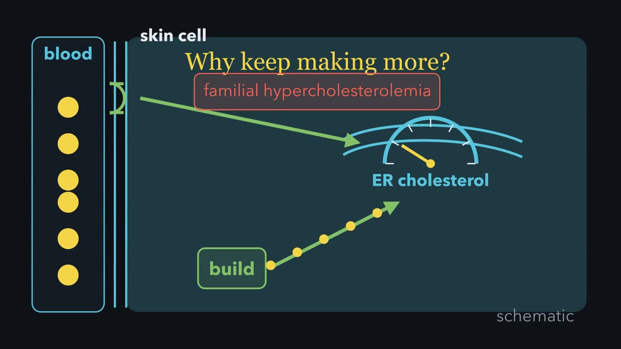
Skin cells from children whose blood is flooded with cholesterol keep making more of it. Why would a cell bathed in cholesterol keep building it?
Pause when the reading reaches a film, and offer to play it. Saved on this device.
Watch the film now, or keep listening
Builds on:Metabolism: Basic Concepts and DesignMembranes: The Impermeable Sheet That Builds ItselfFatty Acid Synthesis: Building the Chain Two Carbons at a TimeWhy these?
 ▶ Watch The cholesterol gauge · 3:15
Read the transcript of “The cholesterol gauge”
Film transcript: The cholesterol gauge
Why would a cell surrounded by cholesterol keep making more? Brown and Goldstein saw this in skin cells from children with familial hypercholesterolemia. LDL crowded the medium, yet the cells behaved as though cholesterol were scarce.
A liver cell gets cholesterol through two supply lines. It can build cholesterol, or pull LDL from blood through LDL receptors. But the cell does not measure LDL outside. It measures cholesterol reaching its endoplasmic reticulum membrane. On the import line, an LDL receptor captures LDL at the cell surface. The particle enters, and its cholesterol joins the cell's internal pool. A working receptor changes what the gauge can see. On the synthesis line, acetyl-CoA feeds the long road toward cholesterol. The main throttle, though not the only one, is HMG-CoA reductase. It makes mevalonate in a rate-limiting, irreversible step, and statins inhibit it.
One ER cholesterol gauge sets both synthesis and LDL uptake. SCAP is the sensor, and SREBP-2 is the passenger. When cholesterol binds SCAP, Insig grips SCAP. The complex stays in the ER. When ER cholesterol falls, SCAP escapes Insig. It carries SREBP-2 to the Golgi, where enzymes release its active piece. That piece enters the nucleus. It raises both HMG-CoA reductase and the LDL receptor. Predict the statin's indirect effect. If synthesis falls, what happens to LDL receptor number? Fewer, more, or unchanged?
A statin lowers cholesterol production, so the ER gauge reads low. SREBP-2 then turns up the LDL-receptor gene. More receptors pull more LDL from blood. The drug closes the making line, and the gauge opens the import line. Familial hypercholesterolemia reveals the same logic from the opposite direction. With too few working receptors, little LDL cholesterol gets inside. In the dish, the skin cells' ER gauge stayed low, so synthesis kept running. In the body, blood LDL rises mainly because clearance is slow. Follow cholesterol into the ER, and the puzzle dissolves. A cell responds to cholesterol it can sense, not cholesterol merely nearby. Low ER cholesterol turns up both making and importing.
From the film
- A liver cell gets cholesterol by making it or importing LDL.
- In liver cells, ER cholesterol controls both supply lines through SCAP and SREBP-2.
- Statins block HMG-CoA reductase and indirectly raise LDL receptors.
- In most familial hypercholesterolemia, too few working LDL receptors slow clearance.
The film above opens with Brown and Goldstein’s familial hypercholesterolemia (FH) puzzle: a cell responds to cholesterol that reaches its interior, not low-density lipoprotein (LDL) merely surrounding it. Their radioactive-iodine measurements supplied the evidence: normal skin cells took up LDL, while FH cells barely did.
(Acetyl-CoA, the two-carbon unit that can burn for energy in the mitochondrion, also becomes storage fat and membrane phospholipids. This lesson follows cholesterol; the other two fates are in Going deeper. Building the fatty acids themselves has its own lesson, fatty acid synthesis.)
The one idea to carry through this lesson. The film above shows how ER cholesterol controls both synthesis and LDL uptake in liver cells through SREBP cleavage-activating protein (SCAP), SREBP-2 and Insig. A statin, cholesterol-absorption blocker or bile-acid resin lowers liver-cell ER cholesterol and therefore tends to raise LDL receptors.
Liver cells carry most of the body’s LDL receptors. This rule covers the sterol arm and has four limits, listed after the drugs.
The spine: condense, assemble, cyclize
All 27 of cholesterol’s carbons come from acetyl-CoA, across roughly thirty enzymatic steps, as Konrad Bloch and his colleagues proved with labeled acetate (Stories & asides has the shark-tank detour).
Condense (in the cytosol, ending on the ER membrane). Two acetyl-CoA make acetoacetyl-CoA, and a third gives 3-hydroxy-3-methylglutaryl coenzyme A (HMG-CoA), a six-carbon molecule. Then HMG-CoA reductase uses two NADPH to make mevalonate. The reductase sits in the ER membrane, its working end facing the cytosol. It runs the main rate-limiting, irreversible step, and it is the most important enzyme in this lesson.
Assemble. Three ATP phosphorylate mevalonate, and it loses one CO₂ to become isopentenyl pyrophosphate (IPP), the universal five-carbon unit. Molecules built from these C5 units are called isoprenoids. Each C5 unit has a pyrophosphate “head” and a branched “tail”. IPP and its isomer DMAPP join head to tail, releasing pyrophosphate each time: C5 plus C5 gives geranyl-PP (C10), and one more C5 gives farnesyl-PP (C15). Squalene synthase then joins two farnesyl-PP at their heads, the pyrophosphate ends, releasing both pyrophosphates and using one NADPH. The product is squalene (C30), and because the heads were joined, it has a tail at each end. (Textbooks name this one reaction both ways, “head to head” and “tail to tail”; it is the same coupling.) Hydrolysis of the released pyrophosphate pulls each coupling forward.
Cyclize. Squalene monooxygenase uses O₂ and NADPH to put an epoxide on one end of squalene, making 2,3-oxidosqualene. Oxidosqualene cyclase then builds all four fused rings and sets the stereochemistry in a single turnover, one of the most elegant enzyme steps in biochemistry. The product is lanosterol. Roughly nineteen more steps, many using O₂ and NADPH, trim it to cholesterol. (The film splits the same road slightly differently: its first stage runs all the way to the isoprene unit.)
Predict. Eighteen acetyl-CoA go into one cholesterol, bringing 36 carbons. Cholesterol has 27. Where do the other nine go?
Check your prediction
Six leave as CO₂, one from each mevalonate as it becomes IPP, so six IPP carry 30 carbons. Squalene and lanosterol keep all 30, and the trimming removes three methyl groups, leaving 27. The ledger:
| Stage | Carbons | Spent on the way |
|---|---|---|
| 18 acetyl-CoA | 18 × 2 = 36 | — |
| 6 mevalonate | 6 × 6 = 36 | 6 × 2 = 12 NADPH at the reductase |
| 6 IPP | 6 × 5 = 30 (6 CO₂ lost) | 6 × 3 = 18 ATP |
| squalene, 2,3-oxidosqualene, lanosterol | 30 | 1 NADPH at squalene synthase, 1 at squalene monooxygenase |
| cholesterol | 27 (3 methyls trimmed) | many more NADPH and O₂ in the trimming |
So making one cholesterol costs 18 acetyl-CoA, 18 ATP and 14 NADPH (12 + 1 + 1) just to reach the epoxide, with more to come in the trimming.
Walk the spine below. Its meter counts each step once; the ledger multiplies by six where a step runs six times.
regulated step − used up · + produced
Two two-carbon acetyl-CoA units condense head-to-tail, releasing one CoA. This is where a 27-carbon molecule literally begins counting up from two-carbon pieces.
A third acetyl-CoA is added to make 3-hydroxy-3-methylglutaryl-CoA (HMG-CoA), the six-carbon substrate one step before the throttle. The cytosolic pool feeds cholesterol; a separate mitochondrial pool feeds ketone bodies.
The main rate-limiting, irreversible step, and the one that commits carbon to the mevalonate pathway. Two NADPH reduce HMG-CoA to mevalonate. This one enzyme is controlled at four levels (SREBP transcription, translation, AMPK phosphorylation, sterol-triggered degradation); in cultured cells the combined feedback spans more than 200-fold.
Regulation. Sterol end-product feedback plus SREBP and AMPK give this step its enormous dynamic range.
Clinical. Statins are nanomolar competitive inhibitors that mimic the reaction intermediate. Blocking the enzyme lowers cell cholesterol, which turns SREBP-2 on, so the liver makes more LDL receptors and clears even more serum LDL.
Three ATP phosphorylate mevalonate, then a decarboxylation loses CO₂ to make IPP, the universal five-carbon isoprenoid building block (it interconverts with its isomer DMAPP). Cost is 3 ATP per IPP, and six IPP go into one cholesterol (18 ATP).
Clinical. Nitrogen-containing bisphosphonates, the main osteoporosis drugs of this class, inhibit farnesyl-PP synthase downstream of here, in osteoclasts.
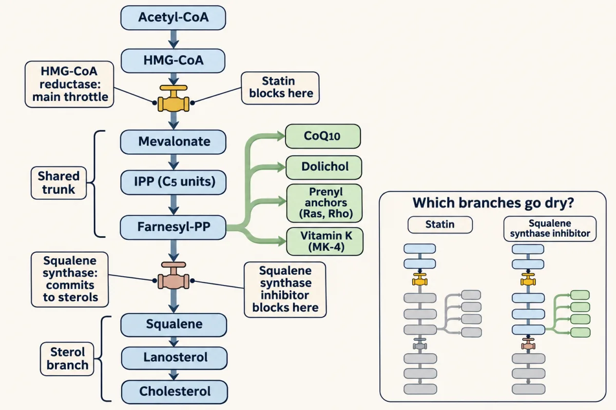
Five-carbon units add head-to-tail: DMAPP + IPP gives the ten-carbon geranyl-PP, then another IPP gives fifteen-carbon farnesyl-PP. Each coupling releases pyrophosphate, hydrolyzed to pull the reaction forward. Farnesyl-PP is the branch point: CoQ10, dolichol and the prenyl anchors leave the trunk here, above squalene synthase, so a statin cuts them and a squalene synthase inhibitor does not.
Clinical. Farnesyl-PP anchors Ras and geranylgeranyl-PP anchors Rho-family GTPases — prenyl lipid tethers for signaling proteins, one reason statins have effects beyond cholesterol.
Two fifteen-carbon farnesyl-PP units join at their pyrophosphate ends, releasing both pyrophosphates and consuming NADPH (textbooks call this coupling both head-to-head and tail-to-tail), to make the thirty-carbon linear hydrocarbon squalene. This is the committing step to sterols: everything past here is locked into the sterol branch, which ends mostly in cholesterol.
Clinical. Squalene is abundant in shark liver — the model Bloch first chose to chase this intermediate before switching to rats.
O₂ and NADPH install a single epoxide oxygen on one end of squalene, priming the linear chain for cyclization. A second regulated point in the pathway.
Regulation. Sterols speed its degradation; with the reductase it is one of the pathway's two irreversible control points.
One enzyme, one turnover, builds all four fused rings and sets the stereochemistry in a single cascade of ring closures and shifts — arguably the most elegant single-enzyme step in metabolism. A linear chain becomes a sterol nucleus.
Roughly nineteen steps, many of them using O₂ and NADPH, remove three methyl groups and adjust the double bonds to yield the final 27-carbon cholesterol. ER cholesterol is the gauge's reading: through SCAP and SREBP-2 it turns down the reductase gene and the LDL-receptor gene together; lanosterol-type intermediates and oxysterols also speed the reductase's destruction.
Clinical. Cholesterol is the scaffold for bile salts (CYP7A1), steroid hormones (P450s including aromatase), and vitamin D — one scaffold, many identities.
Where the throttle sits
Mevalonate is not only the road to cholesterol. Its C5 units also feed CoQ10, dolichol, the side chain of one form of vitamin K, and the prenyl anchors (farnesyl-PP, geranylgeranyl-PP) that tether signaling proteins such as Ras to membranes. So the pathway is a trunk that branches: everything up to farnesyl-PP is shared, and carbon turns into the sterol branch only at squalene synthase.
The cell puts its main brake at the reductase, so it stops carbon before any ATP is spent on the phosphorylation steps. A step is called committed when carbon that passes it has left the general pool for one pathway: the reductase commits carbon to the mevalonate trunk (which still branches), and squalene synthase commits it to sterols alone. The reductase is the main throttle, not the only one. Squalene monooxygenase is a second regulated point, and the gauge raises nearly every enzyme of the pathway.
Predict. One drug blocks 3-hydroxy-3-methylglutaryl coenzyme A (HMG-CoA) reductase (a statin). A research compound blocks squalene synthase instead. Which one also lowers CoQ10?
Check your prediction
The statin. CoQ10 branches off above squalene synthase, so blocking squalene synthase spares it. A statin acts at the top of the trunk and cuts the whole isoprenoid supply, CoQ10 and the prenyl anchors included. That branching is why statins have effects beyond cholesterol.
How LDL gets in
The second supply line starts in the blood, so follow the particle the liver’s receptors pull back in. Cholesterol does not dissolve in blood, so it rides in lipoproteins: a shell of phospholipid, free cholesterol and apolipoprotein around a core of triacylglycerol and cholesteryl ester. ApoB-100 is the address label the LDL receptor reads. The liver secretes VLDL (very-low-density lipoprotein); lipoprotein lipase strips its triglyceride to make IDL (intermediate-density lipoprotein); hepatic lipase finishes the job to make LDL. LDL is made in the blood, not secreted as such.
Cells take LDL up by receptor-mediated endocytosis. ApoB-100 binds the receptor, and the receptors cluster into a clathrin-coated pit that pinches off as a vesicle. In the acidic endosome the LDL lets go of its receptor. The receptor recycles to the cell surface, while the LDL goes on to the lysosome, which breaks it down and releases free cholesterol. HDL runs the traffic in reverse, pulling cholesterol back to the liver.
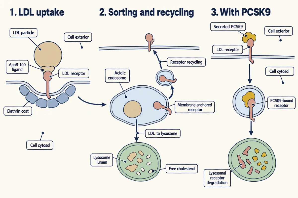
If the receptor is missing or broken, LDL stays in the blood. That is familial hypercholesterolemia, an inherited disorder usually caused by an LDL-receptor mutation, found in roughly one person in 250, and the disease that cracked the whole system open. Brown and Goldstein proved FH is a disorder of uptake, not synthesis. Even a receptor that binds LDL fails if it never gathers into a pit (Stories & asides, “Binding is not enough”).
One gauge, two supply lines
Making cholesterol is expensive and importing it is cheap; the reductase sits in the ER membrane. The film above shows how cholesterol in that membrane controls both supply lines. Here are the molecular parts. SREBP-2 (sterol regulatory element-binding protein 2) is a transcription factor made in an inactive form stuck in the ER membrane; it is the passenger. SREBP cleavage-activating protein (SCAP) is its escort, and SCAP’s sterol-sensing domain is what actually reads cholesterol. Insig is an ER membrane protein that grabs SCAP once SCAP has bound cholesterol.
| ER cholesterol low | ER cholesterol high | |
|---|---|---|
| SCAP | has no cholesterol bound, so Insig leaves it alone | binds cholesterol, changes shape, and is grabbed by Insig |
| The SCAP-SREBP-2 pair | is packed into COPII vesicles bound for the Golgi | is held in the ER |
| SREBP-2 | is cut twice in the Golgi, first by the protease S1P and then by S2P; the freed piece enters the nucleus and binds the sterol regulatory element | never reaches the proteases and stays inactive |
| Genes switched on | the reductase and the other synthesis enzymes, and the LDL receptor | none of them, so their expression falls |
The bottom row is the point: one switch turns on both supply lines.
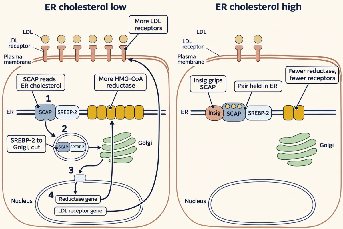
The film above keeps the wiring explicit: cholesterol binds SCAP, and Insig binds SCAP; SREBP is the passenger. Oxysterols such as 25-hydroxycholesterol bind Insig instead of SCAP; either way SCAP is held in the ER.
The gauge works over hours, because it changes how much protein is made. The reductase also has faster controls, each reading something specific.
| Control on the reductase | What it reads | What it does | How fast |
|---|---|---|---|
| Transcription (SREBP-2) | ER cholesterol | changes how much reductase is made | hours |
| Degradation | sterols, chiefly lanosterol-type intermediates and oxysterols, helped by nonsterol isoprenoids | when they are high, Insig sends the reductase for destruction in the ER | tens of minutes to hours |
| Translation | nonsterol products of mevalonate | slows reading of the reductase mRNA | minutes to hours |
| Phosphorylation (AMPK) | the cell’s energy charge, not sterols | switches the enzyme off when ATP is low | minutes |
Three of the four read the pathway’s own products; only AMPK reads fuel, a famine brake on an expensive molecule. These layers overlap: sterols act through Insig at two of them. Of the four, only the transcription control, the gauge itself, also sets the low-density lipoprotein (LDL) receptor.
Predict. A statin blocks the reductase, so the liver makes less cholesterol. What happens to the number of LDL receptors on a liver cell, and to LDL in the blood?
Check your prediction
Receptors go up and blood LDL goes down. With less synthesis, ER cholesterol falls, the gauge reads low, and freed SREBP-2 turns on the LDL-receptor gene. The statin closes one supply line and the gauge opens the other, so a drug that blocks synthesis ends up working largely by boosting uptake. Statins lower blood LDL on average by 30 to 50 percent.
Statins are nanomolar competitive inhibitors shaped like the reaction intermediate. The low reading also turns the reductase gene up, so a treated cell makes extra reductase for the drug to block. That compensation is part of the “rat paradox” Akira Endo ran into (Stories & asides).
Predict. A child has no working LDL receptors. Her blood LDL is several times normal. Brown and Goldstein grew her skin cells in a dish with LDL in the medium. Do those cells make more cholesterol than normal cells, or less?
Check your prediction
More. No LDL gets in, so the cell’s gauge reads low however much LDL is in the medium, and SREBP-2 keeps synthesis running. That is what Brown and Goldstein saw in the skin cells at the top of this lesson: the cells’ chemistry worked, but nothing told them to stop. (In the whole body her liver still gets cholesterol by other routes, so her blood LDL is high mainly because LDL is cleared slowly, not because her liver makes much more.)
Try both cases in the thermostat below: set how much cholesterol reaches the cell, toggle the statin, and follow the chain from ER cholesterol to blood LDL.
ER cholesterol is above the set point: SCAP is held by Insig in the ER, so SREBP-2 is never cut free, the reductase is shut down and degraded, and LDL receptors are sparse. With few receptors clearing it, blood LDL drifts up — the state a high-cholesterol diet pushes you toward. Tick the statin box to see what the drug changes.
One ER gauge (SCAP reading ER cholesterol) sets both supply lines: the reductase that makes cholesterol and the LDL receptor that imports it. HMG-CoA reductase is the committed, rate-limiting step, so the cell guards it four ways — SREBP-2-driven transcription, translational control, AMPK phosphorylation, and sterol-triggered degradation — and a statin adds a fifth, a competitive block. The clinical twist lives in the loop: when a statin starves the cell of cholesterol, the gauge reads low and the cell floods its surface with LDL receptors. Those receptors pull LDL out of the blood — which is how a synthesis inhibitor ends up lowering the number on your lab report. Tick the FH box and that second step disappears.
Same gauge, more drugs
SREBP-2 also turns on the gene for PCSK9, a secreted protein that binds the low-density lipoprotein (LDL) receptor and sends it to the lysosome to be destroyed, so fewer receptors return to the surface. A statin therefore raises PCSK9 along with the receptors, and gives back part of its gain.
Predict. A patient already on a statin adds an antibody that blocks PCSK9. Why does her LDL fall further?
Check your prediction
With PCSK9 blocked, more of the receptors the statin ordered survive. PCSK9 antibodies lower LDL by about 55 percent, alone or on top of a statin; inclisiran instead silences PCSK9’s mRNA in the liver.
Two more drugs act from the gut side. Ezetimibe blocks cholesterol absorption, so less cholesterol reaches the liver from the gut. Bile-acid resins trap bile acids in the gut, so the liver converts more of its own cholesterol into new bile acids. Either way the liver cell’s ER cholesterol falls, and the gauge answers with more receptors.
Where the rule stops.
- The energy brake is separate. AMP-activated protein kinase (AMPK) (last row of the table) also switches off fatty-acid synthesis, and it acts mainly on the enzyme itself, not through the sterol gauge.
- The gauge brakes itself through PCSK9, as above.
- It needs working receptors. Without them, as in the familial hypercholesterolemia (FH) child, the gauge calls for receptors that cannot be added, so a statin loses most of its LDL-lowering step.
- Fat synthesis follows a different lead. A sister factor, SREBP-1c, also escorted by SREBP cleavage-activating protein (SCAP), drives fatty acid synthase and answers mainly to insulin.
What cholesterol becomes
Most of a cell’s cholesterol stays in its membranes, and excess is stored as cholesteryl ester (the liver also exports it in lipoproteins). In particular tissues, cholesterol, or for vitamin D a late precursor, is also the scaffold for three kinds of product, each tailored by known enzymes:
- Bile salts. CYP7A1, the 7alpha-hydroxylase, commits cholesterol to bile salts.
- Steroid hormones (glucocorticoids, mineralocorticoids and sex steroids), tailored by tissue-specific enzymes such as the P450 aromatase, which makes estrogens, and 5alpha-reductase.
- Vitamin D, a secosteroid hormone made in skin from 7-dehydrocholesterol, a late precursor of cholesterol, then activated in liver and kidney.
Many drugs in this lesson, from statins to finasteride (a 5alpha-reductase inhibitor) to aromatase inhibitors, work by blocking one known enzyme in the pathway. In the chapter on integration of metabolism, these same switches answer to the fed and fasted state.
Going deeper
The other two fates of acetyl-CoA: the phosphatidate hub
Storage fat and membrane phospholipids share a backbone. GPAT adds a fatty acid to glycerol 3-phosphate at the sn-1 position, making lysophosphatidate; AGPAT adds the second at sn-2, making phosphatidate. The positions show up in downstream lipids: sn-1 tends to carry a saturated chain, sn-2 an unsaturated one. Mind the name. Phosphatidate is diacylglycerol-3-phosphate, not diacylglycerol; the phosphate is the whole point.
Phosphatidate is the fork, and it splits two ways:
- Remove the phosphate. PAP activity, carried by the lipin enzymes (Lipin 1 is the best studied of three), gives diacylglycerol. Diacylglycerol is acylated a third time into triacylglycerol, the storage form. But it also feeds membranes: the two most abundant membrane lipids, phosphatidylcholine and phosphatidylethanolamine, are made by handing diacylglycerol an activated head group from CDP-choline or CDP-ethanolamine.
- Add CTP. Phosphatidate becomes CDP-diacylglycerol, the activated donor for phosphatidylinositol, phosphatidylglycerol and cardiolipin (and, in bacteria and yeast, phosphatidylserine).
The CDP steps use the activated-intermediate logic you have seen with UDP-sugars: making the activated head group releases pyrophosphate, whose hydrolysis pulls the reaction forward.
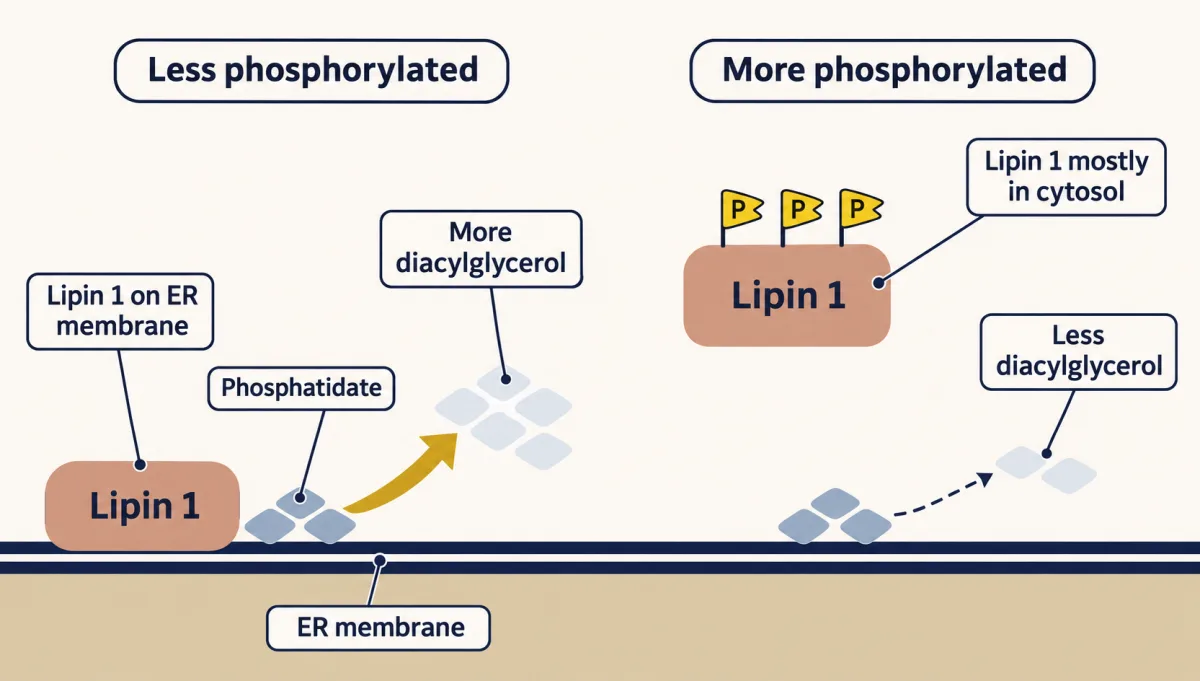
Lipin 1 localization in cultured fat cells
PAP is also controlled by where it sits. Less phosphorylated Lipin 1 binds the ER membrane, where its substrate lives, and makes diacylglycerol. More heavily phosphorylated Lipin 1 stays mostly in the cytosol, away from its substrate, and makes less. The effects depend on the lipin paralog, its phosphorylation and the cell type. In cultured fat cells, epinephrine and the fatty acid oleate dephosphorylate Lipin 1 and move it onto the membrane, while insulin, surprisingly, phosphorylates it and shifts more of it into the cytosol. Losing Lipin 1 causes lipodystrophy in mice and recurrent muscle breakdown (rhabdomyolysis) in children. Where an enzyme sits can matter as much as how much of it there is.
Sphingolipids and Tay-Sachs disease
Sphingolipids run on a different backbone entirely. Palmitoyl-CoA condenses with serine, and after a few steps you reach ceramide, the sphingolipid hub. Add phosphocholine and you get sphingomyelin; add sugars and you get cerebrosides and then gangliosides. The clinical anchor is Tay-Sachs disease: loss of hexosaminidase A blocks breakdown of the ganglioside GM2, which builds up in the lysosomes of neurons, and the result is fatal neurodegeneration.
More branches of the mevalonate trunk
The shared trunk above squalene synthase is why statins reach beyond cholesterol, and three details fill it in. The vitamin K made from mevalonate is MK-4, whose side chain is built on a ring taken from dietary vitamin K. Nitrogen-containing bisphosphonates, the main osteoporosis drugs of this class, inhibit farnesyl-PP synthase in osteoclasts, below mevalonate on the shared trunk. And while farnesyl-PP anchors Ras, geranylgeranyl-PP anchors Rho-family GTPases.
The numbers behind HMG-CoA reductase control
The four controls in the reductase table come with sizes. In cultured cells, sterol and nonsterol feedback together can change the amount of enzyme more than 200-fold. AMP-activated protein kinase (AMPK) phosphorylates the enzyme at Ser872. Sterol-triggered degradation can cut the enzyme’s half-life more than tenfold.
Your notes
Select any text in the lesson to highlight it or add a note to it. Your highlights and notes are saved in this browser and collected in your notebook.
This browser is not letting the site save anything (a private window or strict privacy settings), so highlights and notes will be lost when you leave the page.
How we measure it
Isotopic precursor tracing
Feed an animal acetate labeled with deuterium or carbon-14 and follow the atoms into the product. This is how Bloch and Rittenberg proved animals build cholesterol from two-carbon acetate, and how Bloch and Langdon later caught squalene as an on-pathway intermediate. Every one of cholesterol's 27 carbons traces back to acetyl-CoA, and the label is what proved it.
Microbial natural-product screening
Grow thousands of fungal strains, extract their broths, and assay each for the ability to shut down a target enzyme. Endo screened more than 6,000 strains to pull compactin from Penicillium citrinum; Alberts built a sensitive HMG-CoA reductase assay at Merck and pulled lovastatin from Aspergillus terreus. The principle: organisms make potent enzyme inhibitors as chemical weapons, and you can mine them.
Radioligand binding and receptor-mediated endocytosis assays
Tag LDL with radioactive iodine and measure how much a cell binds and internalizes. Brown and Goldstein showed normal fibroblasts gobble LDL while familial-hypercholesterolemia cells barely take it up — pointing to a defect in the receptor, not in cholesterol chemistry. Electron micrographs of one patient's cells then showed receptors that bind LDL but never cluster into clathrin-coated pits, so binding alone is not enough; location matters.
P450 monooxygenase chemistry
Cytochrome P450 enzymes install single oxygen atoms onto inert carbon scaffolds: R-H plus O2 plus NADPH yields R-OH plus water. Working with hydroxysteroid dehydrogenases, tissue-specific P450s turn the same cholesterol-derived backbone into cortisol, aldosterone, testosterone, and estradiol: CYP11A1 cuts the side chain to make pregnenolone, CYP17A1 cuts it again on the way to androgens, and aromatase converts androgens to estrogens. A P450 also strips a methyl group off lanosterol on the way to cholesterol. Aromatase (CYP19A1) and CYP7A1, the 7alpha-hydroxylase that commits cholesterol to bile salts, are both P450s. Because each isoform runs particular steps, and each gland expresses its own set, a drug such as an aromatase inhibitor can block one branch without shutting down the rest.
Practice questions on this lesson (3) → MCAT-style, original, each with an explanation
Where this lesson fits
Before thisReview first
- Metabolism: Basic Concepts and DesignThis course · Lesson 15
You need the energy economy of ATP and NADPH from there to count the cost of building cholesterol.
- Membranes: The Impermeable Sheet That Builds ItselfThis course · Lesson 12
You need membrane structure from there to see where the reductase and SCAP sit and why cholesterol matters.
- Fatty Acid Synthesis: Building the Chain Two Carbons at a TimeThis course · Lesson 26
You need acetyl-CoA as the cytosolic two-carbon building block and NADPH as the reducing power from there.
After thisStudy next
- Integration of Metabolism: How the Body Switches FuelsThis course · Lesson 30
It shows how fed and fasted hormones switch these cholesterol and fat pathways up or down across the body.
Elsewhere on the siteFilms, simulations and method films in other courses
- A Loop and a One-Way TripFilm · Cell Biology
It shows the LDL receptor looping back while its LDL goes one way to the lysosome.
- Binding or entry?Simulation · Cell Biology
It lets you separate LDL binding from LDL entry, which is the defect in familial hypercholesterolemia.
- An Enzyme Guards the ReceptorFilm · Physiology
It follows cholesterol into the adrenal steroid hormones and the enzymes that shape them.
- Where the Reader WorksFilm · Cell Biology
It shows how a signal sequence sends a protein to its compartment, as SCAP sends SREBP-2 to the Golgi.
Suggested by an AI model from the lessons themselves, then checked. How we use AI
Finish line
Test yourself
8 questions
Practice and MCAT-style, with explained answers.
Next · Lesson 28
Protein Turnover and Amino Acid Catabolism: Tag, Recognize, Destroy, Recycle
A protein's lifespan runs from minutes to a lifetime, set in large part by tags the cell reads and acts on.
Continue →← Lesson 26 · Fatty Acid Synthesis: Building the Chain Two Carbons at a Time