Blips from a Silent Nerve
Read the transcript of “Blips from a Silent Nerve”
Film transcript: Blips from a Silent Nerve
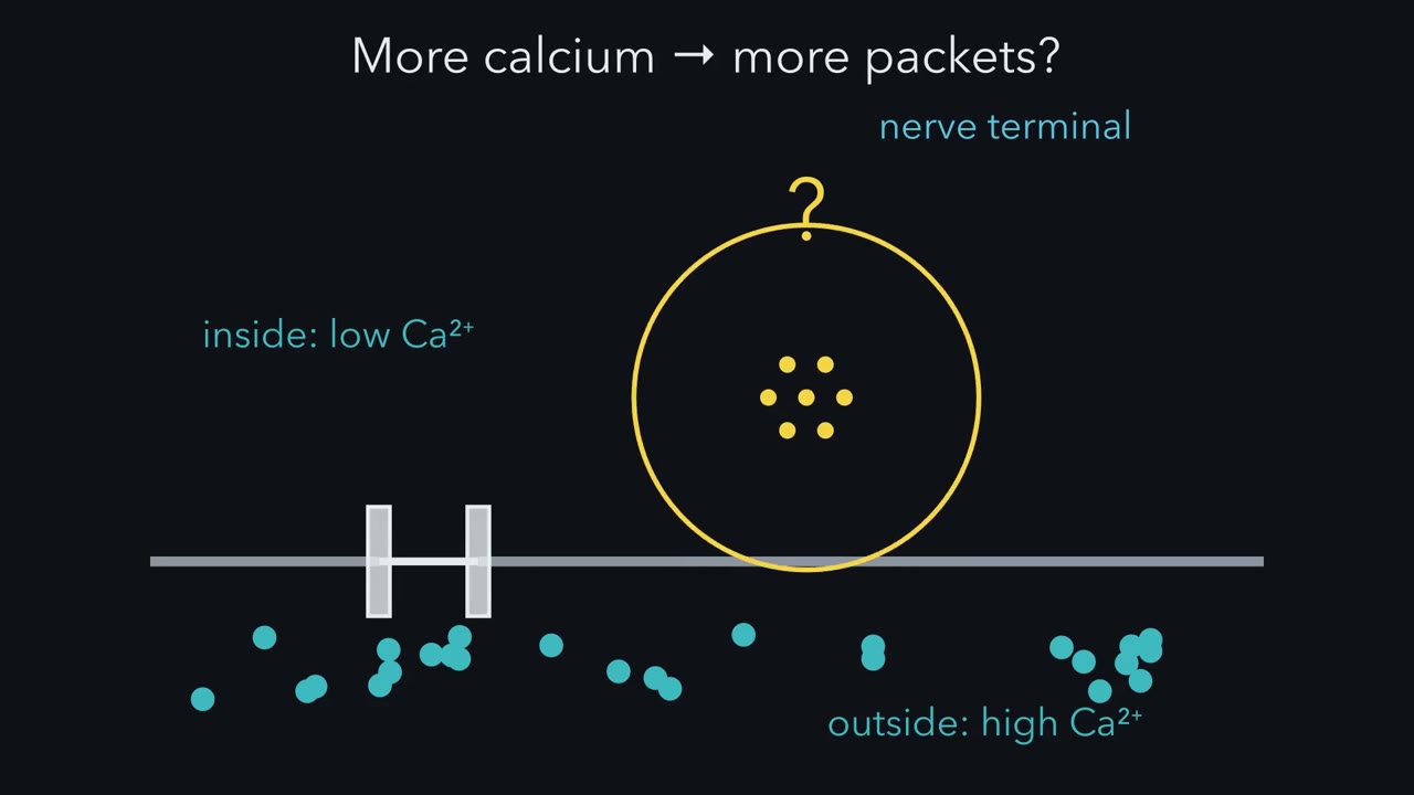
How does a nerve hand a signal to a muscle? And what is that signal made of? A synapse is a site where one neuron passes a signal to another cell. At a chemical synapse, the terminal releases a transmitter. It crosses a gap and acts on receptors. The best-studied example is the junction between a motor nerve and a skeletal muscle fibre. The signal crosses in a relay of five steps. For now, the names are enough. First, an action potential arrives at the nerve terminal. Second, calcium flows into the terminal. Third, vesicles fuse with the membrane and release their transmitter, acetylcholine. Fourth, acetylcholine crosses the gap. Fifth, it opens receptor channels on the muscle fibre, and the fibre depolarizes.
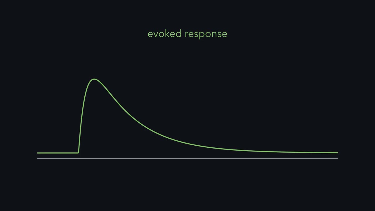
That depolarization is the endplate potential. The endplate is the patch of fibre under the terminal. Now the record that started the story. Paul Fatt and Bernard Katz put a fine glass electrode inside a frog muscle fibre. Voltage runs up the side. Time runs along the bottom. They did not stimulate the nerve at all. So the trace should be flat. Mostly it is. But small blips rise from it at irregular moments. Each blip is about the same size. Each is far too small to fire the fibre. Move the electrode away from the endplate, and the blips shrink and fade. So Fatt and Katz attributed them to acetylcholine released spontaneously by the terminal.
They called them miniature endplate potentials, and reported them in 1952. Each miniature looked like a scaled-down copy of the response the nerve evokes. Here is the puzzle. Is the nerve's real response built from blips like these?
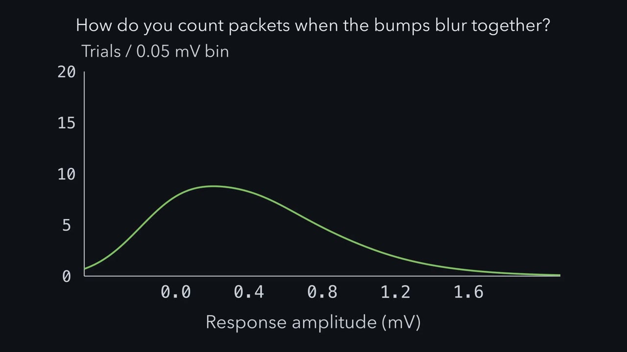
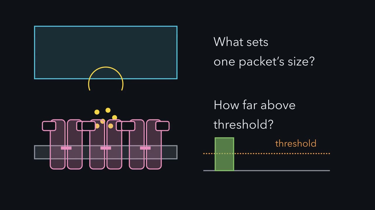
It is. The evidence comes in the next chapter. That answer raises more questions. How do you count units that merge together? Why does calcium matter so much? What sets the size of one unit? And how can a recording tell which side of a synapse is at fault? The miniature is the unit. Every evoked response at this junction is built from it.
A resting frog muscle fibre shows small voltage blips at its endplate with no nerve impulse at all. Those blips are the units of the evoked response at this junction.
Continue in the lesson: Synaptic Transmission and Quantal Release →
2:38 · Free, with captions and a transcript. The lesson has the full text, the questions and the sources.
More films from this lesson

How this film was made. Narration: an AI narrator voice (Kokoro, af_heart) reading narration written from this lesson's text. Animation: AI-generated in code (Manim) by Claude, a schematic with no drawn molecular structures. Captions come from the narration script. See something wrong? Report it.