Developmental Neuroscience · Unit 1: The problem and the tools
The Growth Cone: The Tip That Steers the Axon
Rewritten on : new text, flashcards and film. Your notes and highlights stay, but the text around them may have moved.
Actin at the front of a growth cone often streams backward even while the tip moves forward, and in the standard assay it turns toward a cue only 5 to 10% stronger on one side. How?
Pause when the reading reaches a film, and offer to play it. Saved on this device.
Watch the film now, or keep listening
Builds on:Seeing Neurons: Golgi, Cajal and the Neuron DoctrineWhy these?
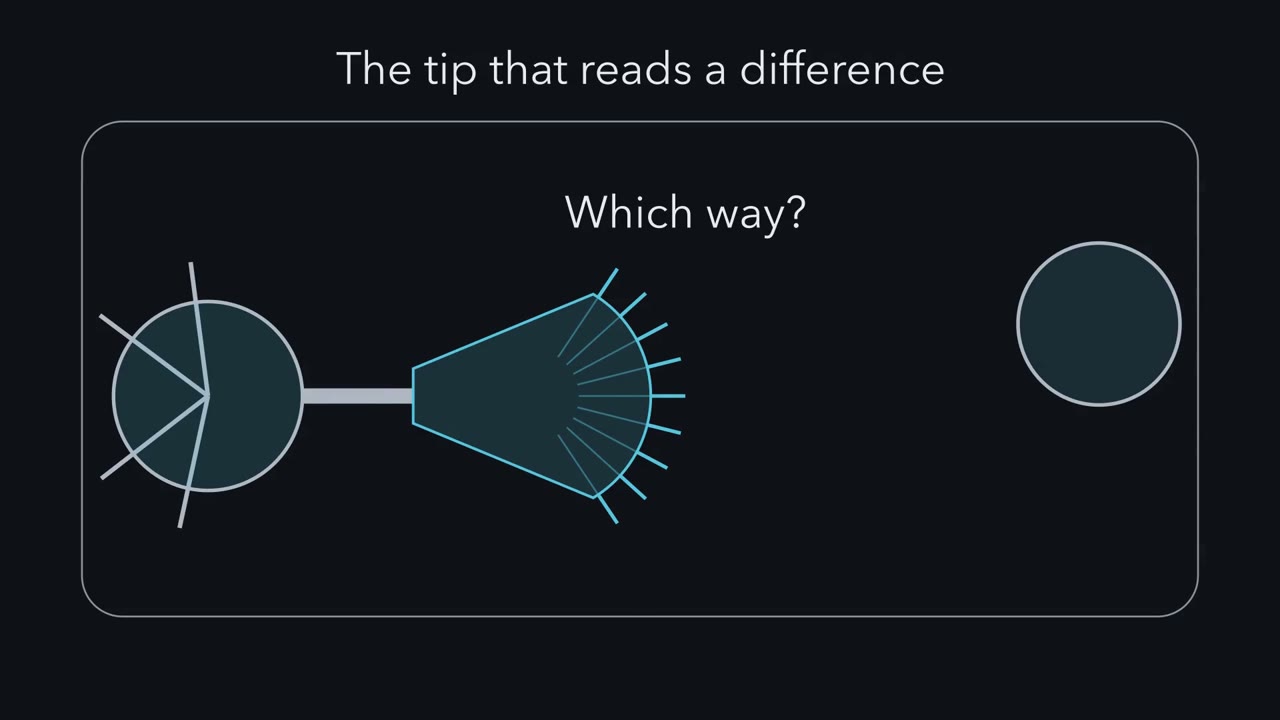 ▶ Watch The Tip That Reads a Difference · 2:12
Read the transcript of “The Tip That Reads a Difference”
Film transcript: The Tip That Reads a Difference
How does the tip of a growing axon know which way to turn? An axon is the long fibre a nerve cell sends out to reach its partner. At its end is a flat, moving fan called the growth cone. Microtubules fill the centre of the fan. Actin fills its edge, in thin spikes and flat sheets. The spikes are filopodia. They are feelers and feet at once, sampling the ground and gripping it. Molecules outside the cell, called guidance cues, attract or repel the growth cone. In a classic test, a fine pipette releases a cue beside a living growth cone. The cue thins with distance. So the near side meets a little more than the far side. How much more? In the standard assay, only about five to ten percent.
Now flood the whole dish with ten times more cue. Do growth cones turn harder? No. Across many cones there is no net turn. There is more cue, but no difference between the sides. So the difference across the width gives the direction. The overall amount changes how strongly cones respond. Here is the rule, for a growth cone on a flat surface in a dish. The edge moves forward by the actin added there, minus the actin that slides back. Grip on the ground, the clutch, holds back part of that slide. A turn sticks where microtubules move in and build new axon. Microtubules can also start the turn themselves. Three questions follow. How can a tip move forward while much of its actin streams backward?
How does so small a difference become a turn of the whole axon? And can the tip make that decision on its own? Carry one idea. A cue gives a direction by making the two sides of the growth cone differ.
From the film
- A growth cone steers by sensing guidance cues that attract or repel it.
- In the standard assay, the difference in cue across the width of the growth cone gives the direction, not the overall amount.
- The tip has microtubules in its centre and actin at its edge, in filopodia and lamellipodia.
A growing axon finds its partner because the growth cone at its tip senses guidance cues around it and steers toward or away from them.
An axon is the long fibre a nerve cell sends out to reach its partner. The growth cone is the flat, moving fan at its end. Guidance cues are molecules outside the cell that attract or repel axons. When a cue reaches one side of the growth cone more strongly than the other, it changes the machinery on that side, and the growth cone turns. Turn by turn, the growth cone leads the axon to the right target.
The layout
We know the axon is drawn out from its tip because someone watched one grow alive. In 1907 Ross Harrison kept a piece of frog embryo alive in a drop of clotted lymph and watched fibres grow out of it while their swollen ends changed shape: an axon is drawn out from one cell by movement at its tip. Santiago Ramón y Cajal had already named that swollen end the growth cone, from fixed tissue.
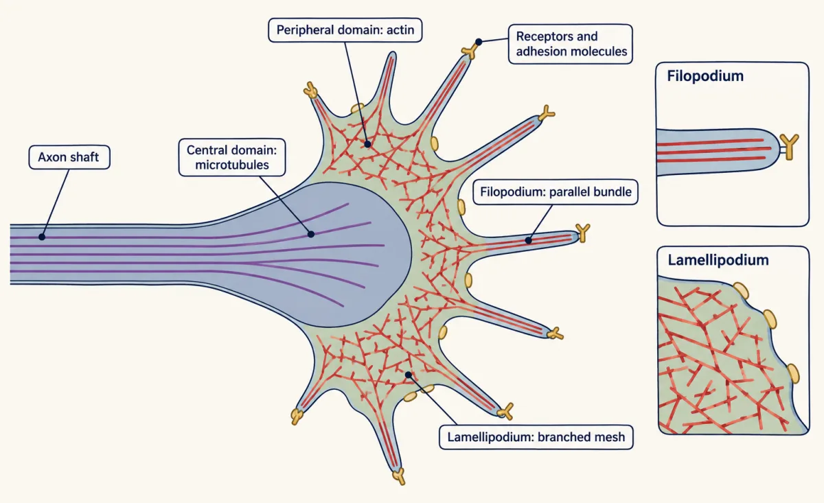
In culture, the fan’s parts divide the labour.
- The central domain sits where the axon shaft widens into the fan. It is packed with microtubules, hollow tubes about 25 nanometres wide built from dimers of alpha and beta tubulin.
- The peripheral domain is the fan itself, built of actin filaments, which are much thinner (roughly 6 to 10 nanometres). In filopodia, the thin spikes at the edge, actin filaments are bundled in parallel. In lamellipodia, the sheets between the spikes, they form a branched mesh.
- The surface carries receptors for guidance cues and adhesion molecules that grip the substrate (the surface it grows on) or other cells.
The filopodia are both feelers and feet: they carry receptors and adhesion molecules out ahead of the growth cone, sample what is there, and attach. The same tip senses its surroundings and moves through them.
The layout raises three puzzles.
- The actin in the fan streams backward even while the edge advances. How does the edge move forward?
- In the standard pipette assay, a cue is only 5 to 10% stronger on one side of the growth cone than the other. How does so small a difference turn the axon?
- The tip is far from the nucleus. Can it make the decision on its own?
The one idea to carry through this lesson. On a flat, adhesive surface, a growth cone’s edge moves forward by however much actin is added there minus however much the network slides back (retrograde flow). Adhesion to the surface, the clutch, holds back part of that slide. A turn sticks on the side where microtubules move in and turn the fan into axon, usually the side protruding more; microtubules can also start the bias themselves. A guidance cue gives a direction by making the two sides differ. The overall amount of cue gives no direction, though it changes whether and how strongly the cone responds.
Puzzle 1: how the edge advances
Read the transcript of “Engine and Clutch”
Film transcript: Engine and Clutch
How can the edge of a growth cone move forward while its actin network streams backward? Look inside the fan. New actin joins the filaments at the front edge. Myosin II pulls the network back toward the centre. Its own assembly pushes it back too. Near the centre, the filaments are taken apart. This backward movement is called retrograde flow. In 1988, Paul Forscher and Stephen Smith filmed this in a sea slug with giant, flat growth cones. They added cytochalasin, a drug that stops actin adding at the front edge. Addition at the front stops. What happens to the actin just behind the edge?
An empty band opens at the front and widens. The rest of the network keeps moving back. So the flow was there all along. Addition at the front had been hiding it. Then the microtubules push forward into the emptied band. The actin had been holding them back.
So why does the edge ever advance? In 1988, Tim Mitchison and Marc Kirschner proposed a clutch. Think of the actin network as a running engine, and the ground as the road. If actin is added and carried back at the same rate, the engine runs, and the edge goes nowhere. Adhesion molecules can link the network to the ground. Then the clutch engages, and the backward flow slows. If building stays the same, it now pushes the edge forward.
Put it as bookkeeping. Advance equals actin added at the edge, minus actin carried back. Suter and colleagues made the grip themselves. They held a bead coated with an adhesion molecule against the edge. Behind the bead, the backward flow slowed.
With illustrative numbers, flow falls from three and a half to half a micrometre a minute. Building stays the same, so the edge moves seven times faster. Now make the grip, or the building, unequal across the two sides of the cone.
Carry one idea. Grip can speed the edge just by holding back the flow, with no faster building.
From the film
- On a flat surface, the edge advances by the actin added at the front minus the actin carried back.
- Adhesion to the surface, the clutch, holds back part of the backward flow.
- Grip can speed the edge just by slowing the flow, without faster building.
Actin streams backward
Inside the fan, actin is never still. New subunits join the filaments at the end that faces the leading edge. The network is pulled back toward the centre by motor proteins (myosin II) and pushed back by its own assembly against the membrane (Medeiros and colleagues, 2006), and near the centre the filaments are taken apart. This backward movement is called retrograde flow. When addition and disassembly balance, it keeps roughly the same size while its parts stream backward.
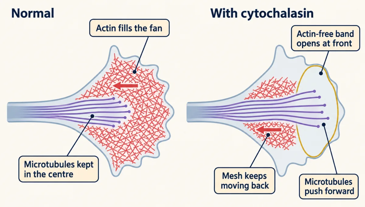
How do we know the flow is there, and not just a still mesh? Paul Forscher and Stephen Smith filmed growth cones of Aplysia, a sea slug with large, flat growth cones, and in 1988 added a cytochalasin, a drug that stops actin subunits adding at the leading edge.
Predict. The drug blocks addition at the front. What happens to the band of actin just behind the edge? And what do the microtubules in the centre do?
Check your prediction
A band free of actin opened at the front and widened, while the remaining network kept moving back. A still mesh would have stayed in place; a widening band means it was moving. So the actin had been flowing backward all along; addition at the front had been hiding it. Microtubules, normally kept in the centre, then pushed forward into the emptied band. The actin mesh had been holding them back.
The clutch
How does the edge move forward against that flow? Tim Mitchison and Marc Kirschner proposed an answer in 1988: the clutch hypothesis. Think of the actin network as a running engine and the substrate as the road. Suppose actin is added at the front and carried back at the same rate. Then the engine runs, the clutch is disengaged, and the edge goes nowhere. When adhesion molecules link the network to the substrate, the clutch engages and retrograde flow slows. In the simplest case, where addition stays the same, the same polymerization now pushes the edge forward.
rate of advance = rate of actin addition at the edge − rate of retrograde flow
This is bookkeeping at the edge, with both rates measured along the same direction: whatever is added and not carried back is the distance the edge gains. The grip on the surface runs through immunoglobulin-family adhesion molecules such as L1 and through integrins, receptors for the extracellular matrix.
The prediction was testable: grip should slow the flow and speed advance. In 1995 Forscher’s laboratory (Lin and Forscher) reported that where an Aplysia growth cone made a firm contact, flow slowed behind it and the growth cone advanced that way; across conditions, advance was inversely related to retrograde flow. Suter and colleagues (1998) then made the grip themselves. They held a bead coated with an adhesion molecule against the edge of an Aplysia growth cone, and backward flow slowed in line with the bead.
Worked problem: grip from a bead. Use illustrative values. Actin is added at the edge at 4.0 micrometres per minute, and the network flows back at 3.5 micrometres per minute. Under a restrained adhesive bead, flow falls to 0.5 micrometres per minute, and addition stays at 4.0.
Step 1: before the bead. Advance = 4.0 − 3.5 = 0.5 micrometres per minute. Most of the addition is carried straight back.
Step 2: under the bead. Advance = 4.0 − 0.5 = 3.5 micrometres per minute.
Step 3: compare. 3.5 / 0.5 = 7. The edge moves seven times faster, with no change in how fast actin is built.
Predict. Step 2 assumed addition holds steady when the clutch engages, and that need not be true. Suppose grip also slows addition by a fifth. Does the edge still speed up a lot, a little, or not at all?
Check your prediction
A lot. Addition falls to 0.8 × 4.0 = 3.2 micrometres per minute, so advance = 3.2 − 0.5 = 2.7 micrometres per minute, still more than five times the 0.5 before the bead. Grip would stop speeding the edge only if it cut addition to 1.0 micrometre per minute, a quarter of 4.0, or less. The conclusion, that grip speeds advance by slowing flow, does not hang on addition being exactly fixed.
Puzzle 2: how a small difference turns the axon
Read the transcript of “A Small Difference, a Turn”
Film transcript: A Small Difference, a Turn
How does a cue only five to ten percent stronger on one side turn a whole axon? In 1979, Gundersen and Barrett placed a pipette releasing nerve growth factor near chick sensory axons. Within about twenty minutes, the growth cones turned toward the pipette.
One growth cone wanders. So the evidence is the spread of turning angles across many cones. In a control dish with a uniform bath of cue, the spread centres on zero. Now apply the rule to each side. In one common picture, an attractant lets the near side gain more. It may add actin faster there, ease the backward flow, or engage more clutch. A repellent does the reverse.
The gaining side puts out more filopodia and sheets. The fan shifts that way. Microtubules from the centre enter the gaining side more often, and stay longer. Behind them the fan narrows, and that stretch becomes axon shaft, pointing the new way. Signalling inside the cell can enlarge the small difference. Repeated biased growth then adds up to a turn.
So stabilize the microtubules on one side, with no cue at all. Which way does the growth cone turn? Toward the drug. Buck and Zheng found this in frog spinal neurons. The near side protruded first, and the turn needed actin. Microtubules can start a turn by working through actin. Break microtubules down on one side, with a high enough dose, and the cone turns away.
Last question. Does the tip need its cell body to decide? In 2001, Campbell and Holt cut frog retinal axons, leaving growth cones with no cell body. The isolated tips still turned in gradients of guidance cues. their assay, turning needed protein made from messenger RNA already at the tip. Not every cue needs new protein. Chick sensory growth cones turned toward nerve growth factor without it.
So the tip can decide on its own, at least in a dish. Carry the rule. A turn sticks where microtubules move in and build axon, and they can start it too. The cues that make the sides differ, netrins, slits, semaphorins and ephrins, come next.
From the film
- A turn sticks on the side where microtubules move in and build new axon.
- Microtubules can start a turn themselves, working through actin.
- Isolated growth cones can turn in a dish without a cell body, though some cues need protein made at the tip.
The record
The test for steering is a pipette. In 1979 Gundersen and Barrett placed a micropipette releasing nerve growth factor (NGF) near growing axons of chick sensory neurons. The growth cones turned toward the pipette within about 20 minutes (Science 206:1079; characterized further in 1980, J Cell Biol 87:546). The pipette makes a gradient: the side of the growth cone nearer the source meets more of the cue than the far side. A growth cone that turns toward the source is attracted, and one that turns away is repelled. Either way, the cue can steer the axon.
In the standard form of the assay used today, a fine pipette about 100 micrometres from a growth cone releases a cue in brief, steady pulses, usually at 45° to the direction the axon is heading. The experimenter photographs the growth cone every few minutes for about an hour. Then they draw the axon’s path and measure the angle between its starting heading and the line its new stretch has taken.
One growth cone’s path bends toward or away from the pipette, but a single growth cone wanders, so steering is read across many: their turning angles form a distribution shifted to one side of zero. In a control dish, with a uniform bath of the same cue or a pipette with no cue, the distribution centres on zero.
How big is the difference?
Predict. The cue spreads out from the pipette tip and thins with distance. In these assays the growth cone is about 10 micrometres wide and about 100 micrometres away. Does its near side meet twice as much cue as its far side, a little more, or the same?
Check your prediction
A little more. Treat the pipette as a small source releasing steadily into still fluid in three dimensions; then concentration falls roughly as one over the distance. With the pipette side-on, the near edge is about 95 micrometres away and the far edge about 105, so the near edge meets about 105/95 ≈ 1.1 times as much: roughly 10% more. At the usual 45° it is about 7%. Real gradients depend on the cue and how it is delivered, and estimates for the standard assay are about 5 to 10% across the growth cone’s width. A uniform bath of ten times the concentration contains far more cue but no difference, so it offers no direction: across many growth cones, no net turn.
Try it: raise the level of the cue and then its steepness, and watch which one sets the direction.
- Attractive cue, side away from source
- 5.0
- Attractive cue, side nearer source
- 7.0
- Difference across the cone
- 2.0
In this sketch, each side’s response is assumed proportional to the cue it meets, so only the difference across the cone matters. The lesson says turning is a difference in balance between the two sides, not how that balance depends on cue; these numbers and the path are invented.
From a difference to a turn
Now apply the rule to each side. Each side of the edge advances by its rate of actin addition minus its rate of backward flow; call that its margin. In one common picture, an attractive cue raises the near side’s margin, by adding actin faster, by easing the myosin-driven flow, by engaging more clutch, or by a mix. A repellent lowers it, and some repellents collapse that side or, at high enough exposure, the whole growth cone. Signalling inside the cell can turn a 5 to 10% difference outside into a larger one between the sides (the guidance-receptor lessons).
The gaining side puts out more filopodia and lamellipodia, and the fan’s mass shifts that way. Microtubules are the stable scaffold of the axon shaft, so where they settle, the axon goes. Microtubules from the central domain enter the gaining side more often and stay longer. Behind them the fan narrows, and that stretch becomes axon shaft pointing in the new direction. Repeated growth biased toward one side adds up to a turn.
Microtubules steer as well as follow
If microtubules matter for the turn, could biasing them alone turn the cone with no cue at all? Buck and Zheng (2002) tested this in Xenopus spinal neurons with drugs released from a pipette onto one side. Zhou, Waterman-Storer and Cohan (2002) did the matching test on actin, removing the actin bundles from one side of the periphery.
Predict. Fill in the last column before you open the answer.
| Applied to one side | What it does there | Turn? |
|---|---|---|
| Uniform bath of a cue that attracts in a gradient | exposes both sides equally | ? |
| Attractant from a pipette | near side gains more | ? |
| Repellent from a pipette | near side gains less; some repellents collapse it | ? |
| Local loss of actin bundles | near side loses its actin bundles | ? |
| Low-dose taxol from a pipette | stabilizes microtubules on the near side | ? |
| Nocodazole from a pipette, enough to break down microtubules | near side loses microtubules | ? |
Check your prediction
No net turn; toward; away; away; toward; away.
Removing actin bundles on one side turned growth cones away from that side: microtubules withdrew from the region that lost its bundles and extended into regions where bundles remained, and the axon bent that way (Zhou, Waterman-Storer and Cohan, 2002). The outcome depends on bundle–microtubule interactions as well as the edge balance.
The two microtubule rows are Buck and Zheng’s results. Local taxol made the near side protrude more, and the cone turned toward the drug; that turn needed actin assembly and Rho-family GTPase activity. So microtubules can start a turn, working through actin, not only lock in one that actin began. Nocodazole that broke down microtubules on the near side turned the cone away; a tenfold lower dose in the pipette gave no turn. No cue gradient was imposed, so local changes to microtubules can set direction downstream of the cue. Some cues also act on microtubules directly. Actin and microtubules work together, and either one can decide which side wins.
One caution for the design problem: a turning angle means little if the cone barely advanced. In every arm, measure how far the treated cone grew, set a minimum before reading its direction, and record stalls and collapse separately.
What the rule covers. It is the cytoskeletal account of advance and turning for a growth cone on a flat, adhesive surface in culture, over minutes to about an hour. Three limits matter. First, it says how a difference between the sides becomes a turn, not which signalling route makes the difference (calcium, cyclic nucleotides, Rho GTPases, local translation) or what makes a cue attract rather than repel; those belong to the guidance-receptor lessons. Second, “advance equals addition minus flow” accounts for the leading edge only; turning the edge’s gain into axon also needs microtubules and organelles to move up behind it. Third, in the embryo axons also change course by collapsing and re-extending and by sprouting branches that are kept or pruned. In three-dimensional gels some central nervous system axons have been reported to extend with little dependence on adhesion (Santos and colleagues, 2020, Cell Reports); how much of the clutch account carries over to three dimensions is an open question.
Puzzle 3: can the tip decide on its own?
The growth cone can sit far from the cell body and its nucleus. Does it wait for instructions from them?
In 2001 Campbell and Holt cut the axons of retinal neurons from Xenopus frog embryos, leaving growth cones with no cell body attached. These are the axons of retinal ganglion cells, the retina’s only output, on their way to form the optic nerve (see the retina). Then they ran the turning assay on the isolated tips.
Predict. Does a growth cone with no cell body still turn in a gradient of a guidance cue? If it does, what could it still need?
Check your prediction
It turns. The isolated growth cones still turned in gradients of netrin-1 or Sema3A. In their assay the growth cones also needed to make protein: blocking protein synthesis in the growth cone itself blocked turning to these cues. The cell sends messenger RNA down the axon ahead of time, and the growth cone translates it at the tip.
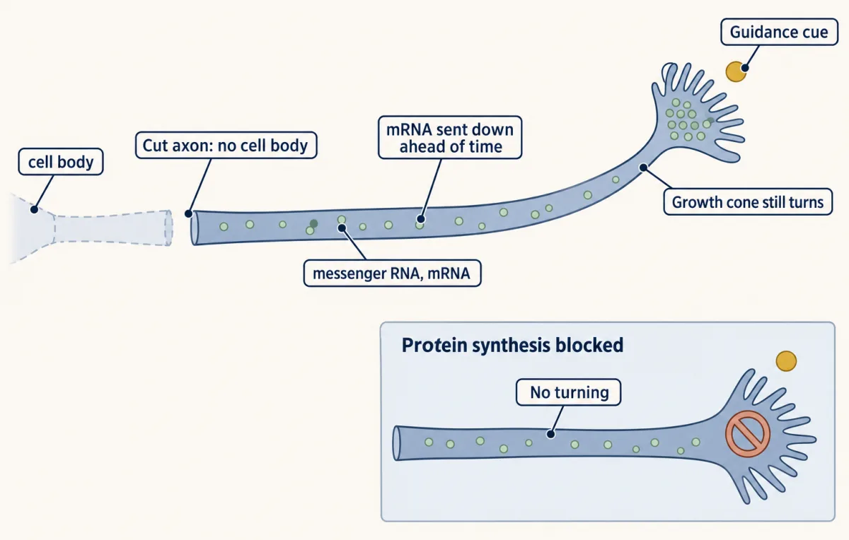
So the tip can decide on its own, within minutes, without waiting for its cell body. That shows the tip is enough to turn in a dish; it does not show that the cell body never contributes in the embryo. Whether the tip must also make new protein depends on the cue and the neuron.
The rule from the start organizes the first two puzzles: the edge gains what is added minus what slides back, and a turn sticks where microtubules build axon. The severing experiment answers the third. The cues that make the two sides differ, among them netrins, slits, semaphorins and ephrins, come in the next lessons.
Design problem
A growth cone from a chick sensory neuron turns toward a pipette releasing nerve growth factor (NGF). Design manipulations to test whether turning needs (a) actin dynamics on the near side, (b) microtubules on the near side, and (c) the cell body. Say what result each would give if the part is needed.
Saved in this browser only.
One way to do it
(a) Use a second pipette to release a low dose of an actin-assembly blocker, such as cytochalasin, onto the side facing the NGF. If turning toward NGF depends on new actin there, the growth cone should no longer turn toward the source; it may turn away, as growth cones did after a related manipulation, removing actin bundles from one side. Repeat with the drug on the far side: if the cone now turns toward NGF more sharply, the result fits actin balance between the two sides.
(b) Release nocodazole, which breaks down microtubules, on the near side. If microtubules must invade the near side for the turn to stick, the growth cone may still put out filopodia toward the NGF but fail to turn the axon. As a positive control, release a little taxol on one side with no NGF. In Xenopus spinal neurons this turns the cone toward the taxol; check that chick sensory growth cones do the same.
(c) Cut the axon behind the growth cone and repeat the assay at once. If isolated growth cones still turn, as cut growth cones do in several cue assays, the tip can decide on its own. Add a protein synthesis inhibitor to the isolated growth cone to ask whether the tip must also make protein. For chick sensory growth cones turning toward NGF, Roche and colleagues (2009) found that it did not.
Going deeper
Fixed tissue against a living fibre
By the late 1800s two pictures of how an axon formed were in play. In one, the axon grows out from a single nerve cell, as an extension of that cell. In the other, the nerve forms in place along its route, from a chain of cells or from strands of living material (one version, associated with Victor Hensen, called them protoplasmic bridges) already joining the nerve centre to its destination.
Wilhelm His and Santiago Ramón y Cajal argued for the first picture from dead tissue. Cajal used Golgi’s silver chromate stain, which blackens only a few cells in a sample but blackens each of them almost entirely. In the spinal cords of chick embryos a few days old, he found young nerve fibres of different lengths, each ending in a swollen club with spikes and fine projections. In 1890 he named it the growth cone and argued that it was the front of an axon on the move.
The trouble was that every one of his specimens was fixed. A row of fibres of different lengths can be read as one fibre growing, or as fibres that were always there becoming visible as they mature. A still picture cannot show movement, and more stained sections would not settle it.
Harrison’s preparation was built to make the answer unambiguous. He removed a piece of the neural tube (the embryo’s first nervous system, a tube of cells along its back) from frog embryos at a stage when no nerve fibres could yet be seen. He placed it in a drop of fresh lymph from an adult frog on a coverslip. The lymph clotted quickly into a soft mesh that held the tissue. Then he inverted the coverslip over a hollow slide and sealed it. This is an explant: a piece of tissue kept alive outside the body.
Each feature of the design closed off an alternative. The tissue was taken before any fibres existed, so the fibres that grew were not old fibres exposed by dissection. The clot contained no living chain of cells for a nerve to form from. And in some cultures a fibre could be seen arising from a single isolated cell. He reported this in 1907, and a longer paper in 1910 drew mostly on later preparations he judged more convincing.
Harrison also left a question open. His fibres grew into an unorganized clot, with nothing to point them anywhere. In the embryo an axon has to reach a particular partner, and the pipette assay was the tool that later tested steering.
Harrison’s 1914 test: does a cell need a solid?
In 1914 Harrison asked whether cells need a solid surface or can move through fluid alone. He gave cultures three kinds of support: clotted plasma, smooth glass, and threads of spider web. He also suspended some pieces of tissue in large drops so that they touched nothing. Cells from frog embryos wandered along the web threads, but cells in unsupported drops generally did not move. Pieces of forebrain from five-day chick embryos produced nerve fibres in all four clotted-plasma preparations, and none on spider web or in unsupported drops. Harrison concluded that cells move only on a solid. His chick cultures also suggested that not every solid suits nerve fibres, but he said they were too few to be conclusive, and the web could hold only small drops.
Adhesion: preference is not speed
Grip also shapes the path an axon takes. Later culture experiments put growth cones on dishes patterned with surfaces of different adhesiveness. In Paul Letourneau’s 1975 study with chick sensory neurons, growth cones given a choice tended to follow the more adhesive surface. Preference is not speed. In culture, how adhesive a surface is turns out to be a poor predictor of how fast axons grow on it (Lemmon and colleagues, 1992). Motor-clutch models, in which clutch bonds load and break under force, give one reason the relationship need not be simple.
What sets addition and grip
The Rho-family GTPases Rho, Rac and Cdc42 control where actin is built and how hard myosin pulls (see that lesson); like most small GTPases, each is a switch that is on when bound to GTP (that lesson). Calcium, cyclic nucleotides and local translation are other routes, covered in the guidance-receptor lessons.
Why a growth cone is not a car
A vehicle steered by the speeds of its two sides, like a tracked vehicle, has both sides joined through its rigid body. If its near side moves faster, it swings toward its far, slower side. Growth cones in an attractive gradient turn toward the side that gains. The difference is that a growth cone is not rigid: it changes shape, the gaining side protrudes more, and microtubules and organelles move into selected regions and make them axon. That is why the engine-and-clutch picture in this lesson is used for advance only.
From protrusion to axon
Advance at the edge is only the first step in making axon. The edge protrudes; the peripheral actin network is then remodelled so that organelles and microtubules can move up into the new space, with remaining actin bundles guiding the microtubules; and the stretch behind narrows into shaft: protrusion, engorgement and consolidation (Goldberg and Burmeister, 1986). These steps are regulated separately, so more protrusion on one side does not by itself fix where the axon goes.
When does a growth cone need to make protein?
Turning without a cell body and turning without new protein are separate tests, and the need for local translation found by Campbell and Holt is not true of every cue or every neuron. In the same study, one kind of collapse did not need new protein, and Roche, Marsick and Letourneau (2009) found that chick growth cones, including sensory growth cones turning toward nerve growth factor (NGF), still responded to cues with protein synthesis blocked.
The molecules of the clutch
Later work traced the linkage in growth cones themselves. In rat hippocampal growth cones a protein called shootin1 links L1 to the flowing actin (Shimada and colleagues, 2008). Similar clutches work in other migrating cells (reviewed by Case and Waterman, 2015). At the tips of filopodia, Ena/VASP proteins keep filament ends from being capped, so the spike can keep lengthening.
Your notes
Select any text in the lesson to highlight it or add a note to it. Your highlights and notes are saved in this browser and collected in your notebook.
This browser is not letting the site save anything (a private window or strict privacy settings), so highlights and notes will be lost when you leave the page.
How we measure it
Explant culture in a hanging drop
A piece of embryonic tissue is placed in a drop of nutrient medium on a coverslip, which is inverted over a hollow slide and sealed. Because the tissue is alive and visible, you can return to the same preparation and see what grows out of it.
Time-lapse imaging of living growth cones
A camera records the same growth cone at short intervals, often with contrast-enhancing optics or fluorescent labels on actin and tubulin. Movement of the edge, of filopodia and of the actin network can then be measured rather than inferred from fixed shapes.
Cytoskeletal drugs, applied in the bath or on one side
Cytochalasins block actin filament growth, nocodazole breaks down microtubules and taxol stabilizes them. Adding them to the whole dish tests what a structure needs; releasing them from a fine pipette onto one side tests what decides direction.
The pipette turning assay
A micropipette near a cultured growth cone releases a candidate cue, making a gradient across it. If the growth cone turns toward or away from the pipette, the cue can steer the axon. Because a single growth cone wanders, the evidence is the distribution of turning angles across many growth cones, compared with a control dish.
Practice questions on this lesson (3) → MCAT-style, original, each with an explanation
Where this lesson fits
Before thisReview first
- Seeing Neurons: Golgi, Cajal and the Neuron DoctrineThis course · Lesson 2
Cajal's Golgi-stained fibres and the idea that neurons are separate cells set up the growth cone's discovery and Harrison's test here.
After thisStudy next
- Pioneers, Guideposts and the Logic of Guidance CuesThis course · Lesson 22
It asks which cues the growth cone reads and how pioneer axons use them, taking the steering idea into the embryo.
- Netrin and DCC: One Cue, Two AnswersThis course · Lesson 23
It shows how one cue attracts or repels depending on the receptor, the signalling step this lesson leaves open.
- Topographic Maps: Sperry's Eye and Eph GradientsThis course · Lesson 26
It uses the gradient-sensing growth cone to explain how axons find their place in an ordered map.
Elsewhere on the siteFilms, simulations and method films in other courses
- Tight Ends, Loose EndsFilm · Cell Biology
It explains how filament ends grow or shrink, which sets the actin and microtubule behaviour at the tip.
- How Internal Motion Becomes MovementFilm · Cell Biology
It shows polymerization minus backward flow at a leading edge, with integrin coupling cutting slip, as in the clutch.
- A Grip Switched On From InsideFilm · Cell Signaling
It shows how an integrin grip switched on from inside links the cell to its surface, as in the clutch.
Suggested by an AI model from the lessons themselves, then checked. How we use AI
Finish line
Test yourself
8 questions
Practice and MCAT-style, with explained answers.
Next · Lesson 4
Tools for Following Cells and Genes
A neuron has no label saying when it was born or where it came from. So how do you find out?
Continue →← Lesson 2 · Seeing Neurons: Golgi, Cajal and the Neuron Doctrine