Neuroscience · Unit 3: Sensory systems
Smell and Taste
Rewritten on : new text, figures, flashcards and film. Your notes and highlights stay, but the text around them may have moved.
One mouse olfactory neuron, one receptor gene out of about a thousand. One bitter cell, one job. Why do the two chemical senses code so differently?
Pause when the reading reaches a film, and offer to play it. Saved on this device.
Watch the film now, or keep listening
Builds on:Principles of Sensory CodingSynaptic Transmission and Quantal ReleaseWhy these?
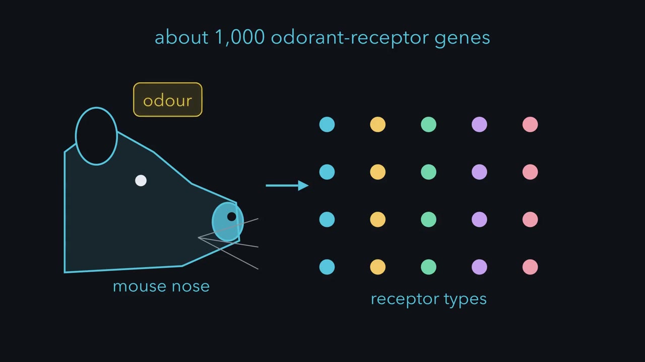 ▶ Watch A Cell Cannot Name a Molecule · 2:14
Read the transcript of “A Cell Cannot Name a Molecule”
Film transcript: A Cell Cannot Name a Molecule
A mouse has about a thousand odorant-receptor genes. It reads an odour from a pattern across its receptors. It also has dozens of bitter-receptor genes. Yet bitter is one taste. Why the difference? Start with one receptor cell. Its output is its activity: how strongly it is active. Two different molecules arrive, each at a concentration that activates it equally. Can this cell tell which molecule it met?
No. A receptor cell reports how strongly it is active, not which molecule activated it. Vision calls this univariance. A cone cannot tell a dim light near its peak from a brighter one far from it. To tell molecules apart when their concentrations vary, a sense needs more than one line. A line is a set of cells whose activity reaches the brain as one channel. In smell, a line is all the neurons with one receptor, and the glomeruli they converge on. In taste, a line is one kind of receptor cell: the sweet cells, the bitter cells, and so on. A pattern is the relative strength of activity across lines, not just which lines are on. The rule: a sense can tell two stimuli apart only if their patterns across separate lines differ.
Smell keeps about a thousand receptor types on separate lines. So an odour is read as a pattern across many lines. Taste pools its receptors into a few kinds of cell. So a taste is read mainly from which kind of cell is active. A line whose activity always means one quality, whatever turned it on, is called a labeled line. Keep the scope in view. The rule is about the periphery, mostly in mice, and it ignores spike timing. Gene count alone does not set the number of lines. What matters is whether receptor identity is kept separate or pooled.
From the film
- A receptor cell reports how strongly it is active, not which molecule activated it.
- To tell molecules apart, a sense needs separate lines, and a stimulus is read from the pattern of activity across them.
- Smell keeps about a thousand receptor types on separate lines, while taste pools its receptors into a few kinds of cell, mostly in mice.
Sweet, umami and bitter taste cells in mice all pass their signal through one enzyme, PLCβ2. In 2003 Zhang and colleagues deleted it, and the taste nerves stopped responding to the tested sweet, amino-acid and bitter stimuli all at once.
One enzyme served three tastes, yet a normal mouse does not mistake a sweetener for a bitter compound. The same mouse has dozens of bitter-receptor genes, yet bitter is one taste. Its nose, with about a thousand odorant-receptor genes, reads an odour from a pattern across its receptors. Why do the two chemical senses code so differently?
One coding problem, two answers
Smell and taste both turn chemicals into spikes. Both start with receptor cells that carry receptor proteins, and both have to tell the brain what arrived. That raises four puzzles.
- Identity. A receptor cell’s output is its activity. How can the brain learn which molecule arrived?
- Wiring. Most mature mouse olfactory neurons express one odorant-receptor gene at high abundance, and neurons sharing that receptor usually converge on about two glomeruli per bulb. What does that arrangement achieve?
- Shared machinery. If sweet, umami and bitter cells use the same enzyme, what keeps the three tastes apart?
- Pooling. Why is bitter one taste when there are dozens of bitter receptors?
One rule makes sense of all four, and it needs two terms. A line is a set of cells whose activity reaches the brain as one channel. In smell, a line is all the neurons that express one receptor, together with the glomeruli (knots of nerve endings in the olfactory bulb) they converge on. In taste, a line is one kind of receptor cell: the sweet cells, the bitter cells, and so on. A pattern is the relative strength of activity across lines, not just which lines are on.
The one idea to carry through this lesson. A receptor cell reports only how strongly it is active, not which molecule activated it. So a chemical sense can tell two stimuli apart only if they produce different patterns of activity across its separate lines. Smell keeps about a thousand receptor types on separate lines, so an odour is read as a pattern across many lines. Taste sorts its receptors into a few kinds of cell, with bitter cells pooling many, so a taste is read mainly from which kind of cell is active.
A line whose activity always means one quality, whatever turned it on, is called a labeled line. The taste section tests whether taste cells are labeled lines.
The first sentence of the rule is not new. In vision it is the principle of univariance: a cone’s response cannot say whether it met a dim light at the wavelength it is most sensitive to or a brighter light far from it (the retina). Reading a stimulus from many lines at once is the population code of sensory coding.
Keep the rule’s scope in view. It is about the periphery (the receptor cells and the nerves leaving them) and mostly about mice. It is a first approximation that ignores spike timing. It says what information survives to the first relay in the brain; it does not count how many odours an animal can tell apart, and circuits further in can recombine lines.
Smell keeps its lines apart
Read the transcript of “Smell Keeps Its Lines Apart”
Film transcript: Smell Keeps Its Lines Apart
How does the nose keep a thousand receptor types on separate lines? Most mature olfactory neurons in a mouse express one receptor gene at high abundance. That receptor can still bind many odorants. So all the neurons with one receptor form one line. Where do their axons go? Mombaerts and colleagues made one receptor's neurons fill their axons with a marker. The labelled cells were scattered across the epithelium. Their axons gathered and ended in two fixed glomeruli in each bulb. Each glomerulus collects mainly from neurons with one receptor type. So the glomerular layer is largely a map of lines. What does each line detect? Malnic and colleagues imaged single mouse neurons as odorants were applied.
Then they identified the receptor in each responding cell. One receptor recognized several odorants. One odorant was recognized by several receptors. And concentration could change which receptors an odorant activated. Together, these make a combinatorial code. Now practise reading such a code. This matrix is invented for illustration. Four receptors, A to D, each simply on or off. Odorant one turns on A and B. Odorant two, B and C. Odorant three, A, B and C. Odorant four, D alone. Remove receptor A. Which two odorants now give the same pattern?
Odorants two and three. Both now read B and C, so the model predicts they cannot be told apart. Now put receptor A back, and raise odorant one to tenfold concentration. Receptor C has a lower affinity for it, so it responds only at the higher concentration. What happens to odorant one's pattern?
C is recruited. Odorant one now reads A, B and C, the pattern of odorant three. The model predicts it could be mistaken for odorant three. Quality can change with concentration. This toy counts patterns, not smells. What a mouse can tell apart needs a behavioural test. Smell keeps its lines apart. So an odour is read as a pattern across many of them.
From the film
- Most mature olfactory neurons in a mouse express one receptor gene at high abundance, so all neurons with one receptor form one line.
- Neurons with the same receptor are scattered across the epithelium, but their axons converge on two fixed glomeruli in each bulb.
- One receptor can recognize several odorants and one odorant can activate several receptors, so odours are read as a combinatorial code that can change with concentration.
Olfactory sensory neurons sit in the olfactory epithelium high in the nasal cavity. Each sends cilia into the mucus, where odorants bind, and an axon through the bone above to the olfactory bulb. There the axons end in glomeruli, round knots of neuropil (tangles of axons and dendrites) where receptor-neuron axons meet the dendrites of the bulb’s output neurons, the mitral and tufted cells.
The receptors are G-protein-coupled receptors, with seven transmembrane segments. They come from a family of roughly a thousand genes in the mouse and a few hundred intact genes in humans. Linda Buck and Richard Axel found the first candidate members in 1991; how the family grew and shrank belongs to molecular evolution.
One receptor gene per neuron. The evidence is single-cell sequencing (Hanchate and colleagues, 2015), which found one abundant receptor transcript in most mature mouse neurons.
Predict. A neuron expresses only one receptor gene. Can it respond to more than one odorant?
Check your prediction
Yes, if its receptor binds more than one. One receptor per neuron is a statement about genes, not about odorants, and the next section shows that a single receptor does bind many. What the gene rule fixes is that each neuron speaks for one receptor type, so all the neurons with that receptor form one line.
Where do the axons of one line go? Mombaerts and colleagues (1996) engineered mice so that neurons expressing one chosen receptor also made a marker that filled their axons. Picture the result. The labelled cell bodies are scattered across the epithelium. Their axons gather as they travel, and they end in two fixed glomeruli out of the roughly 1,800 in each bulb. To test whether the receptor itself helps choose the target, they replaced the coding sequence of the receptor P2 with that of another receptor, M12. The axons moved to new glomeruli, distinct from both P2’s and M12’s usual targets: the receptor helps guide its own axon but is probably not the only cue, since the swapped-in receptor did not simply carry the axons to its own site.
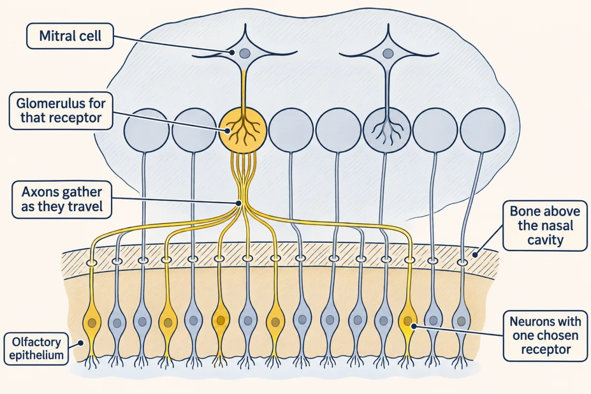
Each glomerulus therefore collects mainly from neurons expressing one receptor type. The glomerular layer is largely a map of lines: receptor identity laid out as position. How that map is built is covered in partners, layers and self-avoidance; olfactory neurons are also replaced throughout life (adult neurogenesis).
In the canonical olfactory neuron, every receptor uses the same steps. The bound receptor activates the G protein Golf, which drives adenylyl cyclase 3 to make cAMP. cAMP opens cyclic-nucleotide-gated channels that let in sodium and calcium, and a calcium-activated chloride current amplifies the depolarization. The molecular steps are in senses and cGMP, G proteins and cAMP and PKA. Because the cascade is shared, it does not attach a chemical name to the response. Which odorant arrived is carried by the pattern of response strengths across lines (in real neurons, timing can contribute too).
From the bulb, mitral and tufted cells project to the piriform cortex and other olfactory areas without first relaying in the thalamus, unlike vision, hearing and touch, although a route through the thalamus to frontal cortex also exists.
Reading the pattern
If each receptor type defines one line, what does that line detect? Malnic, Hirono, Sato and Buck (1999) loaded dissociated mouse olfactory neurons with a calcium indicator and applied a panel of odorants. Then they identified the receptor in each responding cell by single-cell RT-PCR. Picture the result as a grid, with receptors down the side, odorants across the top, and a filled square wherever a cell responded.
The grid showed three things.
- One receptor recognized several odorants.
- One odorant was recognized by several receptors.
- Changing an odorant’s concentration could change which receptors it activated.
Odour identity, on this view, is a combinatorial code: a pattern across receptors rather than one receptor per odour.
A grid of filled and empty squares is a simplification. Real responses are graded, so two odorants that activate the same set of receptors can still be told apart if the relative strengths differ. The exercise below uses on-or-off squares only to keep the counting simple.
Worked problem: a made-up receptor matrix. Suppose you imaged mouse olfactory neurons and found four receptor types, A to D. These data are invented for illustration. At a low concentration the odorants activate these receptors:
| Odorant | A | B | C | D |
|---|---|---|---|---|
| 1 | on | on | off | off |
| 2 | off | on | on | off |
| 3 | on | on | on | off |
| 4 | off | off | off | on |
Assume the brain can tell two odorants apart only if their patterns differ.
Step 1: remove receptor B. Odorant 1 becomes A; odorant 2 becomes C; odorant 3 becomes A+C; odorant 4 stays D. All four patterns are still different, so the model predicts no loss of discrimination: B was never the only difference between two odorants.
Predict. Restore B and remove receptor A instead. Which odorants can no longer be told apart?
Check your prediction
Odorants 2 and 3. Odorant 1 becomes B; odorant 2 is B+C; odorant 3 is also B+C; odorant 4 is D. Odorants 2 and 3 now give the same pattern, so the model predicts they can no longer be told apart. Losing a line costs only the distinctions that depended on it.
Predict. Restore all four receptors. Now raise odorant 1 to tenfold concentration. In this invented example, C has a lower affinity for odorant 1: it needs more odorant to reach its response threshold, so it is recruited only at the higher concentration. Which odorant’s pattern does odorant 1 now match, and what does the model predict?
Check your prediction
Its pattern becomes A+B+C, the low-concentration pattern of odorant 3. The model predicts that odorant 1 at high concentration could be mistaken for odorant 3: a change in quality with concentration. This is the third finding of the Malnic grid in miniature: real odorants recruited additional receptors at higher concentrations.
Last step: capacity. More separate lines allow more possible patterns. If you treat each of N receptor types as a switch that is either on or off:
number of distinct non-empty patterns = 2N − 1
Four switches give 24 = 16 patterns. Removing the all-off pattern leaves 15 that could report an odorant.
The 2N figure is the capacity of a toy model, not a count of smells: responses are graded and noisy, and the bulb and cortex transform the pattern. What a mouse can tell apart needs a behavioural test; a matrix alone cannot settle it. Try the model below.
- Capacity (toy model, N = 4)
- 2⁴ = 16 patterns, 15 non-empty
- Distinct non-empty patterns among the 4 odorants
- 4 of 4
Rule to keep: in this sketch a receptor is simply on or off, with no noise and no transformation in the bulb or cortex, so two odorants with the same pattern are predicted to be indistinguishable. 2N − 1 is a toy capacity, not a count of smells; the matrix is invented, and every prediction needs a behavioural test.
Taste pools its receptors into cells
Read the transcript of “Taste: The Cell Decides”
Film transcript: Taste: The Cell Decides
If three kinds of taste cell send the same chemical signal, how does the brain tell them apart? In the taste bud, type II cells detect sweet, umami and bitter. Sweet and umami cells carry T1R receptors. Bitter cells carry T2R receptors. After the receptor, all three use the same steps. An enzyme, PLCβ2, leads to a calcium rise. Calcium opens a channel, and the cell depolarizes. Then ATP flows out through a channel onto the nerve fibre. So the signal is the same. The rule's answer: what differs is which cell the nerve listens to. Zhang and colleagues deleted PLCβ2 from the whole mouse. Nerve responses to the tested sweet, amino-acid and bitter stimuli were lost.
In lick tests, sweet preference was strongly reduced, and bitter avoidance was lost. Sour and salty responses remained, because those cells use other routes. Then they put it back only in cells with bitter receptors. Which responses come back? Bitter only, all three, or none?
Bitter only, in the nerve and in behaviour. Sweet and amino-acid responses did not return. The shared enzyme works only for its own cell's quality. The lines are separate. Does the cell also decide what a line means? Put a receptor in the wrong cell. Mueller and colleagues put a human bitter receptor into the sweet cells of mice. Its ligand is a bitter compound that normal mice ignore. Will these mice avoid it, or drink it?
They drink it, while normal mice stay indifferent. The receptor detects the molecule, but the sweet cell decides what that means. In mouse taste, each kind of cell acts like a labeled line. One consequence: a bitter cell pools many receptors. By its rate alone, that cell cannot say which bitter compound it met. Not every bitter cell carries the same receptors, so the pathway may keep some distinctions. Still, pooling fits a sense that mostly answers: eat it, or reject it? Keep the scope. These are mouse experiments on the periphery. Some taste cells and nerve cells respond to more than one quality. Smell reads a pattern across many lines. Taste reads mainly which kind of cell is active.
From the film
- Sweet, umami and bitter cells run the same chain from receptor to ATP release, so the signal itself is the same and the cell the nerve listens to is what differs.
- Restoring PLCβ2 only in bitter-receptor cells brought back bitter responses and nothing else, and a bitter receptor placed in sweet cells made mice drink a bitter compound.
- In mouse taste each kind of cell acts like a labeled line, and a bitter cell pools many receptors, so by its rate alone it cannot say which bitter compound it met.
Taste starts in taste buds, clusters of specialized epithelial cells, not neurons, on the tongue and palate. Afferent fibres of cranial nerves VII, IX and X carry their signals to the nucleus of the solitary tract in the medulla, which relays through the thalamus (in rodents by way of the pontine parabrachial nucleus) to gustatory cortex in the insula. The textbook map with sweet at the tip and bitter at the back is discredited: taste buds across the tongue detect every quality, with modest regional differences in sensitivity.
The five qualities taught for mammals are sweet, sour, bitter, salty and umami. A taste bud holds glial-like supporting cells (type I) and these receptor cells:
| Cell | Quality | Detector |
|---|---|---|
| Type II | sweet | T1R2 + T1R3 |
| Type II | umami | T1R1 + T1R3 |
| Type II | bitter | many T2Rs |
| Type III | sour | the proton channel OTOP1 |
| ENaC cells (mice; their place among the cell types is still debated) | low salt | the sodium channel ENaC |
Taste has far fewer lines than smell, partly because it has fewer receptor genes but mainly because it pools them. Sweet cells carry one main receptor, the pair T1R2 and T1R3. Umami cells carry T1R1 with T1R3; in mice this pair responds to many L-amino acids, which is why mouse experiments often test “amino-acid” responses. Bitter cells draw on the T2R family, about 25 in humans and roughly 35 in mice, and in rodents a single bitter cell expresses many T2Rs at once (Adler and colleagues, 2000). All three kinds of receptor are G-protein-coupled.
Now look at what happens after the receptor. In all three kinds of type II cell the same steps follow (the names matter less than the fact that the chain is identical):
- the enzyme PLCβ2 makes inositol trisphosphate (IP3);
- IP3 opens IP3R3 channels, which release calcium from the endoplasmic reticulum (PLC, IP3, DAG and PKC);
- calcium opens TRPM5, a channel gated by calcium that passes monovalent cations, sodium and potassium, so the cell depolarizes (in mice TRPM4 contributes alongside it; Dutta Banik and colleagues, 2018);
- the cell has no conventional vesicular synapse, so instead depolarization opens CALHM1/3, a voltage-gated channel, and ATP flows out through it onto the afferent nerve fibres.
If the signal leaving a sweet cell is chemically the same as the signal leaving a bitter cell, what keeps sweet and bitter apart?
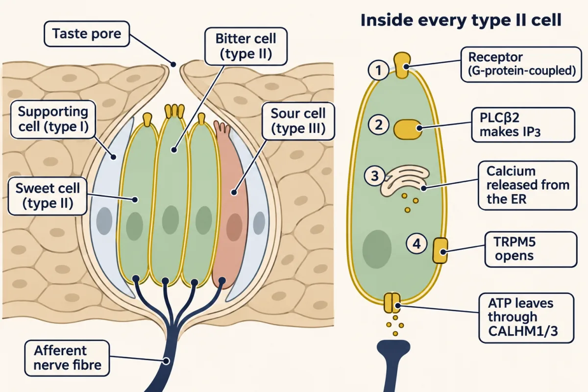
The rule answers it. A cell reports how strongly it is active, not what activated it. So the cascade does not need to differ: what differs is which cell the nerve fibre listens to. If that is right, the identity of the cell, not the receptor or the cascade, should set the quality.
Zhang and colleagues tested this in mice in 2003. First they deleted the gene for PLCβ2 from the whole animal. Responses of the taste nerves to the tested sweet, amino-acid and bitter stimuli were lost. In brief-access lick tests, which count a mouse’s licks at a spout over a few seconds, preference for sweet and amino-acid stimuli was strongly reduced and avoidance of bitter was lost. Sour and salty responses remained, because those cells use other routes. Then they put PLCβ2 back, but only in cells that express T2R bitter receptors.
Predict. PLCβ2 is now present in bitter cells and nowhere else in the taste bud. Which responses come back: bitter only, all three, or none?
Check your prediction
Bitter responses returned, in the nerve and in behaviour. Sweet and amino-acid responses did not. The enzyme is shared, but it works only for the quality of the cell it sits in. Sweet and bitter therefore run in separate cells, each a separate line to the nerve.
That experiment shows the lines are separate. It does not yet show that the cell, rather than the receptor, decides what the line means. For that, put a receptor into the wrong cell. Zhao and colleagues (2003) expressed an engineered receptor, activated only by a synthetic drug the mouse would otherwise ignore, in sweet cells alone. Mueller and colleagues (2005) went further and put a human bitter receptor, T2R16, into mouse sweet cells. Its ligand, phenyl-β-D-glucopyranoside, is a bitter compound that normal mice ignore at the concentrations tested.
Predict. Offered that ligand, do these mice avoid the solution, drink it, or ignore it as normal mice do?
Check your prediction
They drink it. The mice became attracted to the bitter ligand, and the engineered-receptor mice to the synthetic drug, while normal mice stayed indifferent. The receptor detected the molecule, but the sweet cell decided what the detection meant. Mueller and colleagues also put the engineered receptor in bitter cells, and those mice avoided the same drug. In this sense each kind of taste cell acts as a labeled line in the mouse periphery: its activity means one quality, whatever turned it on.
One consequence is easy to miss. Because a rodent bitter cell pools many T2Rs, its rate alone cannot tell the brain which bitter compound it met, only how strongly it was activated. Two different bitter compounds, each at a concentration that activates the cell equally, send the same signal from that cell. Not every bitter cell need carry the same set of T2Rs, so the bitter pathway as a whole may keep some distinctions. Still, pooling fits a sense that mostly answers eat it or reject it, not which molecule it is.
Sour and salt: the channel routes
Type III cells detect sour. They form conventional synapses, releasing amine transmitters such as serotonin by calcium-dependent exocytosis; serotonin is probably not their only signal, since mice lacking the nerve’s ATP receptors, P2X2 and P2X3, lose taste nerve responses to every quality (Finger and colleagues, 2005). Their acid sensor is OTOP1, a proton-selective channel identified by Tu and colleagues in 2018. In Otop1-null mice, gustatory nerve responses to acids were severely and selectively reduced (Teng and colleagues, 2019). PKD2L1, long used to mark sour cells, is not the primary sour receptor.
Two results show why the cell, the nerve and the behaviour have to be kept apart. Zhang and colleagues (2019) put OTOP1 into sweet cells, and those cells then responded to sour stimuli: a result about the cells, not about what the mice tasted. Otop1-null mice show the opposite split. Despite losing OTOP1-mediated proton currents and most of their nerve response to acid, they still avoided acid in two-bottle tests, so avoidance is not a pure measure of sour taste. Some acid sensitivity remained, so this is not evidence that all sour-cell activity was absent.
Salty is the least settled quality. In mice, deleting the ENaCα subunit of the epithelial sodium channel from taste cells abolished the amiloride-sensitive response (the part blocked by the drug amiloride) to low sodium concentrations and the mice’s attraction to low salt (Chandrashekar and colleagues, 2010). High NaCl, which mice avoid, also recruited sour and bitter cells (Oka and colleagues, 2013). In this simplified account, high NaCl in mice with ENaC intact drives three cell classes, ENaC sodium cells plus sour and bitter cells as two additional aversive pathways: a multi-line pattern, which the rule allows. Non-sodium salts such as KCl recruit the aversive pathways without the sodium-selective ENaC pathway. How human salt taste is transduced is unsettled.
Fat is a candidate quality, not an established one, and kokumi, a sense of richness that enhances other tastes, is usually not counted as a quality of its own. Much of what people call flavour is smell: odorants from food in the mouth reach the nose from behind (retronasal smell).
Back to the puzzles, and where the rule stops
Now return to the four puzzles. Identity is carried by which lines are active and how strongly. Smell keeps each receptor type on its own line, because most mature neurons express one receptor gene and like axons converge on about two glomeruli per bulb; that is what the wiring achieves. Taste can share one cascade across sweet, umami and bitter cells because the nerve reads which cell fired, not what happened inside it. And bitter can be one taste because bitter cells signal one quality whatever receptor fired, and each pools many bitter receptors.
Smell meets an open-ended world of molecules with a large, overlapping receptor array: quality is read from a pattern. Taste mostly answers a few questions (eat it or reject it, and what it contains) with cells dedicated to each quality: quality is read mainly from which cell fires, a labeled line at the periphery.
That is the leading view from mouse genetics, and it has limits. Some taste cells and some taste ganglion neurons respond to several qualities, so whether taste is coded by labeled lines or by patterns across fibres, and how strictly the lines stay separate further into the brain, is still argued. These experiments do not show one exclusive wire for each human percept, and one stimulus, such as high NaCl, can drive several cell classes.
Design problem
Is “bitter” a property of the receptor or of the cell carrying it? Design an experiment in mice that tells the two apart.
Saved in this browser only.
One way to do it
Choose a receptor whose ligand the mouse normally ignores, such as an engineered receptor activated only by a synthetic drug, and express it only in sweet cells by driving it from the T1R2 gene’s promoter. Give the drug in a brief-access lick test. If the cell sets quality, the mice should lick the drug eagerly, as for a sweetener; if the receptor did, they should not. Then express the same receptor only in T2R bitter cells: the cell hypothesis predicts avoidance of the same drug. The Zuker and Ryba laboratories reported experiments of this design (Zhao and colleagues, 2003; Mueller and colleagues, 2005).
Controls: confirm by in situ hybridization or antibody staining that the receptor is only in the targeted cells; record the chorda tympani, a taste nerve, to show the drug now drives a response; test wild-type littermates, which should ignore the drug; and run a concentration series. A complementary test is the PLCβ2 rescue in T2R cells described above.
Limits: this is the mouse periphery. Licking is a behaviour, not a reported percept, and brain circuits could in principle re-sort the signal.
Going deeper
How the receptor family was found
In 1991 Linda Buck and Richard Axel described 18 members of a gene family from rat olfactory tissue that encoded proteins with seven transmembrane segments, the shape of G-protein-coupled receptors. Their title kept a hedge: the family may encode odorant receptors. The paper did not show what any receptor binds or where its neurons project.
One copy of the gene, and neurons that have not chosen yet
In the mouse neurons Chess and colleagues examined in 1994, a given receptor gene was transcribed from only one of its two alleles. Hanchate and colleagues also found that many immature neurons express several receptor genes at low levels, so singular expression is something a neuron arrives at as it matures.
Receptor pharmacology
In heterologous cell assays, human T1R2/T1R3 responded to diverse sweeteners and human T1R1/T1R3 to L-glutamate, with 0.2 mM IMP shifting the glutamate half-maximal effective concentration (EC50) (the concentration giving a half-maximal response) about 30-fold lower (Li and colleagues, 2002). Meyerhof and colleagues (2010) screened 25 human bitter receptors against 104 bitter compounds and found that receptors varied greatly in how many compounds activated them. The rodent finding that one bitter cell expresses many T2Rs should not be read as every human bitter cell expressing all of them: human cells express subsets (Behrens and colleagues, 2007).
Worked problem: one TRPM5 channel
Prawitt and colleagues measured a unitary conductance of 25 pS for human TRPM5 expressed in HEK-293 cells. Take outward current as positive, so inward current is negative, as in the action potential lesson. Assume a linear current-voltage relation, a membrane potential of −60 mV and a reversal potential of 0 mV, as for a non-selective cation channel. Then I = g × (Vm − Erev) = 25 pS × (−60 mV − 0 mV) = −1,500 fA = −1.5 pA. The minus sign means inward current of positive charge. This is a stipulated example, not a measured taste-cell current.
Your notes
Select any text in the lesson to highlight it or add a note to it. Your highlights and notes are saved in this browser and collected in your notebook.
This browser is not letting the site save anything (a private window or strict privacy settings), so highlights and notes will be lost when you leave the page.
How we measure it
Calcium imaging with single-cell RT-PCR
Dissociated olfactory neurons loaded with a calcium indicator are exposed to a panel of odorants, and each responding cell is then picked up and its receptor transcript identified by RT-PCR. The result is a matrix linking receptor identity to the odorants that activate it.
Gene targeting to label or swap receptors
A receptor gene is modified so that the neurons expressing it also make a visible marker, or its coding sequence is replaced with another receptor's. Following the labelled axons shows where receptor-defined neurons project and whether the receptor helps decide it.
Knockout with cell-restricted rescue
A gene is deleted from the whole animal, then restored only in one cell type with that cell's promoter. If the function returns, that cell type is enough to carry it in that assay; if a related function does not return while the rescued one does, it probably needs other cells.
Gustatory nerve recording
Electrodes on the chorda tympani or glossopharyngeal nerve record the summed activity of taste afferent fibres while solutions flow over the tongue. It measures what the periphery sends to the brain, separately from what the animal chooses to drink.
Practice questions on this lesson (3) → MCAT-style, original, each with an explanation
Where this lesson fits
Before thisReview first
- Principles of Sensory CodingThis course · Lesson 9
It introduces population codes and labeled lines, which this lesson applies to smell and taste.
- Synaptic Transmission and Quantal ReleaseThis course · Lesson 5
Taste cells release ATP and serotonin onto nerve fibres, so you need how a cell transmits to the next.
Elsewhere on the siteFilms, simulations and method films in other courses
- One in Two ThousandFilm · Sensory Systems
It goes deeper into how one olfactory neuron picks a single receptor gene.
- Which Taste, and How Good?Film · Sensory Systems
It shows how taste identity and value are read out separately from taste cells to cortex.
- What the Bulb SendsFilm · Sensory Systems
It follows the glomerular map of receptor types on to piriform cortex.
Suggested by an AI model from the lessons themselves, then checked. How we use AI
Finish line
Test yourself
9 questions
Practice and MCAT-style, with explained answers.
Next · Lesson 15
Motor Units, Reflexes and Locomotion
A knee tap makes the quadriceps kick and inhibits the hamstrings, yet the sensory axon that starts both only ever excites. How?
Continue →