Biology · Unit 2: Cells
Organelles as a System
Rewritten on : new text, flashcards and film. Your notes and highlights stay, but the text around them may have moved.
The insulin gene never leaves a beta cell's nucleus, yet insulin leaves the cell after a meal. What carries it out, and in what order?
Pause when the reading reaches a film, and offer to play it. Saved on this device.
Watch the film now, or keep listening
Builds on:Respiration and ATPWhat Am I Made Of?Why these?
Where this lesson sits
- Virginia SOL BIO.3b
- Practices BIO.1c, BIO.1d, BIO.1e
Essential question: How do the parts of a cell work together to keep it alive, like the departments of a factory?
-
Life Science (grade 7) LS.2 b and c
Review it at grade 7 level
In Life Science you learned that all living things are made of cells, and that cells have parts with different jobs. You looked at plant and animal cells and saw that plant cells have a cell wall and, in green parts, chloroplasts, while both kinds have a cell membrane, cytoplasm and a nucleus. You learned each part one at a time, like a list. Biology adds how the parts work together as a system: one cell part hands its product to the next, and some parts supply energy that the others use.
-
Biology BIO.3b You are here
This lesson
-
Biology II Biology II: Advanced Survey of Biology Topics, BIOII.2 a
Preview where it goes next
Biology II: Advanced Survey of Biology Topics looks at the cell's membranes as one connected set, often called the endomembrane system: the nuclear envelope, ER, Golgi, vesicles, lysosomes, vacuoles and cell membrane. Mitochondria and chloroplasts are usually counted outside that system. You meet what this lesson left out: smooth ER making lipids, how a protein carries an address that tells the cell where to send it, and how insulin starts as a longer precursor chain that is cut into its final form. You also see evidence that mitochondria and chloroplasts began as bacteria living inside another cell.
-
College
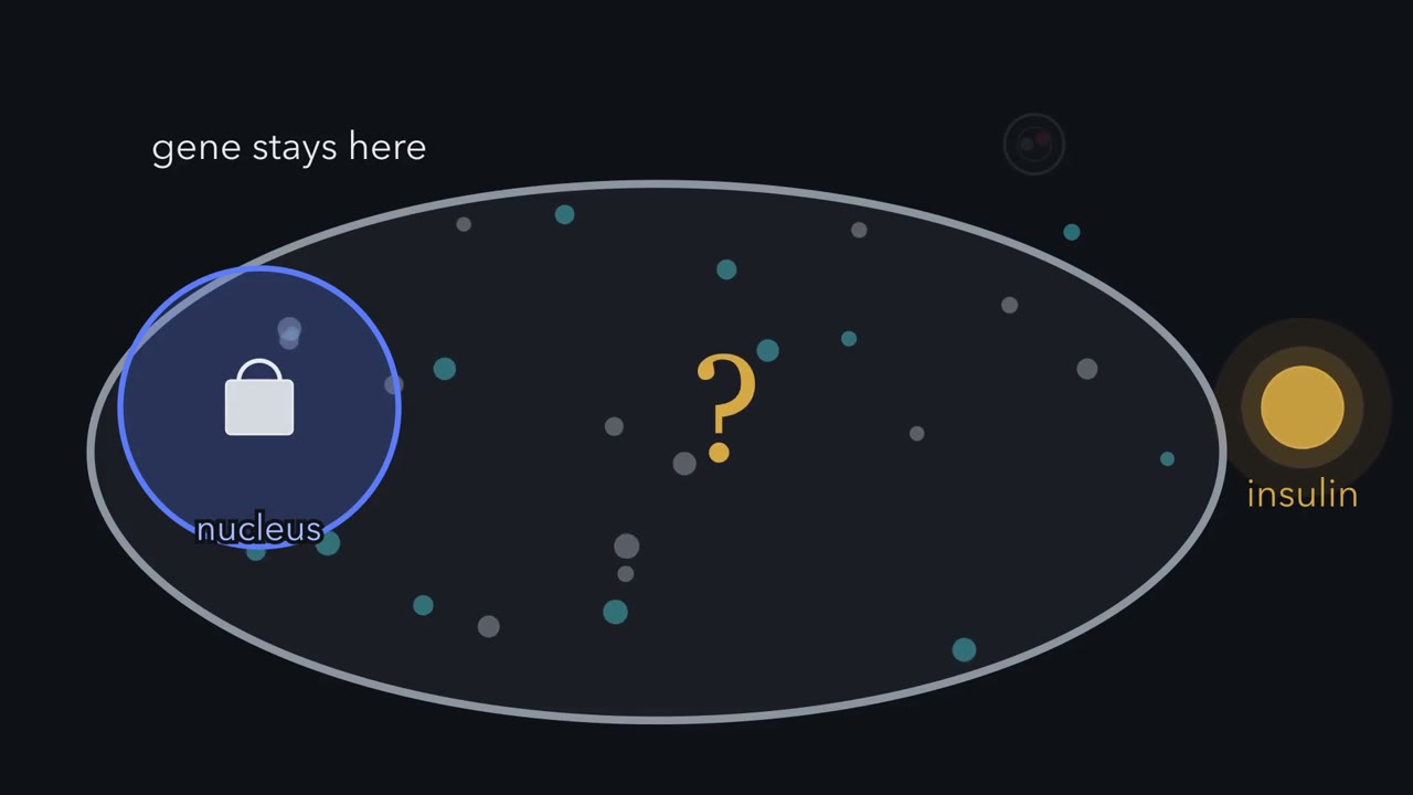 ▶ Watch One Cell, One Shipping Line · 3:12
Read the transcript of “One Cell, One Shipping Line”
Film transcript: One Cell, One Shipping Line
The insulin gene never leaves a beta cell's nucleus. So how does insulin end up outside the cell? After a meal, your blood sugar rises, and beta cells in your pancreas release this protein hormone. Here is the answer in one picture. First a copy of the instructions travels, then the protein itself. Each part can only work on what the part before it hands over. Start in the nucleus. It holds most of the cell's DNA, its instructions. The gene for insulin is one stretch of that DNA. But the gene itself stays put. Here is a puzzle for you. The gene stays locked in the nucleus. So what leaves the nucleus instead, carrying its instructions out?
A copy. The cell copies the gene into messenger RNA. That working copy leaves the nucleus and carries the instructions to the next stop. The next stop is a ribosome on the rough ER. The ER is a set of folded membranes. On the rough ER, ribosomes dot the surface. A ribosome reads the messenger RNA and links building blocks into a protein chain. The chain threads into the ER, where it folds.
Now the protein has to move. A vesicle, a small bubble of membrane, buds off the ER. It carries the protein across to the Golgi apparatus. The Golgi is a stack of flat sacs. It sorts the protein and packs it into new vesicles. There the protein waits until it is needed. At the last stop, a vesicle meets the cell membrane. The two membranes fuse. The vesicle opens to the outside, and insulin is released. None of this is free. Building a protein chain and moving vesicles both use energy. Much of that energy comes as ATP, a small molecule cell parts can use. Mitochondria release energy from sugar and store it in ATP. Now look at the whole route.
The nucleus sends out a copy. Then the protein moves: rough ER, vesicle, Golgi, vesicle, cell membrane. Notice what a route like this means. Each part can only work on what the part before it hands over. So every step depends on the one before it. So how does insulin get out of the cell? No single part does it. A copy of the gene leaves the nucleus, and the protein is handed from part to part.
Mitochondria supply much of the ATP.
From the film
- A beta cell keeps the insulin gene in its nucleus and sends out a messenger RNA copy instead.
- A ribosome on the rough ER reads the messenger RNA and builds the protein, which folds inside the ER.
- Vesicles carry the protein to the Golgi apparatus, which sorts and packs it, and a vesicle fuses with the cell membrane to release it.
- Each part can only work on what the part before it hands over.
- Many steps use energy, much of it as ATP made by mitochondria.
The first film followed insulin from a beta cell’s nucleus to its cell membrane. First, check three ideas you may bring with you.
What do you think? Decide before you read.
-
Scientists disagree: Plant cells have mitochondria too. Chloroplasts capture light energy and store it in sugar. Mitochondria release energy from sugar and store it in ATP, day and night, in leaves, roots and stems. Electron microscope pictures of plant cells show mitochondria, including in root cells that have no chloroplasts.
What scientists think
Scientists disagree: Plant cells have mitochondria too. Chloroplasts capture light energy and store it in sugar. Mitochondria release energy from sugar and store it in ATP, day and night, in leaves, roots and stems. Electron microscope pictures of plant cells show mitochondria, including in root cells that have no chloroplasts.
-
Scientists disagree: A diagram is one frozen picture. In 2017 scientists filmed six kinds of labelled organelles in living cells, taking a picture every 5 seconds. The organelles kept moving and coming into contact with one another.
What scientists think
Scientists disagree: A diagram is one frozen picture. In 2017 scientists filmed six kinds of labelled organelles in living cells, taking a picture every 5 seconds. The organelles kept moving and coming into contact with one another.
-
Scientists disagree: Bacteria have DNA, but it is not wrapped in a membrane. Electron microscopes show bacterial DNA sitting in an open region of the cell. Bacteria do share some parts with our cells, such as a cell membrane, cytoplasm and ribosomes.
What scientists think
Scientists disagree: Bacteria have DNA, but it is not wrapped in a membrane. Electron microscopes show bacterial DNA sitting in an open region of the cell. Bacteria do share some parts with our cells, such as a cell membrane, cytoplasm and ribosomes.
The one idea. Each cell part on a route can only work on what the part before it hands over. When a eukaryotic cell (one with a nucleus) sends out a protein such as insulin, a messenger RNA copy of the gene leaves the nucleus first. Then the protein travels: from a ribosome on the rough endoplasmic reticulum (ER), by vesicle (a small bubble of membrane) to the Golgi apparatus, and out when another vesicle fuses with the cell membrane. Many steps use energy, much of it as ATP (a small molecule that carries energy cell parts can use) made by mitochondria. So if one step is blocked, the protein cannot reach the stops after it.
Scope: the usual route for exported proteins; a few take shortcuts (Going deeper). If a handoff, such as leaving the ER or the Golgi, is blocked, protein already on the route may build up before the block at first. Some may be broken down, and over hours the cell may make less. Blocking the copy or the ribosome instead stops new protein being made. Bacteria, with no nucleus, ER or Golgi, do it differently (below).
What the first film left out
Finding the endoplasmic reticulum (ER). The first stretch of a new insulin chain is an address tag. A helper particle reads the tag and brings the ribosome and its growing chain to a channel in the ER membrane, and the chain grows through it into the ER.
The cut. That chain is an insulin precursor, one chain longer than insulin. In the storage vesicles that leave the Golgi, cuts remove a middle stretch, leaving two short chains held together: insulin, stored until needed.
The trigger. When blood sugar rises, more sugar enters a beta cell, and its mitochondria break the sugar down and make more ATP (Respiration and ATP shows how).
Predict. ATP pays for work. How else could a beta cell use a rise in its ATP?
Check your prediction
As a signal. The rise sets off a chain of events, passed on by calcium acting as a messenger, that makes more storage vesicles fuse with the cell membrane and release insulin. Beta cells also release a smaller amount between meals.
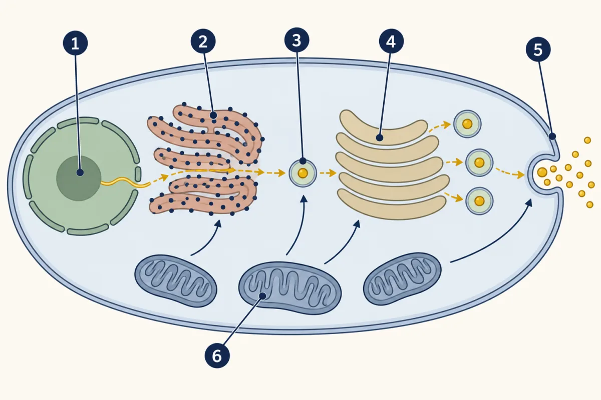
- nucleus
- rough ER
- vesicle
- Golgi apparatus
- cell membrane
- mitochondria
- nucleus
- blank
- blank
- blank
- blank
- mitochondria
Word bank: cell membrane, Golgi apparatus, rough ER, vesicle
- blank
- blank
- blank
- blank
- blank
- blank
Check your labels
- nucleus
- rough ER
- vesicle
- Golgi apparatus
- cell membrane
- mitochondria
Parts on the route, and parts off it
- organelle
- cell membrane
- cell wall
- cytoplasm
- nucleus
- ribosome
- endoplasmic reticulum
- Golgi apparatus
- vesicle
- mitochondria
- chloroplast
- lysosome
- vacuole
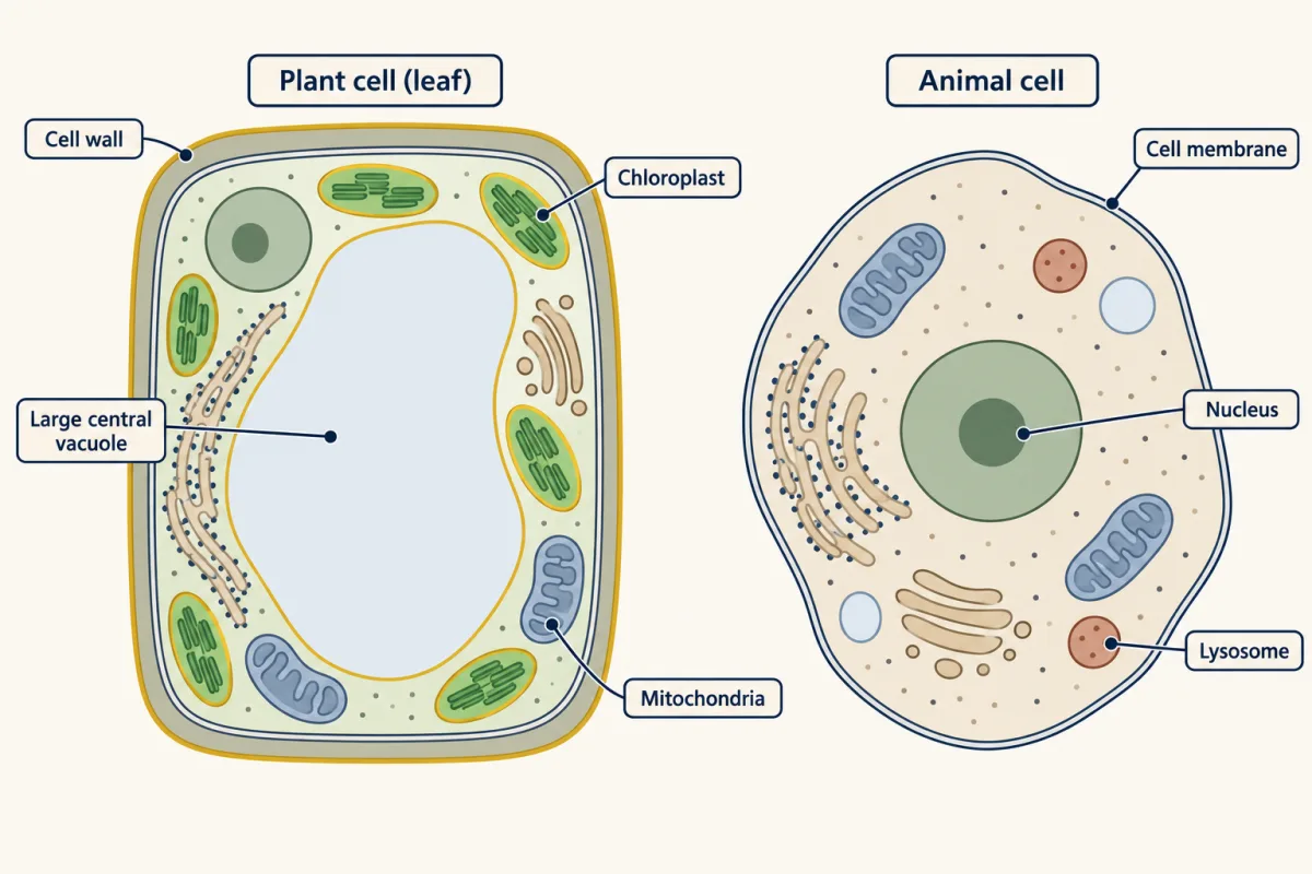
An organelle is a structure inside a cell that does a particular job; most are wrapped in their own membrane. Of the eleven structures on the Virginia list, five are on insulin’s route (nucleus, ribosome, endoplasmic reticulum (ER), Golgi apparatus, cell membrane). Mitochondria supply much of its energy, and the vesicles travel through the cytoplasm. The other four do other jobs (cell wall, chloroplast, lysosome, vacuole).
| Structure | Main job | Plant cells | Animal cells |
|---|---|---|---|
| Cell membrane | Thin outer layer that controls what enters and leaves the cell | Yes | Yes |
| Cell wall | Stiff layer outside the membrane that supports the cell | Yes | No |
| Chloroplast | Carries out photosynthesis, storing light energy in sugar | In green cells, such as leaf cells | No |
| Cytoplasm | Everything inside the membrane but outside the nucleus; many reactions happen here | Yes | Yes |
| Nucleus | Holds most of the cell’s DNA, its instructions | Yes | Yes |
| Ribosome | Builds proteins | Yes | Yes |
| Endoplasmic reticulum (ER) | Folded membranes; rough ER, dotted with ribosomes, folds new proteins for export or for membranes, and smooth ER makes lipids | Yes | Yes |
| Golgi apparatus | Sorts and packs proteins and sends them on | Yes | Yes |
| Lysosome | Sac of digestive enzymes that breaks down worn-out cell parts | Usually not; the vacuole does similar digesting | Yes |
| Mitochondria | Release energy from sugar and store it in ATP | Yes | Yes |
| Vacuole | Sac that stores water and other materials | One large central vacuole that presses on the wall and keeps the cell firm | Small ones, in some cells |
Explore: test the one idea in a model
Switch off any one part of this model beta cell and run the insulin shipment again; a meter shows the ATP supply.
The orange dot is the insulin shipment. Dashed lines show ATP from the mitochondria.
In this sketch the shipment moves one stop at a time, only one part is off at once, and a part on the route that is off stops the shipment before its step, and with the mitochondria off it stops at the rough ER. Rule to keep: a secreted protein goes from the nucleus to a ribosome on the rough ER, by vesicle to the Golgi, and out through the membrane, with mitochondria supplying ATP. This is a sketch of the logic, not a measurement.
Try this.
- Run the route with every part on. First predict how many stops the shipment passes (each vesicle counts as a stop). Then count.
- If the Golgi apparatus is off, where does the shipment get stuck? Predict, then test it.
- Switch the Golgi back on and the mitochondria off. Predict, then run it.
- Switch off the lysosomes, which break down worn-out cell parts. Does insulin still leave? What might build up over days?
A factory model, and where it breaks
In a factory model, the nucleus is the office that keeps blueprints and sends out copies, ribosomes the assembly line, the endoplasmic reticulum (ER) and Golgi finishing and shipping, vesicles delivery trucks, and mitochondria the power plant. It breaks in four places.
- No single manager gives orders; signals such as calcium, and chemistry at each step, set what happens.
- Mitochondria do not create energy; they move energy from sugar into ATP.
- A cell’s parts move around and touch one another all the time.
- Real proteins do not travel as one neat shipment.
How did anyone show that real proteins take this route, in this order? The second film shows the experiment.
Read the transcript of “Catching Proteins on the Move”
Film transcript: Catching Proteins on the Move
How do we know proteins really travel in this order? The first film drew the route, from the rough ER to the Golgi to the cell membrane. But a drawing is not evidence. In 1967, James Jamieson and George Palade found a way to test it. They used thin slices of guinea pig pancreas. The cells they studied make digestive enzymes, not insulin. But they too send proteins out.
Here is the problem. A cell is always making new protein. If every protein carries a mark, the mark is everywhere, and you learn nothing about order. You need to mark just one batch. So they gave the slices a short pulse of a radioactive building block. Only proteins made during those few minutes carried the mark. Then came the chase: plenty of the ordinary building block. Proteins made after that carried no mark. The marked batch was now on its own. They followed the batch in two ways. In one, broken cells were spun in a centrifuge, so their parts settled into layers. In the other, electron microscope pictures showed where radioactive grains sat.
Time for a prediction. The clock reads zero minutes of chase. Right after the pulse, where do you predict most of the marked protein will be? Over the rough ER. At the start of the chase, most grains sat there. That is where ribosomes build new protein. Minutes later, the marked batch had spread out. By seven minutes, Golgi vesicles held almost as many grains as the rough ER. Golgi vesicles are small vesicles at the Golgi's edge. Later still, the mark moved on again. The grains gathered in condensing vacuoles. These are storage granules that are still being formed. At the last time point shown, more than half the grains sat in finished storage granules. These hold the protein until the cell releases it.
Each part's share of the mark peaked in turn. Rough ER, then Golgi vesicles, then condensing vacuoles, then storage granules. The chart shows only four moments of that journey. These were digestive enzyme cells, not beta cells. Still, this experiment gave direct evidence for the order of stops that textbooks still draw. A drawing had become a measurement. So how do we know the order? Mark one batch of new protein, then watch where it goes over time. Each stop's share peaks later than the stop before it.
From the film
- A short pulse of a radioactive building block marks only the proteins made during it.
- Following that marked batch over time shows where it goes in the cell.
- Each part's share of the mark peaked in turn: rough ER, then Golgi vesicles, then condensing vacuoles, then storage granules.
- The marked batch spread out as it moved: by 7 minutes, Golgi vesicles held almost as many grains as the rough ER.
- These cells make digestive enzymes, not insulin, and they gave direct evidence for the order of the stops.
How a marked batch really moves
The second film’s chase times count minutes after a 3-minute pulse ended, and its condensing vacuoles are forming storage (zymogen) granules, not the plant vacuole in the table.
Predict. The film skipped 17 minutes. Which part held the largest share of grains then: rough endoplasmic reticulum (ER), Golgi vesicles or condensing vacuoles?
Check your prediction
Almost a tie. At 17 minutes the rough ER (37.6%) and the Golgi vesicles (37.5%) held nearly equal shares, with 19.5% over condensing vacuoles. A marked batch moves as a spreading wave, not one parcel. Each part’s own share peaks later than the part before it, but the largest share does not hop neatly from part to part.
Values are the share of counted grains over each part (%); a dash means not quoted here.
| Minutes of chase | Rough ER | Golgi vesicles | Condensing vacuoles | Storage (zymogen) granules |
|---|---|---|---|---|
| 0 | 86.3 | — | — | — |
| 7 | 43.7 | 43.0 | — | — |
| 17 | 37.6 | 37.5 | 19.5 | — |
| 37 | — | — | 48.5 | — |
| 57 | — | — | 35.8 | 32.9 |
| 117 | — | — | — | 58.6 |
Source: Jamieson and Palade, Journal of Cell Biology, 1967, paper II, Table II; digestive-enzyme cells, not beta cells. Only some values from the full table are shown, so the rest of each row’s grains were in other parts.
- At 57 minutes of chase, which two parts held most of the counted grains?
- The pulse lasted 3 minutes. How many minutes after the pulse began were the grains counted in the 7-minute row?
The model’s mitochondria switch is based on a 1968 follow-up in the same digestive-enzyme cells. Chemicals that blocked the mitochondria’s ATP making stopped proteins moving from the rough ER to the Golgi. That is the one idea at work for this step: the move needs energy, and without it the protein cannot reach the stops after it.
Same jobs, simpler parts: prokaryotic cells
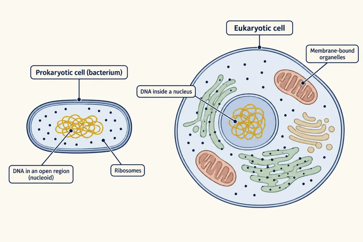
A prokaryotic cell has no nucleus and none of the membrane-bound organelles on this route; bacteria and archaea (another group of single-celled microbes) are prokaryotes. A eukaryotic cell has a nucleus and membrane-bound organelles; plants, animals and fungi are eukaryotes.
| Feature | Prokaryotic cell | Eukaryotic cell |
|---|---|---|
| DNA | In an open region with no membrane, the nucleoid | Mostly inside a nucleus; mitochondria and chloroplasts hold a little |
| ER, Golgi and mitochondria | None | Yes |
| Cell membrane, cytoplasm, ribosomes | Yes | Yes |
| Cell wall | Often | Plants and fungi, not animals |
A single bacterium still carries out every life process with this simpler set of parts.
Predict. A bacterium also sends proteins out, yet it has no endoplasmic reticulum (ER) or Golgi. How do you think it does the job? Hint: think about the membrane, or membranes, around the cell.
Check your prediction
With channels in its membranes. Its ribosomes build the protein in the cytoplasm, and a channel in the cell membrane moves it across. Many bacteria also have a second, outer membrane, which needs another channel or a system that spans both membranes. In bacteria that use oxygen, the cell membrane also makes much of the ATP, the job mitochondria do in your cells. Others make most of theirs in the cytoplasm.
Cells built for their work
Cells that need a large, steady supply of ATP made using oxygen often hold more mitochondria. In one study of mouse muscles, mitochondrial content differed nearly tenfold between muscle types. In normal mouse hearts, which beat without rest, mitochondria filled about 30% of muscle cell volume. These are single studies, and disease can change the amount too (Going deeper).
Chloroplasts follow the work too. Inner leaf cells (mesophyll cells) of the small plant Arabidopsis, which do most of its photosynthesis, held 121 chloroplasts each on average; root cells underground are not green and have none.
Predict. Plants with a change in a nuclear gene called arc5 had only 13 chloroplasts per leaf cell. Does that mean their leaf cells have only a small fraction of the normal amount of chloroplast?
Check your prediction
Not necessarily: each fully grown arc5 chloroplast was about six times larger, so the count alone understates how much chloroplast is there. Count the parts, and measure their size too. The gene also shows one organelle depending on another: it sits in the nucleus, yet changes how chloroplasts divide.
| Cells measured | What was measured | Value |
|---|---|---|
| Heart muscle cells, normal mice (3 mice) | Mitochondria, % of cell volume | 30.2 ± 1.1 |
| Arabidopsis leaf mesophyll cells, normal plants | Chloroplasts per cell (average) | 121 |
| Arabidopsis leaf mesophyll cells, arc5 mutant | Chloroplasts per cell (average) | 13 |
Sources: Rajab and colleagues, Scientific Reports, 2022 (six-month-old mice; value is mean ± standard error); Pyke and Leech, Plant Physiology, 1994.
Claim, evidence, reasoning
Make a claim that answers the essential question: how do the parts of a cell work together? Use both tables.
Start with: The data show that a new protein moves blank, and that cells blank.
Start with: Right after the pulse, blank% of the grains were over the blank. Later the label was in blank. Mitochondria filled blank% of normal heart muscle cells.
Start with: This shows the parts work as a system because blank, and mitochondria matter because many steps on the route use ATP, so cells with a large, steady need for ATP hold blank.
Compare with a model answer
Cell parts pass a protein along in order. Right after the pulse, 86.3% of the grains were over the rough ER. By 7 minutes of chase many were over Golgi vesicles, and by 117 minutes 58.6% were in storage granules. Mitochondria filled 30.2% of normal mouse heart muscle cells, and content differed nearly tenfold between mouse muscle types, so cells with different work can hold different amounts. Limits: the pulse-chase used digestive-enzyme cells from guinea pigs, not human beta cells, and each value comes from one study.
Saved in this browser only.
Design problem
Do leaves grown in brighter light have more chloroplasts per cell? Plan an investigation with a light microscope. Name your variables, constants, control, sample size and safety steps.
Saved in this browser only.
One way to do it
- Independent variable: light level. Grow plants of one species from one seed batch, some in bright and some in dim light.
- Dependent variable: chloroplasts per cell in a thin leaf sample, focusing slowly up and down so no layer is missed.
- Constants: species, plant age, leaf position, watering, temperature, magnification and the counting rule.
- Control: the dim-light plants are the comparison group.
- Sample size: several plants per group, several leaves per plant, at least 20 cells per leaf. Report the mean and the spread.
- Fairness: a partner codes the slides, so the counter does not know the group.
- Limit: chloroplasts can overlap and be missed or counted twice; use one counting rule for every cell.
- Safety: handle slides and cover slips carefully and report broken glass.
Evaluate: check your thinking
Without looking back, explain to a partner where new insulin would first pile up if vesicles could not bud off the endoplasmic reticulum (ER), and why switching off the lysosomes in the model did not stop insulin leaving the cell.
Science practices you used: using a model and naming its limits, reading data tables with means and their standard errors, doing a time calculation from data, and planning a fair test with a large enough sample.
Going deeper
Two more mitochondria numbers, and a fair test
In the same heart study, mitochondria filled 41.1 ± 2.7% of heart muscle cell volume in GENA348 disease-model mice. That does not show amounts fit a cell’s work; it may be a response to the disease. In 5 normal rats, liver-cell mitochondria made up 18% of the whole liver’s volume (Weibel and colleagues, Journal of Cell Biology, 1969). Why is comparing 30.2% in heart cells with 18% in liver not a fair, matched comparison?
One answer
Two things differ: the species (mice, rats) and the measure (share of one cell type’s volume, share of the whole organ’s volume).
Shortcuts and other energy carriers
A few exported proteins skip the endoplasmic reticulum (ER) and Golgi. And ATP is not the only energy carrier on the route: ribosomes and the machinery that buds vesicles also use a close chemical relative of ATP.
Your notes
Select any text in the lesson to highlight it or add a note to it. Your highlights and notes are saved in this browser and collected in your notebook.
This browser is not letting the site save anything (a private window or strict privacy settings), so highlights and notes will be lost when you leave the page.
How we measure it
Pulse-chase labelling
Cells get a short pulse of a radioactive building block, then plenty of the ordinary one, so only proteins made during the pulse are marked. Sampling at set times afterwards shows where that marked group of proteins has moved.
Cell fractionation
Broken cells are spun in a centrifuge so their parts settle into separate layers, such as ER pieces and Golgi pieces. Measuring the label in each layer shows which part held the marked protein at that time.
Electron microscopy and volume measurement
An electron microscope shows organelles far smaller than a light microscope can. Measuring many thin slices, or rebuilding a cell in 3D, gives how much of a cell's volume mitochondria fill.
Where this lesson fits
Before thisReview first
- Respiration and ATPThis course · Lesson 5
Mitochondria make the ATP that pays for steps along the route, and that lesson shows how sugar becomes ATP.
- What Am I Made Of?This course · Lesson 2
You need to know proteins are built from amino acids, since the route makes and moves proteins.
After thisStudy next
- Crossing the MembraneThis course · Lesson 8
The cell membrane is the last stop here, and this lesson shows what it lets in and out.
- From DNA to ProteinThis course · Lesson 18
It takes the messenger RNA copy and ribosome step further, showing how the code is read into a protein.
- One Cell or ManyThis course · Lesson 11
It builds on cells having different parts for different jobs, and on how cells share work.
Elsewhere on the siteFilms, simulations and method films in other courses
- Pulse-chase labellingMethod film
The marked-batch data in this lesson come from labelling proteins briefly and following where they go.
Suggested by an AI model from the lessons themselves, then checked. How we use AI
Finish line
Test yourself
8 questions
Practice and MCAT-style, with explained answers.
Next · Lesson 8
Crossing the Membrane
Why does a slice of cucumber left in salty water get lighter and go limp, while a slice in plain water gets heavier?
Continue →