Cell Signaling · Unit 2: The parts — switches and wiring
Lipids as Anchors and Addresses
Rewritten on : new text, flashcards and film. Your notes and highlights stay, but the text around them may have moved.
How does a protein with no membrane-spanning segment end up stuck to a membrane, and how does a lipid label tell it where?
Pause when the reading reaches a film, and offer to play it. Saved on this device.
Watch the film now, or keep listening
Builds on:Membranes: The Impermeable Sheet That Builds ItselfGTPase Switches: The TimerProtein Modules: SH2, SH3, PTB, PDZ and PHWhy these?
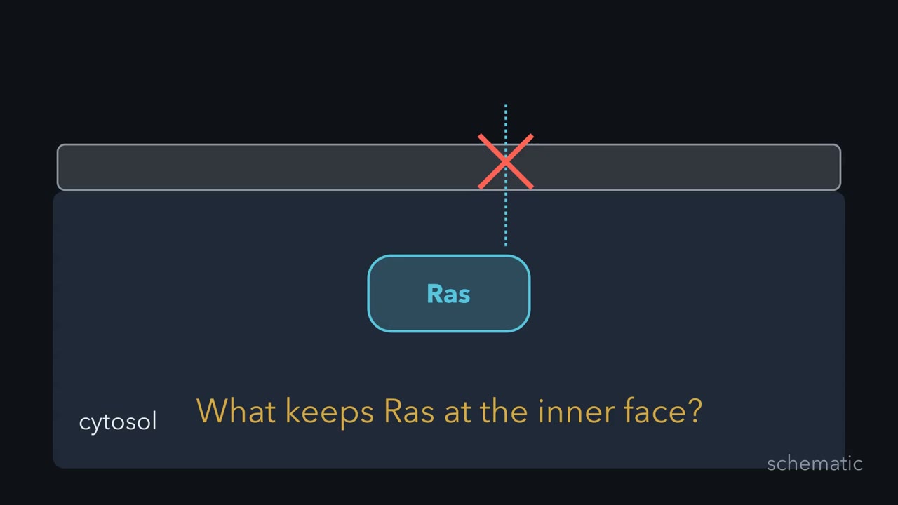 ▶ Watch Why One Farnesyl Tail Is Usually Not Enough · 3:39
Read the transcript of “Why One Farnesyl Tail Is Usually Not Enough”
Film transcript: Why One Farnesyl Tail Is Usually Not Enough
Ras has no segment crossing the membrane, yet it works at the membrane's inner face. What keeps it there? The puzzle has two parts. A lipid must join the protein, and that lipid must remain in a bilayer. First, separate two interactions that students often mix up. A covalent bond joins the lipid to the protein. A much weaker, non-covalent contact holds the lipid tail in the bilayer. The covalent bond can last while the tail repeatedly enters and leaves membranes. Permanent attachment to Ras does not mean permanent membrane binding. Watch one farnesyl tail sample the membrane and release. What should happen if Ras gains a compatible second contact?
Will its visits remain brief, or become much longer? The visits become longer. Both contacts must release together before the protein escapes. That coincidence is rarer. A second lipid can provide that contact. So can a positive patch attracted to negative lipids on the membrane's inner face. H-Ras and N-Ras use farnesyl plus palmitate. K-Ras4B uses farnesyl plus a lysine-rich positive patch. In both, farnesyl supplies the first contact. These are not identical addresses. They are two ways to combine membrane contact with another signal. Why is the change so large? Favorable free-energy contributions add. The equilibrium effect grows exponentially, not step by step.
This is a thermodynamic effect, not a new covalent bond. A computed curve shows how a modest extra contribution can greatly raise the share of time on the membrane. The second contact also creates control. Remove palmitate, or weaken a positive patch, and the strong hold becomes weak again. That is a switch in location. The protein can leave without breaking its permanent farnesyl attachment. Palmitate is especially useful because cells can remove and replace it. It goes on mainly at the Golgi, and it can come off on any membrane.
Farnesyl remains attached while palmitate changes whether H-Ras is tightly trapped or loosely sampling membranes. The same protein changes residence without losing farnesyl. This is the grip half of the lesson's rule. Lipids control signaling protein location by supplying grip, address, or both. The rule applies within this membrane-targeting scope. Here, the second signal converts a brief encounter into regulated membrane residence. So ask three questions about any lipid-modified protein. Which contact puts it on membranes? Which additional signal strengthens or directs that contact? And what can remove or oppose it? The answers predict both localization and the switch that can change it.
From the film
- A second signal turns weak membrane contact into a regulated hold.
- Lipids control signaling protein location by supplying grip, address, or both.
- A permanent lipid-protein bond does not make membrane contact permanent.
- Removing palmitate or neutralizing a positive patch releases the protein without breaking its permanent anchor.
Ras has no membrane-spanning segment, yet most Ras sits on the inner face of the plasma membrane. What holds it there, and how can a membrane tell a protein where to work?
Ras, a small GTP-binding switch that relays a growth signal inward, has to meet the proteins that control and receive its signal.
The one idea. Lipids control signaling protein location by supplying grip, address, or both.
Scope: this rule covers the peripheral membrane proteins and cytosolic lipid readers in this lesson. An anchor need not specify one organelle, not every PH domain reads phosphatidylinositol 3,4,5-trisphosphate (PIP3), and other protein contacts can contribute.
Covalent anchors: what is attached, and where?
The first film left out the bond chemistry, which tells you which contacts a cell can reset.
| Anchor | Size | Where it goes | Enzyme | Can it come off? |
|---|---|---|---|---|
| Myristate | 14-carbon fatty acid | N-terminal glycine, through an amide bond | N-myristoyltransferase | No |
| Palmitate | 16-carbon fatty acid | A cysteine, through a thioester | Palmitoyl acyltransferases (PATs) | Yes, by thioesterases |
| Farnesyl | 15-carbon isoprenoid | Cysteine of a C-terminal CAAX box, through a thioether | Farnesyltransferase | No |
| Geranylgeranyl | 20-carbon isoprenoid | The same CAAX cysteine, when the final residue favors it | Geranylgeranyltransferase I | No |
Myristate goes on a glycine that becomes the first residue after the starting methionine is clipped off, usually while the protein is still being made. Src-family kinases carry one. Prenyl groups contain five-carbon isoprene units: three in farnesyl and four in geranylgeranyl. A CAAX box is a cysteine, two aliphatic amino acids and a final residue that helps determine which prenyltransferase acts. After prenylation, the last three residues are removed and the new end is methylated, hiding its charge. H-Ras, N-Ras and K-Ras all end in a CAAX box.
Amide and thioether bonds are essentially permanent, so myristate and prenyl groups usually remain for the protein’s life; only palmitate’s thioester is readily hydrolyzed, by thioesterases.
Two contacts make residence strong and regulatable
The first film’s Ras pairings are not the only ones: Fyn and Lck carry myristate plus palmitate. A 20-carbon geranylgeranyl can hold some proteins by itself.
The first film’s curve rests on one number. At body temperature, an extra favourable contribution of about 1.4 kcal/mol changes the equilibrium by about tenfold. The exact gain depends on geometry and local concentration, so no universal rule simply multiplies two binding constants.
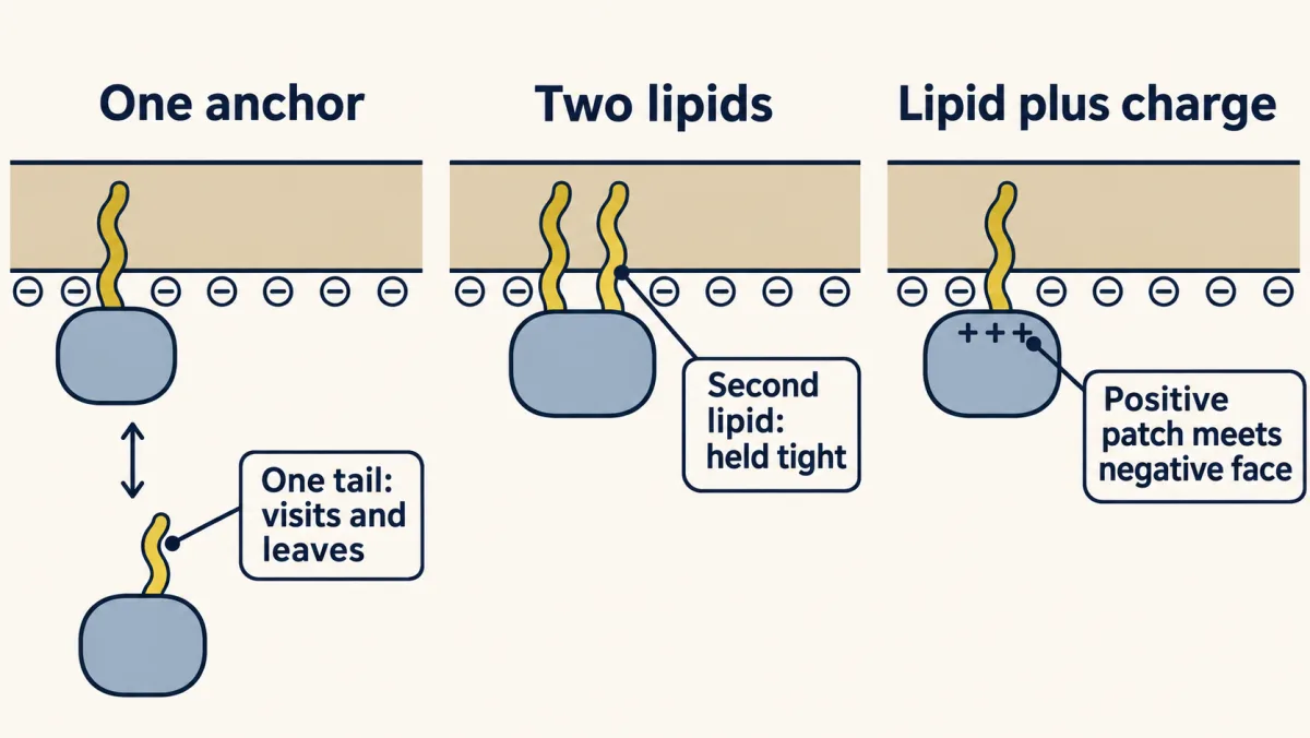
For K-Ras4B the switch is a kinase. Protein kinase C phosphorylates Ser181 beside the lysine patch. The added negative charge weakens attraction to the plasma membrane, so K-Ras4B tends to move to internal membranes, including mitochondrial membranes. This is a farnesyl-electrostatic switch. PDEδ, a carrier with a hydrophobic pocket, can shield a farnesyl group and help Ras cross the cytosol between membranes.
Palmitate turns a hold into a cycle
The first film carried H-Ras from the Golgi to the plasma membrane but left out where palmitate comes off. Thioesterases can remove palmitate wherever H-Ras sits, which weakens its grip and lets it sample internal membranes and return toward the Golgi for another round. Which thioesterases act, and where they are most active, is not fully settled; the robust point is that palmitate goes on mainly at the Golgi and can come off anywhere.
This is a kinetic bilayer trap. No plasma-membrane-specific receptor for H-Ras is required: the rates of trapping, vesicle transport, release and recapture create steady-state enrichment.
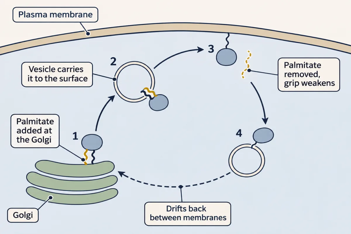
Predict. A drug blocks the thioesterase that normally resets this cycle. Does H-Ras become soluble, collect ever more tightly at the plasma membrane, or lose its focused distribution?
Check your prediction
It should lose its focused distribution and spread across membranes. Palmitate still provides membrane binding, so H-Ras does not become soluble. But without release, return and recapture at the Golgi, the cycle that maintains plasma-membrane enrichment stalls; the farnesyl group stays attached.
Can blocking an anchor block cancer signaling?
If Ras requires farnesylation, a farnesyltransferase inhibitor (FTI) should keep newly made Ras from the membrane, and a Ras that cannot reach the membrane cannot signal. Ras genes are mutated in roughly one in five human cancers, and one FTI, tipifarnib, reached Phase III clinical trials.
Predict. Farnesyltransferase is blocked, but geranylgeranyltransferase I remains active. Which Ras proteins might keep a lipid anchor, and what observation would support that explanation?
Check your prediction
K-Ras and N-Ras can receive geranylgeranyl on the same CAAX cysteine and remain membrane-associated; H-Ras is not rescued this way. Membrane localization that falls only when both prenyltransferases are inhibited supports a backup enzyme.
This backup helps explain why the drugs worked biochemically yet disappointed against the Ras that matters most. K-Ras is by far the most commonly mutated of the three Ras genes; H-Ras is the least. The logic still has uses: tipifarnib has been tested in mutant H-Ras cancers, and lonafarnib is approved for progeria, where a farnesylated nuclear-envelope protein is the culprit.
An FTI keeps newly made Ras from acquiring its farnesyl grip; it does not strip farnesyl that is already attached. A cell can also move a protein without changing its anchor, by rewriting the address on the membrane it lands on.
Read the transcript of “How a Membrane Writes an Address”
Film transcript: How a Membrane Writes an Address
A drug that blocks anchor installation leaves newly made proteins without grip. But a cell can also move a protein without changing its anchor. How does a protein choose one membrane region over another? The membrane itself can carry a changing address.
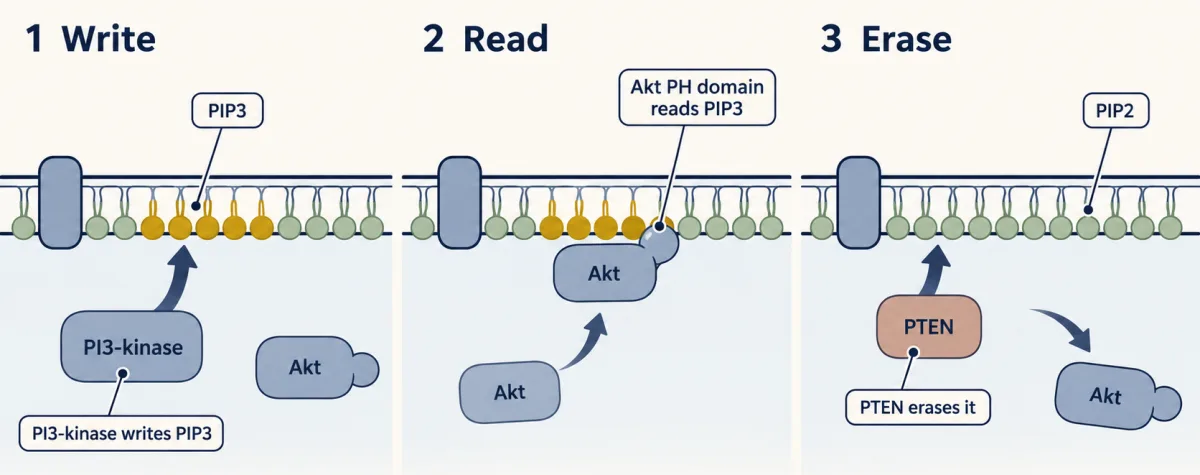
Phosphoinositides are membrane lipids with head groups facing the cytosol. Enzymes can add or remove phosphates from those heads. The tails stay buried, while the exposed head-group pattern changes what cytosolic proteins can recognize. In this pathway, PI3-kinase is the writer. When recruited and activated, it converts membrane PIP2 into PIP3. Production can be local, so one membrane region gains the address before another. PIP3 does not stay under one enzyme.
Lipids diffuse sideways, while phosphatases remove the mark. The visible pattern reflects production, spread, and removal together. The address can therefore move. It is a dynamic address, not a fixed postal code. A blue reporter now floats beneath two membrane regions. Only one region contains PIP3. Where should a matching reader accumulate? Keep watching the reporter, not just the lipid.
The reporter accumulates at the PIP3 region. Its reader is a pleckstrin homology domain, shortened to PH domain. Drawn as a cup, it grips a compatible lipid head group and recruits the attached protein. Akt uses such a domain. Before its matching lipid appears, Akt can remain in the cytosol. When the address rises, Akt is recruited to that membrane region. A green fluorescent PH-domain fusion makes this movement visible. PTEN is an eraser. It removes the phosphate that distinguishes PIP3 from PIP2. As the address fades, the matching readers lose that contact and return toward the cytosol. The reporter is evidence, not a direct photograph of one lipid.
Its binding preference, competing contacts, and cell shape can affect the glow. Controls must test the binding pocket, the writer, and imaging geometry. This is the address half of the lesson's rule. Lipids control signaling protein location by supplying grip, address, or both.
Writing and erasing the lipid pattern changes which readers gather at a membrane. So read a lipid signal as a process. Ask where the writer acts, which domain recognizes the product, and which eraser removes it. Those three answers predict the changing recruitment pattern.
From the film
- Writers, readers, and erasers turn membrane lipids into changing addresses.
- PIP3 production, spread and removal together set where the address sits.
- PI3-kinase writes PIP3, matching PH domains read it, and PTEN erases it.
- A PH-domain reporter measures its own recruitment, so controls establish what that glow means.
Phosphoinositides make dynamic addresses
The second film followed one address, phosphatidylinositol 3,4,5-trisphosphate (PIP3), and left out the other phosphoinositides, the other erasers and the controls a reporter needs. Phosphoinositides carry phosphates at positions 3, 4 and 5 of an inositol ring, and cells make seven combinations. They are enriched differently: PI4P at the Golgi, PI3P on early endosomes and PI(4,5)P2, called phosphatidylinositol 4,5-bisphosphate (PIP2), at the plasma membrane.
Predict. Replace a PIP3-reading domain in a fluorescent reporter with a domain that selectively reads PI4P. Which compartment should become most prominent?
Check your prediction
The expected answer is the Golgi, the classic PI4P-rich compartment. In practice, sensitive PI4P probes also light up the plasma membrane and late endosomes, and several Golgi PI4P readers also need the small GTPase ARF. A reader’s pattern reflects its ligand plus any coincident contacts, not PI4P alone.
The second film’s writer is class I PI3-kinase, which adds the 3-phosphate to PIP2 to make PI(3,4,5)P3, or PIP3. A local source can mark the front of a crawling cell, whereas a growth factor bathing the cell can raise PIP3 over much of the plasma membrane.
Readers and erasers
The second film’s PH domain (pleckstrin homology domain) is a fold of roughly 100 amino acids. Only some PH domains have a pocket for a particular phosphoinositide head group, and many do not read phosphatidylinositol 3,4,5-trisphosphate (PIP3). Akt’s does, and it also binds PI(3,4)P2, formed when PIP3 loses its 5-phosphate.
Phosphatase and tensin homolog (PTEN), which removes the 3-phosphate, is not the only eraser: SHIP1 and SHIP2 remove the 5-phosphate instead, producing PI(3,4)P2.
Predict. If SHIP activity rises while PI3-kinase activity stays the same, must Akt recruitment fall in direct proportion to PIP3?
Check your prediction
No. SHIP lowers PIP3 but makes PI(3,4)P2, which Akt’s PH domain can still bind. Recruitment can therefore outlast or differ from the PIP3 pattern.
Without PTEN, PIP3 stays high longer and PH-domain proteins keep arriving. Loss of PTEN is common in human cancers.
Turn recruitment into a measurement
The second film’s PH domain–green fluorescent protein (GFP) reporter named three controls without giving their results.
Predict. A growth factor recruits a PH-domain–GFP reporter. What should happen if its lipid-binding pocket is mutated? What should a PI3-kinase inhibitor do?
Check your prediction
The pocket mutant should remain cytosolic after stimulation, showing that recruitment requires lipid binding. A PI3-kinase inhibitor such as wortmannin should block recruitment, showing that the relevant lipid depends on PI3-kinase.
Design problem
Build a reporter that lights up the plasma membrane only where and when PI3-kinase is active. What would you fuse to GFP, and what controls show that the signal is specific?
Saved in this browser only.
One way to do it
Fuse GFP to a PH domain that binds PIP3 tightly and selectively. Akt’s PH domain also binds PI(3,4)P2, so a PIP3-preferring domain such as the GRP1 PH domain is cleaner. It stays cytosolic until local PI3-kinase activity recruits it to a bright membrane region.
Use the pocket mutant and PI3-kinase inhibitor above. Cells lacking PTEN should retain more membrane reporter even at rest, showing that localization follows lipid level, not the stimulus alone. Finally, image a free red fluorescent protein and report the membrane green-to-red ratio: a thick ruffle can brighten both channels without any chemical change.
Going deeper
Why membrane location matters
A membrane reduces a search from three dimensions to two, raising encounter frequency for partners confined there. Ras must meet SOS, the exchange factor that loads GTP, and Raf, the kinase active Ras recruits. Grb2 recruits SOS to the membrane. The inner plasma-membrane face also differs from the outer face and the Golgi: it is rich in negatively charged lipids. A hydrocarbon chain favours the bilayer interior over the watery cytosol.
The discovery path
In the early 1950s Mabel and Lowell Hokin followed radioactive phosphate while studying RNA in acetylcholine-stimulated pigeon pancreas slices. The radioactivity lost during purification was in phospholipid they had been discarding. In 1975 Robert Michell connected this lipid turnover to receptors.
Lewis Cantley’s group later followed a lipid kinase that came down with viral oncoproteins such as v-Src and polyoma middle-T antigen. Cantley recalled that its product ran about a millimetre from the known 4-phosphate lipid on thin-layer chromatography. With Peter Downes, the group mapped the phosphate to the 3 position. Their 1988 work identified phosphatidylinositol-3-phosphate, a lipid with no precedent in the literature; later work connected the enzyme to phosphatidylinositol 3,4,5-trisphosphate (PIP3) in stimulated cells.
Rafts and reporter limits
Some models place palmitoylated proteins in small cholesterol-rich lipid rafts. Living membranes contain small, short-lived ordered domains, but their size, lifetime and importance for sorting particular proteins remain debated.
A PH-domain reporter can also reflect other lipids, general charge and coincident protein contacts. Membrane shape changes apparent intensity.
Your notes
Select any text in the lesson to highlight it or add a note to it. Your highlights and notes are saved in this browser and collected in your notebook.
This browser is not letting the site save anything (a private window or strict privacy settings), so highlights and notes will be lost when you leave the page.
How we measure it
Labeling with radioactive phosphate (32P)
Cells or tissue slices take up 32P-phosphate, which ends up in any molecule that gets a phosphate. Counting radioactivity in purified fractions shows which molecules took up new phosphate. Turning counts into true rates also needs to know how heavily the precursor pools were labeled.
Thin-layer chromatography of lipids
A drop of lipid extract is run up a coated plate in a solvent. Lipids with different head groups travel different distances, so a new spot a small distance away from a known standard is evidence for a new lipid.
PH domain–GFP imaging in live cells
A PH domain fused to green fluorescent protein moves to the membrane when its lipid ligand appears there. Watching where the glow goes shows where and when PIP3 builds up.
Mutating the lipid-attachment site
Changing the N-terminal glycine or the cysteines that receive a lipid to residues that cannot be modified, then tracking the mutant protein, shows which anchor a protein depends on.
Practice questions on this lesson (3) → MCAT-style, original, each with an explanation
Where this lesson fits
Before thisReview first
- Membranes: The Impermeable Sheet That Builds ItselfBiochemistry · College: going further
It explains why a long hydrocarbon chain sits happily in a bilayer, which is the basis of every lipid anchor here.
- GTPase Switches: The TimerThis course · Lesson 7
Ras is the GTP-binding switch whose membrane position this lesson explains, so you need how it cycles first.
- Protein Modules: SH2, SH3, PTB, PDZ and PHThis course · Lesson 9
PH domains are introduced there as readers of marks, and this lesson uses them to read PIP3.
After thisStudy next
- PI 3-Kinase, Akt and InsulinThis course · Lesson 20
It continues from PIP3 and the Akt PH domain to what Akt does once it reaches the membrane.
- Ras to MAPK: The Kinase CascadeThis course · Lesson 19
It uses Ras sitting on the inner membrane face to recruit Raf and start the kinase cascade.
- PLC, IP3, DAG and Protein Kinase CThis course · Lesson 14
It follows the PIP2 lipid cut by PLC, whose product DAG recruits kinase C to the membrane.
Elsewhere on the siteFilms, simulations and method films in other courses
- Which Side Is Outside?Film · Cell Biology
It shows the two faces of a membrane, where lipid-anchored proteins sit on the cytosolic side.
Suggested by an AI model from the lessons themselves, then checked. How we use AI
Finish line
Test yourself
8 questions
Practice and MCAT-style, with explained answers.
Next · Lesson 12
GPCRs and Trimeric G Proteins
Adrenaline strengthens the heartbeat but relaxes the airways. How can the same hormone give opposite orders?
Continue →