Cell Biology · Skim version
Protein Sorting into Compartments
Read the full lesson → Skim the whole course →
The guiding idea
Made in a test tube with no membranes, a secretory protein comes out longer than the one the cell secretes. Why?
Almost every protein in a eukaryotic cell is made in the cytosol, yet thousands of them end up inside the ER, mitochondria, chloroplasts or peroxisomes.
From the film: Where the Reader Works
- A protein's address, its signal sequence, is a stretch of its own chain; moved onto a suitable passenger, it can carry that passenger to the same place.
- For many secretory proteins, SRP catches the ER signal at the ribosome's exit, the chain enters while still being made, and signal peptidase cuts the signal off; some ER proteins enter after release by other routes.
- TOM sits on the mitochondrion and PEX5 works in the cytosol, and most mitochondrial and peroxisomal proteins are read after the chain is released.
- To enter the mitochondrial matrix a protein must be at least partly unfolded, as methotrexate-locked DHFR shows; peroxisomes can import cargo that stays folded.
- An exposed address helps specify where a protein goes; where and how its reader works helps decide when it is read, as the chain grows or after release.
From the film: How Do You Know It Went In?
- The added protease cannot cross an intact microsome membrane, so a protected protein is inside; if detergent removes that protection, the membrane was the shield.
- In 1975 an antibody light chain was trimmed and protected only when ER vesicles were present while it was made; globin was not protected.
- Moving a stretch onto a passenger that stays in the cytosol tests whether it is sufficient; deleting it from its own protein tests whether it is necessary.
- A signal that works for one passenger may not work for every passenger, and when a protein carries two signals, only an experiment can say where it goes.
-
The address
-
Into the ER while being made
-
After release: mitochondria, chloroplasts, peroxisomes
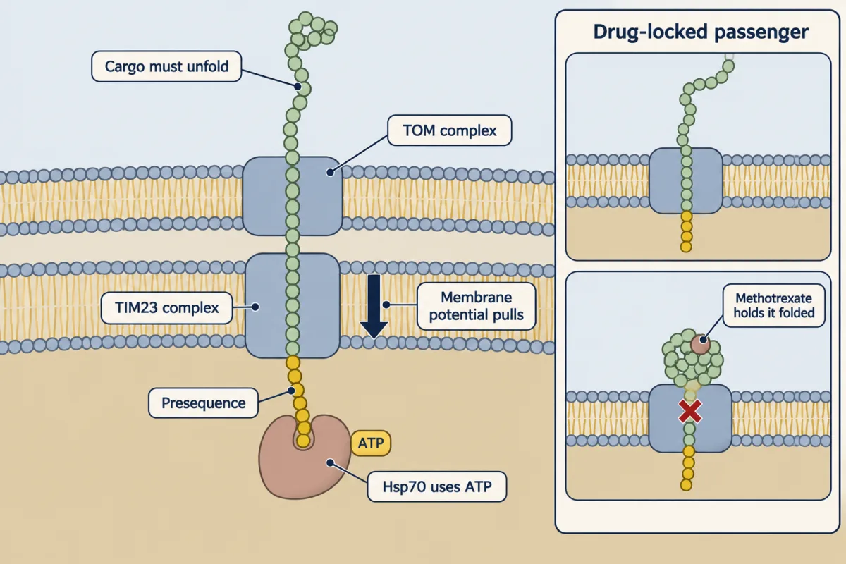
A matrix protein crosses both membranes as an at least partly unfolded chain, drawn in by the membrane potential and held by ATP-using Hsp70. Hold the DHFR passenger folded with methotrexate, and import stops (Eilers and Schatz, 1986). AI-generated illustration 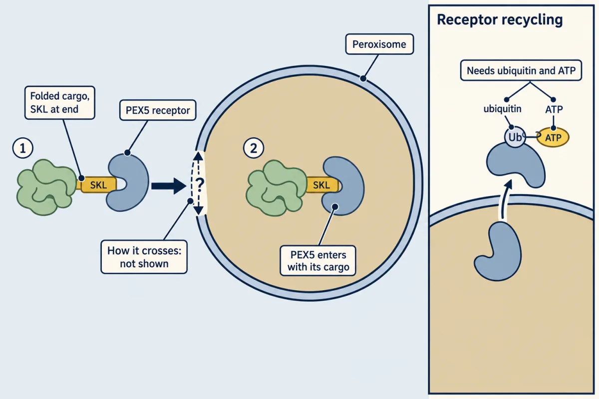
Peroxisomes can take up cargo that stays folded. The crossing is not drawn because how the membrane lets such cargo through is still open. AI-generated illustration -
How do you know where a protein went?
-
Going deeper
Where this lesson fits
Before thisReview first
- Organelles as Distinct EnvironmentsThis course · Lesson 6
It shows what each compartment is like inside, which explains why proteins need addresses to reach the right one.
- Membrane Organization and TopologyThis course · Lesson 4
You need how proteins are inserted and oriented in a membrane to follow the ER signal and anchor cases here.
- Experiments That Distinguish MechanismsThis course · Lesson 3
Protease protection and fractionation logic from there is used here to decide whether a protein went inside vesicles.
After thisStudy next
- The Nuclear Envelope and Nuclear TransportThis course · Lesson 8
Nuclear import reads a different signal and crosses through pores, so it extends the idea of sequence addresses.
- The Secretory PathwayThis course · Lesson 12
Proteins that enter the ER here continue through the Golgi, so it follows their route after translocation.
- Protein Quality Control and Organelle TurnoverThis course · Lesson 9
Proteins that fail to fold or sort correctly after entering compartments are handled by the quality control described there.
Elsewhere on the siteFilms, simulations and method films in other courses
- Centrifugation and subcellular fractionationMethod film
Microsomes used in the protection experiment are made by centrifuging broken cells into fractions.
- From Gene Copy to Protein ChainFilm · Biology
It shows how a ribosome reads mRNA to build the chain whose signal sequence is read here.
- One Cell, One Shipping LineFilm · Biology
It follows insulin from ribosome on the rough ER to secretion, the route entered here.
Suggested by an AI model from the lessons themselves, then checked. How we use AI