Genetics and Molecular Biology · Unit 4: From gene to protein, and its control
New to this? Review it at high school level: From DNA to Protein
The Genetic Code and Translation
Rewritten on : new text, flashcards and film. Your notes and highlights stay, but the text around them may have moved.
How can one added base regroup every codon that follows, while one substituted base may change no amino acid at all?
Pause when the reading reaches a film, and offer to play it. Saved on this device.
Watch the film now, or keep listening
Builds on:Transcription: From DNA to RNARNA Processing: Caps, Tails and SplicingComplementation and Gene Function: From Beadle and Tatum to BenzerWhy these?
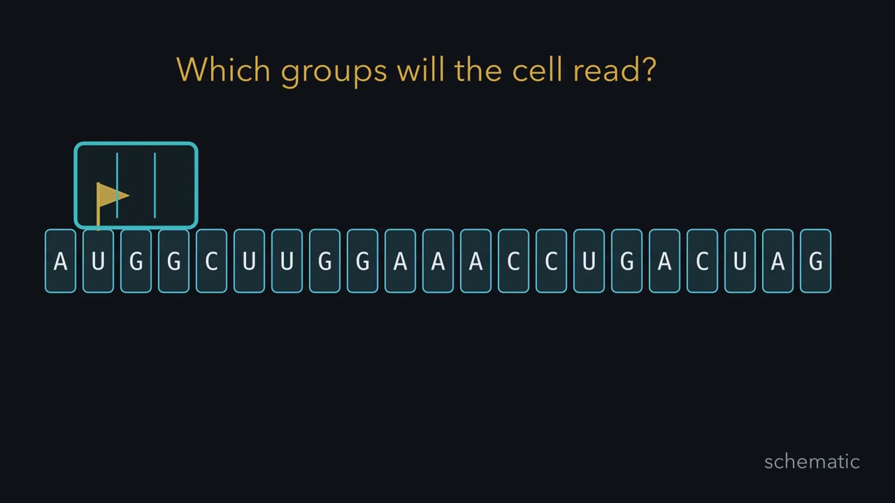 ▶ Watch The Frame Sets the Message · 3:21
Read the transcript of “The Frame Sets the Message”
Film transcript: The Frame Sets the Message
How can an added base regroup every downstream codon? The answer lies in how a cell divides a continuous message. Watch the same ribbon throughout this film. Its letters will stay fixed until we deliberately change them.
A ribosome needs a starting place. Once initiation selects that site, a teal reading window locks onto the message. The window covers a triplet, then advances to the next nonoverlapping triplet. Each window position is a codon. The frame is the alignment between that window and the ribbon. Start at a different base, and every box contains different letters. Start at the same base, and the boxes remain fixed as the window moves. A fixed start makes each next triplet a codon. One base or two bases make too few words for all the amino acids. Triplets make enough. So fixed-length words must be at least triplets. Only experiments could test the real reading unit.
Now replace a base inside a codon. The surrounding boundaries do not move. That codon may keep the same amino acid, specify another amino acid, or become a stop. Later codons keep their original grouping. Predict what happens if a base is inserted after the start. Will only the nearest codon be regrouped, or will every downstream codon be regrouped? Hold the reading window still and follow the ribbon.
The new base joins the ribbon. Every downstream letter slides relative to the fixed window. New triplets appear all the way along, until a stop ends translation. This is a frameshift: the grouping changed, not merely a letter. Researchers called the shift classes plus and minus. A nearby plus and minus often restored function after a disrupted stretch. Same-sign shifts did not restore the frame until a complete triplet's worth accumulated. The later message then returned to its earlier grouping. In phage experiments, those patterns often restored gene function. They supported a fixed reading point and a triplet, or a multiple. Genetics supported a fixed frame, but did not assign the dictionary.
Cell-free translation and ribosome-binding tests linked defined RNA messages to amino acids. Together, the evidence turned moving boxes into codons with assignments. A substitution can alter a codon without moving later boundaries. An insertion or deletion that is not a multiple of three moves the downstream frame. The lasting lesson is simple. Keep the starting register, and every later codon keeps its place.
From the film
- A fixed start makes each next triplet a codon.
- A substitution changes one codon; an insertion or deletion that is not a multiple of three moves the downstream frame.
- Frameshift genetics supported a fixed frame; cell-free biochemistry assigned the codons.
How can one added base regroup every codon that follows, while one substituted base may change no amino acid at all?
A codon is a group of three bases in messenger RNA (mRNA) that specifies one amino acid or a stop. You know the twenty side chains from amino acids; mRNA comes from transcription and, in eukaryotes, RNA processing.
Finding the size and frame of a codon
The first film counted the words four bases can spell: 4, 16 and 64 for one, two and three bases. If every codeword has the same length, it needs at least three bases; that is arithmetic, not evidence that cells use triplets.
The first film’s plus-and-minus pattern came from 1961 work by Francis Crick, Leslie Barnett, Sydney Brenner and Richard Watts-Tobin on the rIIB gene of phage T4, the gene you met in complementation and pathways. A frameshift, an insertion or deletion that is not a multiple of three, moves the downstream codons out of register until a stop or a compensating change. Three pluses, or three minuses, restored wild or nearly wild growth in the combinations tested.
They concluded that the code is read from a fixed starting point, in nonoverlapping groups of three bases or a multiple of three, with no punctuation between words. The case against overlapping codons came from others’ work on tobacco mosaic virus. The phage work assigned no codon and did not claim to prove that the unit was exactly three.
Assigning the codon dictionary
In 1961 Marshall Nirenberg and J. Heinrich Matthaei, at the National Institutes of Health, made an E. coli cell-free system: broken cells that still make protein when given an RNA. They added poly(U), a synthetic RNA made only of uridine, plus radioactive amino acids. Phenylalanine entered an acid-insoluble product with many properties of polyphenylalanine. The reaction needed ribosomes and the soluble fraction.
Predict. Could that experiment alone distinguish a one-base word U, a two-base word UU, and a three-base word UUU?
Check your prediction
No. Every grouping of poly(U) contains only U, so the experiment linked U runs to phenylalanine but could not determine codeword length. Combined with the frame evidence, UUU was the likely codon for Phe.
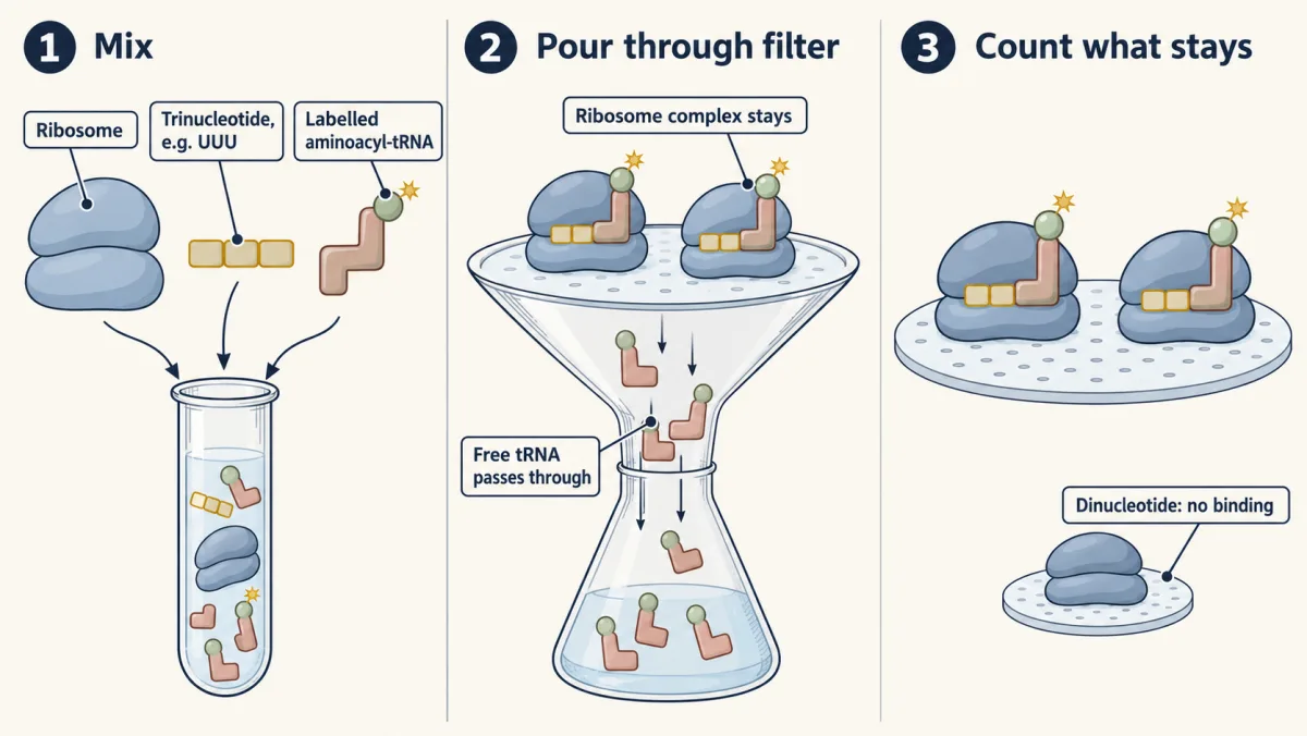
Random copolymers, RNAs with two bases in random order, revealed codon composition but not order; two methods supplied it. In 1964 Nirenberg and Philip Leder mixed one trinucleotide with ribosomes and an aminoacyl-tRNA. UUU held Phe-tRNA on the ribosome, AAA held Lys-tRNA, and CCC held Pro-tRNA. Ribosome complexes stick to a filter while free tRNA passes through, so each triplet could be tested separately.
Har Gobind Khorana’s laboratory made and translated RNAs of exact repeating sequence. Poly(UC), read as UCU CUC UCU CUC, gave an alternating peptide of serine and leucine. That linked the two codons to the two amino acids; other evidence said which was which.
The finished standard table has 64 codons: 61 sense codons for amino acids and three stops, UAA, UAG and UGA. AUG means methionine both at a start and inside a gene. Most amino acids have several codons, a property called degeneracy. Synonymous codons usually differ at the third base. Crick’s 1966 wobble hypothesis explains why: the third codon base can pair loosely with the first anticodon base, so one tRNA can read more than one codon.
The standard table is nearly universal, not universal. Vertebrate mitochondria read UGA as Trp, some ciliates read UAA and UAG as Gln, and a special mRNA signal can make UGA encode selenocysteine.
Use the frame before changing it
A single RNA strand has three reading frames, beginning at base 1, 2 or 3. Double-stranded DNA has six, three on each strand. Initiation picks the frame that is translated.
Worked problem. An mRNA reads 5’-GCCAUGGCUUGGAAACCUGACUAGCC-3’. Assume translation begins at the first AUG.
- The first AUG occupies bases 4 to 6.
- Split from there: AUG GCU UGG AAA CCU GAC UAG CC.
- Look up the complete codons: Met-Ala-Trp-Lys-Pro-Asp, then stop. Six residues; the stop adds none.
Predict. Read from base 2 instead. What are the codons, and does this frame meet a stop in the bases shown?
Check your prediction
CCA UGG CUU GGA AAC CUG ACU AGC gives Pro-Trp-Leu-Gly-Asn-Leu-Thr-Ser, with no stop here. Base 3 gives CAU GGC UUG GAA ACC UGA, or His-Gly-Leu-Glu-Thr-stop.
Now mutate the translated region.
- GCU to GCC still gives Ala, so Met-Ala-Trp-Lys-Pro-Asp is unchanged: silent.
- GCU to ACU gives Met-Thr-Trp-Lys-Pro-Asp, Ala to Thr: missense.
- UGG to UGA creates a stop after Met-Ala, a nonsense change.
- Inserting G after AUG gives AUG GGC UUG GAA ACC UGA: Met-Gly-Leu-Glu-Thr, then stop. This is a frameshift because every downstream codon is regrouped, and a formerly out-of-frame stop is now early.
- original protein
- edited protein
Read from the first AUG in nonoverlapping triplets: a base change alters at most one codon, but one inserted or deleted base shifts every codon after it. The AUG and the 5′ bases before it are held fixed here, and the message has no exon junctions, so nonsense-mediated decay is not shown.
Silent means unchanged amino-acid sequence, not necessarily harmless. In human cell lines, synonymous changes can alter mRNA stability.
Every answer above came from a lookup table. A cell has none, and a codon cannot recognize an amino acid by shape. So what makes UGG mean Trp?
Read the transcript of “How Codons Become a Chain”
Film transcript: How Codons Become a Chain
The first film fixed the codon boxes. How does each box choose an amino acid? Keep the same blue message, gold start, and teal reading window. Now add the adaptor that connects sequence to protein. A transfer RNA has two working ends. Its anticodon can pair with a codon. Its other end carries an amino acid. The adaptor keeps both identities physically linked as it moves. Before the adaptor reaches a ribosome, an aminoacyl transfer RNA synthetase usually loads it. The enzyme selects an amino acid and the transfer RNA meant to carry it. Some synthetases also remove mistakes. Translation therefore depends on separate checks. The synthetase chooses the cargo.
At the ribosome, the anticodon tests the codon. The growing chain receives whatever cargo arrived on the accepted adaptor. Charged transfer RNAs connect codons to the growing protein. Predict this case. A transfer RNA carries the wrong amino acid, but its anticodon matches the codon. Will the ribosome reject the adaptor, or accept it?
The pairing check passes. The ribosome does not inspect the amino acid itself. The wrong cargo can enter the chain. Accuracy therefore depends on charging before the ribosome and pairing at the ribosome. Now follow a cycle. The P site holds the transfer RNA carrying the growing chain. The A site receives a charged adaptor whose anticodon matches the next codon. The E site is the exit. The large subunit transfers the chain onto the amino acid in the A site. Then the ribosome advances by a codon. The chain-bearing adaptor moves into the P site, while the empty adaptor reaches the E site and leaves. The ribosome is not merely a track. Its large-subunit RNA positions the two transfer RNA ends and catalyzes peptide-bond formation. The reaction occurs in an RNA-lined pocket.
That is why the ribosome is called a ribozyme. The same blue ribbon moves through the teal window. Each accepted adaptor adds a green block, so the chain grows in codon order. The ribosome repeats the cycle without changing the frame. When a stop enters the A site, a release factor arrives instead of a transfer RNA. The completed chain leaves, and the ribosome can be recycled. The stop adds no amino acid. The codon never touches its amino acid directly. A charged adaptor makes the connection, while the ribosome preserves the order. The adaptor carries identity from the code to the chain.
From the film
- Charged tRNAs connect codons to the growing protein.
- Once a start site fixes the reading frame, the ribosome reads nonoverlapping codons in order, and charged tRNAs connect those codons to amino acids.
- For most tRNAs, a synthetase chooses the amino acid; the ribosome checks codon-anticodon pairing, not the amino acid.
- Elongating tRNAs move from A to P to E; a release factor, not a tRNA, reads a stop.
Two checks connect codons to amino acids
The second film showed the adaptor Crick had argued for, transfer RNA (tRNA), and its two checks: for most tRNAs an aminoacyl-tRNA synthetase chooses the amino acid, and the ribosome tests the tRNA’s anticodon, three bases that pair with a codon. What it left out is the chemistry. The amino acid rides on the tRNA’s 3’ CCA end. There is usually one synthetase per amino acid, and it charges its tRNA or tRNAs in two steps:
amino acid + ATP → aminoacyl-AMP + PPi
aminoacyl-AMP + tRNA → aminoacyl-tRNA + AMP
Pyrophosphate is hydrolyzed, so charging spends two high-energy phosphate bonds. Some synthetases edit, hydrolyzing a wrong amino acid before or after it reaches the tRNA. Editing matters because the ribosome checks codon-anticodon pairing, not the amino acid.
The ribosome sets the start, extends the chain and stops
A ribosome has two subunits: bacterial 30S and 50S make 70S; eukaryotic cytosolic 40S and 60S make 80S. Svedberg values measure sedimentation, so they do not add. Its three tRNA-binding sites span the two subunits: the A site (aminoacyl), P site (peptidyl) and E site (exit).
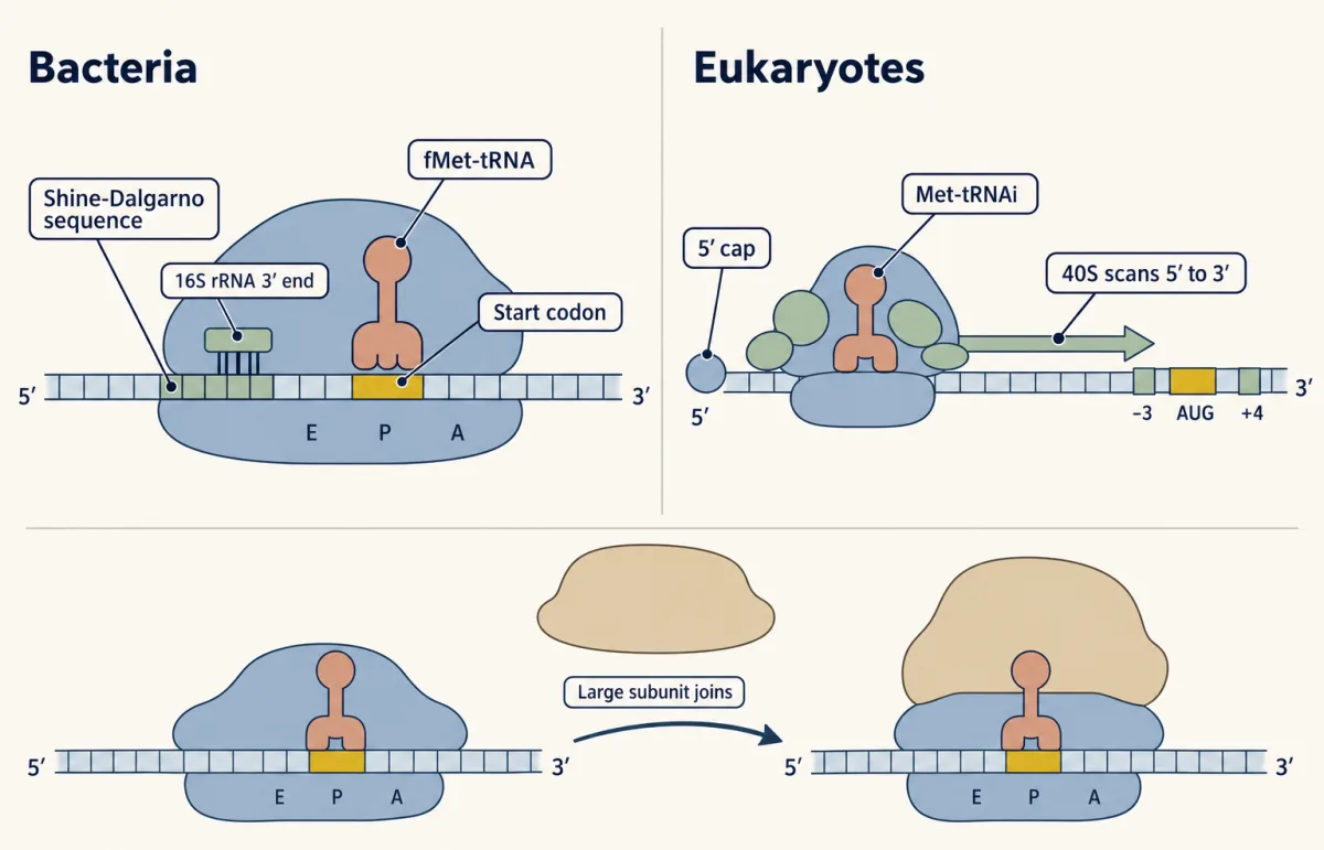
Initiation places an initiator tRNA in the P site and fixes the frame, but bacteria and eukaryotes often find the start differently.
- In many bacterial mRNAs, a purine-rich Shine-Dalgarno sequence a few bases upstream of the start codon pairs with the 3’ end of 16S rRNA, which places the start codon on the small subunit. Most bacteria use an fMet-tRNA (formylmethionine-tRNA) initiator, and GUG can serve as a start as well as AUG. Other bacterial mRNAs, including leaderless ones, begin without Shine-Dalgarno pairing.
- In most eukaryotic mRNAs, the 40S subunit and initiation factors load at the 5’ cap and scan 5’ to 3’. They usually start at the first AUG in good Kozak context, with a purine at position -3 and G at +4 when the A of AUG is +1. The initiator is Met-tRNAi.
Predict. Two AUGs lie near the 5’ end of a mammalian mRNA. The first has poor Kozak context and the second has good context. Must every scanning ribosome start at the first?
Check your prediction
No. Many scanning ribosomes can pass an AUG in poor context and begin at the next AUG in good context. This is leaky scanning, so start selection depends on more than finding the first AUG.
Either way, the large subunit then joins.
The second film’s elongation cycle omitted its driving factors. EF-Tu (eEF1A in eukaryotes) uses GTP to deliver aminoacyl-tRNA to A; if the anticodon matches, GTP is hydrolyzed. The large subunit transfers the chain from P to the amino group of the A-site amino acid. EF-G (eEF2 in eukaryotes) uses another GTP to move the ribosome one codon. The chain grows from N to C terminus. In bacteria the catalytic RNA is the large subunit’s 23S rRNA.
Predict. Counting charging plus one delivery and one translocation, how many high-energy phosphate bonds are spent per peptide bond?
Check your prediction
Charging costs two, because ATP is split to AMP; EF-Tu delivery and EF-G translocation cost one GTP each. The total is about four high-energy phosphate bonds per peptide bond.
At termination, normally no tRNA reads the stop in the A site; a release factor promotes hydrolysis of the bond to the P-site tRNA. In bacteria RF1 reads UAA and UAG, while RF2 reads UAA and UGA; eukaryotic eRF1 reads all three stops. Translation speed and control continue in mTOR and AMPK; folding in protein 3D structure.
A premature stop can also destroy the message
In mammalian cells, splicing leaves an exon junction complex just upstream of most exon-exon junctions. A ribosome on the first round of translation displaces these complexes.
Predict. A human gene has six exons. One premature stop lies in exon 2, well upstream of later junctions; another lies in exon 6, the last exon. Which mRNA is more likely to be degraded?
Check your prediction
The exon 2 mRNA is usually targeted for nonsense-mediated decay (NMD) because translation stops while exon junction complexes remain downstream. A stop in the last exon usually escapes because no exon junction complex remains after it.
More precisely, a stop more than about 50 to 55 bases upstream of the last exon-exon junction usually triggers nonsense-mediated decay (NMD); one in the last exon, or within about 50 bases of the last junction, usually escapes and may make a truncated protein. This is a tendency with exceptions, not a rule for yeast or bacteria. For how such mutations arise, see mutation.
Design problem: assign codons from repeating RNAs
In an E. coli cell-free system, poly(AG), AGAGAG…, directs a peptide of alternating Arg and Glu. Poly(AAG), AAGAAG…, directs three homopolymers: polyLys, polyArg and polyGlu. Assign AGA, GAG, AAG and GAA as far as the data allow. State each assumption and name the experiment that settles what remains.
One way to do it
Assume codons are nonoverlapping triplets, translation can begin in any frame in this system, and each codon means one amino acid.
- Poly(AG) gives alternating AGA and GAG; poly(AAG) gives repeated AAG, AGA or GAA, one per frame.
- Poly(AG) says {AGA, GAG} = {Arg, Glu}. AGA is shared by both polymers, as are Arg and Glu, so the data cannot assign it.
- If AGA is Arg, GAG is Glu and {AAG, GAA} = {Lys, Glu}. If AGA is Glu, GAG is Arg and {AAG, GAA} = {Lys, Arg}.
- AAA was already Lys from poly(A). Similar first two bases favor AAG as Lys, but that is a guess, not data.
The Nirenberg-Leder binding test settles each trinucleotide with labelled aminoacyl-tRNAs: AGA Arg, GAG Glu, AAG Lys and GAA Glu.
Going deeper
How the rIIB mutants were made
The starting rIIB mutant was made with proflavin, a dye Crick and colleagues argued adds or removes a base or bases. Most suppressor mutants that followed arose spontaneously.
Glutaminyl-tRNA and the Kozak reporter
Most bacteria and all archaea lack a glutaminyl-tRNA synthetase and make Gln-tRNA indirectly. In Kozak’s preproinsulin reporter, changing bases around AUG changed protein yield over a 20-fold range.
Where the exon junction complex sits
Splicing leaves an exon junction complex about 20 to 24 bases upstream of most exon-exon junctions.
Your notes
Select any text in the lesson to highlight it or add a note to it. Your highlights and notes are saved in this browser and collected in your notebook.
This browser is not letting the site save anything (a private window or strict privacy settings), so highlights and notes will be lost when you leave the page.
How we measure it
Cell-free translation with radiolabel
Broken cells supply ribosomes, tRNAs and enzymes; you add an RNA template and radioactive amino acids. Acid precipitates most proteins but not free amino acids, so counts in the precipitate tell you which amino acids the template directed into polymer. Some products, such as polylysine, stay soluble and need another assay.
Frameshift suppression in phage
Single-base insertion or deletion mutants are combined by recombination, and the double or triple mutant is tested for growth on a host that needs the gene. Restored growth shows that the reading frame has been put back in register downstream.
Trinucleotide binding to ribosomes
A defined triplet, ribosomes and one labelled aminoacyl-tRNA are mixed and passed through a filter. Ribosomes and anything bound to them stay on the filter, so retained label means that triplet recruits that tRNA, with no protein synthesis needed.
X-ray crystallography of the ribosome
Ribosomal subunits or whole ribosomes are crystallized, often with tRNAs, mRNA or substrate analogs, and the diffraction pattern is turned into an atomic model. It shows which parts, RNA or protein, touch the reacting groups.
Practice questions on this lesson (6) → MCAT-style, original, each with an explanation
Where this lesson fits
Before thisReview first
- Transcription: From DNA to RNAThis course · Lesson 16
You need to know how mRNA is made from a DNA template before reading it as codons.
- RNA Processing: Caps, Tails and SplicingThis course · Lesson 17
Mature mRNA with its exon junctions is the message translated here, and the premature-stop section uses spliced exons.
- Complementation and Gene Function: From Beadle and Tatum to BenzerThis course · Lesson 10
The T4 rIIB gene and its mutants come from there, and the frameshift work builds on them.
After thisStudy next
- The lac Operon: How Bacteria Regulate GenesThis course · Lesson 19
It shows how bacteria control which genes are transcribed and translated into protein, using the codon-to-protein flow learned here.
- Cancer as a Genetic DiseaseThis course · Lesson 21
Nonsense and frameshift mutations that destroy tumor suppressor proteins are applied there.
- Forward and Reverse Genetics: Screens, RNAi and CRISPRThis course · Lesson 24
Repair of a Cas9 cut often leaves small insertions or deletions that shift the reading frame and knock out a gene.
Elsewhere on the siteFilms, simulations and method films in other courses
- From Gene Copy to Protein ChainFilm · Biology
It follows mRNA codons to an amino acid chain and shows how one changed letter alters a protein.
- Transcribe, translate, mutateSimulation · Biology
You can transcribe, translate and mutate a gene to see silent, missense and frameshift effects.
- One String, One ShapeFilm · Biochemistry
It shows how a changed amino acid in the chain, as in missense mutations, can change protein shape and job.
Suggested by an AI model from the lessons themselves, then checked. How we use AI
Finish line
Test yourself
11 questions
Practice and MCAT-style, with explained answers.
Next · Lesson 19
The lac Operon: How Bacteria Regulate Genes
Give E. coli glucose and lactose together and it uses the glucose first, pauses, then starts on lactose. How does one cell choose which genes to express, and when?
Continue →