Physiology · Unit 3: The heart and circulation
Blood Cells, Hematocrit and Clotting
Rewritten on : new text, figures and flashcards. Your notes and highlights stay, but the text around them may have moved.
An endurance athlete can have a lower hematocrit than an untrained friend and more red cells. How?
Pause when the reading reaches a film, and offer to play it. Saved on this device.
Watch the film now, or keep listening
Builds on:Body Water and Its CompartmentsWhy these?
An endurance athlete can have a lower hematocrit than an untrained friend and more red cells. Here is the second puzzle: a cut on your finger seals in a few minutes, while the same clotting proteins are flowing through every healthy vessel in your body, where nothing happens at all.
Both puzzles have the same shape: in blood, the number we measure is global and the control is local. Hematocrit is a concentration for the whole circulation, while the kidney cell that orders new red cells reads the oxygen in its own patch of tissue; a clotting time is measured on a tube of plasma, while the enzymes that seal a wound are made on the membrane of the activated platelets packed into the plug at the breach.
Hematocrit is a ratio, not a cell count
The rule for the first half. Hematocrit is a ratio, so it answers to its numerator and its denominator separately. Nothing in the body regulates the ratio. The kidney regulates the numerator, and it does so by reading the oxygen in its own interstitium — not the red cell count, and not the ratio.
Blood’s components are in blood vessels, blood and lymph. The hematocrit is the fraction of whole blood occupied by packed red cells after centrifugation.
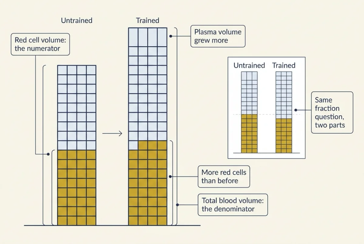
hematocrit = red cell volume / (red cell volume + plasma volume)
Made-up round numbers for an untrained adult, taken as a venous sample: 2.2 L of red cells and 2.8 L of plasma give 2.2 / 5.0 = 44 percent. (A hematocrit spun or counted on a venous sample runs a little above the whole-body value; see Going deeper.)
Predict. Training raises this person’s red cell volume by 10 percent and their plasma volume by 20 percent. Does the hematocrit go up or down, and what has happened to the number of red cells?
Check your prediction
Red cell volume becomes 2.42 L and plasma volume 3.36 L, so hematocrit falls to 2.42 / 5.78 = 42 percent — with 10 percent more red cells than before. Reviews describe this pattern in trained athletes, so “sports anemia” is not anemia. The ratio fell because the denominator grew faster than the numerator.
Pregnancy also expands plasma more than red cells (pregnancy, birth and lactation). In the first days at altitude it runs the other way: diuresis shrinks plasma, and hematocrit rises before red cell mass has changed much (control of breathing). A concentration cannot give a mass: for that you need plasma volume by indicator dilution (body fluid compartments) or hemoglobin mass by carbon monoxide rebreathing (exercise physiology).
The kidney senses oxygen, not red cells
The numerator has a controller: erythropoietin (EPO), a glycoprotein hormone made mostly by the adult mammalian kidney, in fibroblast-like peritubular interstitial cells that sit between the tubules of the cortex and outer medulla rather than in them. The evidence is in situ hybridization on anemic mouse kidneys (Lacombe and colleagues, 1988), which found Epo mRNA in peritubular cells and not in glomerular or tubular cells; those authors’ guess at the cell type was later corrected (Going deeper).
These cells read local tissue PO2. They sit in a relatively hypoxic zone, so a small fall in blood oxygen content, from anemia or low arterial PO2, lowers their oxygen enough to matter. The switch is HIF-2 alpha, a subunit of a hypoxia-inducible transcription factor: prolyl hydroxylases that require oxygen, iron and 2-oxoglutarate mark it for destruction in well-oxygenated cells, and when oxygen falls it accumulates and turns on the EPO gene (ubiquitin and proteolysis). In rodent kidneys, hypoxia raises EPO mainly by recruiting more producing cells; human data are limited.
In lowlanders exposed to hypoxia, plasma EPO rises within the first day or two, then falls while staying somewhat above baseline. EPO acts mainly by preventing the death of late erythroid progenitors, which then divide and mature, so release of reticulocytes (young red cells that still hold RNA) rises over the following days. That is why the cell count lags the hormone.
Two things here are not EPO’s work. Platelets answer to thrombopoietin, the main regulator of megakaryocyte development. And HIF-2 alpha itself directly raises duodenal iron absorption; hypoxia lowers hepcidin too, but mostly indirectly, because the erythropoiesis EPO drives sends signals, such as erythroferrone, that suppress it.
Red cells wear out and their iron comes back
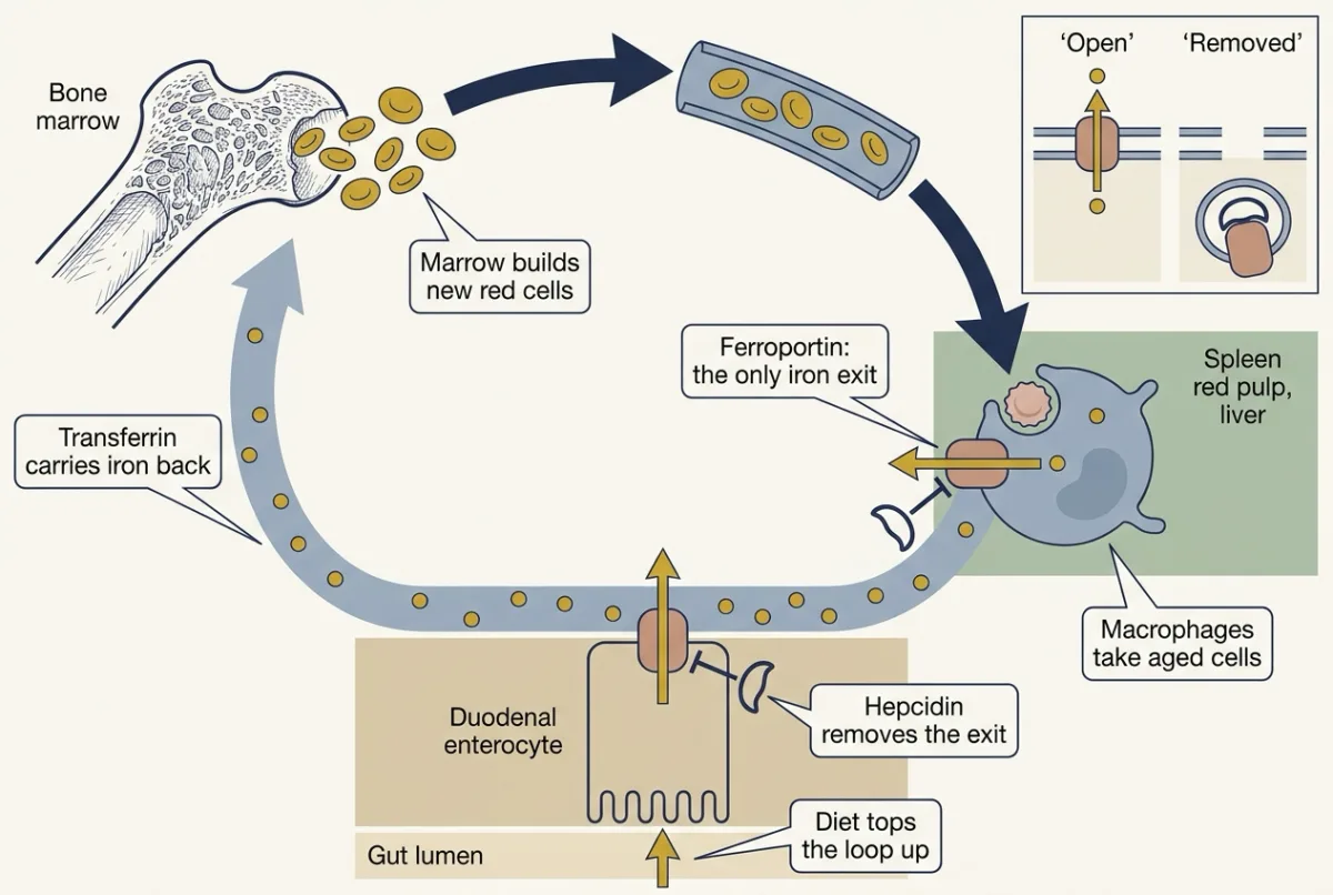
Human red cells circulate about 120 days on average, with real variation between people. Aged cells are removed by macrophages, mainly in the red pulp of the spleen and in the liver; how red pulp macrophages choose which cells to take is not settled. Heme yields iron and bilirubin (absorption and the liver).
The iron leaves through ferroportin, the only known cellular iron exporter, and returns to the marrow on plasma transferrin; duodenal enterocytes export absorbed iron the same way. Hepcidin, a 25-amino-acid liver peptide secreted when iron stores rise and in inflammation, binds ferroportin: in cultured cells this drives its internalization and degradation, and structural work shows hepcidin also occludes the exporter’s central cavity.
Predict. A patient with a chronic inflammatory illness has iron in their macrophages, a low plasma iron concentration, and a marrow behaving as though iron were scarce. Where is the iron stuck?
Check your prediction
Inflammation raises hepcidin, hepcidin removes ferroportin from the surface, and iron recovered from old red cells cannot leave the macrophage. Stores full, plasma empty: the plasma number reports one gate, not the total — the global-and-local theme again. This is the anemia of inflammation; too little hepcidin does the opposite and lets iron flood in (overload).
Worked problem. Made-up round numbers for an adult human at steady state: blood volume 5.0 L; 5.0 x 10¹² red cells per L; hemoglobin 150 g per L; mean lifespan 120 days; 3.46 mg iron per g hemoglobin (4 x 55.85 = 223.4 g of iron per mol of a tetramer of about 64,500 g per mol). Assume production equals removal, every cell lives the same time, no blood loss, and all hemoglobin iron is recycled.
- Total red cells = 5.0 x 10¹² per L x 5.0 L = 2.5 x 10¹³ cells.
- Production = 2.5 x 10¹³ / 120 days = 2.08 x 10¹¹ cells per day; divided by 86,400 s per day, 2.4 x 10⁶ cells per second.
- Total hemoglobin = 150 g per L x 5.0 L = 750 g; turnover = 750 g / 120 days = 6.25 g per day.
- Iron moved = 6.25 g per day x 3.46 mg per g = 21.6 mg per day. Reviews give 20 to 25 mg a day recycled by macrophages against 1 to 2 mg absorbed: over 90 percent of the marrow’s iron is recycled.
- Iron in circulating hemoglobin = 750 g x 3.46 mg per g = 2,600 mg, about 2.6 g.
- Perturbation: if hemolysis shortened lifespan to 30 days with hemoglobin held constant, production must rise fourfold, to 8.3 x 10¹¹ cells per day, and hemoglobin iron flux to 750 / 30 x 3.46 = 86.5 mg per day. If reticulocytes stay recognizable in blood for about one day, their share rises from about 1/120 = 0.8 percent to 1/30 = 3.3 percent.
Read step 4 back as physiology: that recycling is why 1 to 2 mg absorbed a day can keep 2.4 million new cells a second in hemoglobin. The diet tops up a loop; it does not fill the tank.
Two highland peoples, two answers to thin air
In a lowlander at altitude, hemoglobin mass rises only gradually, about 1 percent per 100 hours of exposure in pooled data from athletes, so the early hematocrit change is mostly the denominator moving. And raising hemoglobin is not the universal human answer to thin air: Beall and colleagues (1998) measured Tibetans and Bolivian Aymara at 3,800 to 4,065 m with the same technique, and Tibetan values sat close to a sea-level sample while Aymara values sat 3 to 4 g per dL higher. In 2010, variants at EPAS1, the gene for HIF-2 alpha, were associated with lower hemoglobin in Tibetans. These are associations in two populations; the numbers and the other caveats are in Going deeper.
Why might more hemoglobin not be better? Hemoglobin carries about 1.34 mL of oxygen per g (oxygen transport), so more red cells per litre means more oxygen per litre — but red cells also thicken blood, and viscosity rises steeply and nonlinearly with hematocrit, most of all at low shear (deformability offsets part of the cost in microvessels). Delivery is flow times content, so a high hematocrit raises content while tending to lower flow. In the excessive erythrocytosis of some Andean residents, then, predict hematocrit, viscosity, oxygen content per litre at a given saturation, and resistance to flow all to rise together.
A cut seals while the circulation does not
So the question is not how fibrin forms. It is where, and how much.
The one idea to carry through this half of the lesson. Thrombin is made on surfaces, not in solution. A small, self-limiting trickle is made on the tissue factor-bearing cells that the broken wall exposes. The large burst is made only on the membrane of the activated platelets that the plug has anchored at the breach. Away from that surface, flow carries thrombin off and the brakes on intact endothelium destroy it within a few micrometres. A clot grows where a surface holds the enzymes, and nowhere else.
Scope. This is coagulation in a living vessel. It does not describe the prothrombin time and the aPTT, run in a tube with added phospholipid in place of platelets and no endothelium at all (and, for the aPTT, on an artificial contact activator) — which is exactly why those times and a patient’s bleeding can disagree. “On platelets” means chiefly platelets: any membrane that turns its phosphatidylserine outward will serve, and platelets are the surface that arrives in quantity at a wound. The distance is an order-of-magnitude estimate from diffusion and decay rates, not a measurement.
First, the record the rule has to explain
Add a small amount of tissue factor to plasma that still contains platelets, and follow thrombin over time. Three plasmas, with made-up round numbers in the pattern these experiments give:
| Plasma | Thrombin first appears | Peak thrombin |
|---|---|---|
| Normal | 4 min | 300 nM |
| Factor VIII-deficient (hemophilia A) | 4 min | 60 nM |
| Factor XII-deficient | 4 min | 290 nM |
Read the columns separately: the first is when thrombin starts, the second is how much gets made.
Predict. Hemophilia A causes severe bleeding into joints and muscles. Factor XII deficiency causes no abnormal bleeding at all. From the table alone, which column tracks the bleeding?
Check your prediction
The size. All three plasmas start making thrombin at the same time, so starting is not what is broken in hemophilia. The factor VIII-deficient plasma reaches 60 / 300 = 20 percent of the normal peak; the factor XII-deficient plasma reaches 290 / 300, about 97 percent. The patient who bleeds is the one whose peak collapsed.
So thrombin generation has two separable parts: starting, which neither deficiency touches, and amplifying, which needs factor VIII and not factor XII. That is the shape the mechanism has to have.
The plug builds the surface
How does a procoagulant membrane get held at the breach, against flow? A cut small vessel first constricts, through reflex neurogenic mechanisms and local endothelin and platelet thromboxane A2, which slows the stream. Then von Willebrand factor (VWF) binds exposed collagen and the platelet receptor GpIb, tethering passing platelets to the wall — the step that holds against fast arterial flow. Tethered platelets activate: they release ADP, which acts on P2Y12, a Gi-coupled receptor (G protein-coupled receptors), and they make thromboxane A2 from arachidonic acid through COX-1 and thromboxane synthase. Both recruit more platelets and switch the integrin GpIIb/IIIa to bind fibrinogen, which bridges neighbouring platelets into a plug. Intact endothelium alongside releases prostacyclin, which inhibits aggregation, so the plug does not spread along the wall.
Trickle, then burst: why thrombin is made twice
The cell-based model (Hoffman and Monroe, 2001) places coagulation on cell surfaces in three overlapping stages — initiation, amplification and propagation.
Stage 1: a trickle, on the cells under the wall
Tissue factor is a membrane protein on cells beneath the endothelium, out of contact with blood until the wall breaks. Exposed, it binds plasma factor VIIa, and with calcium that pair cuts factor X to Xa and factor IX to IXa.
Xa with its partner Va makes a little thrombin — nowhere near enough to clot anything, and it does not last, because tissue factor pathway inhibitor (TFPI) shuts the tissue factor-VIIa-Xa assembly down.
So why does the shut-off not end the story? The trickle was never meant to clot blood. It is a signal, and in the short time it exists that thrombin does four things — the amplification stage:
- activates platelets, which turn their phosphatidylserine outward and so become a procoagulant surface;
- cuts factor V to Va;
- cuts factor VIII to VIIIa, releasing it from the von Willebrand factor that carries it;
- cuts factor XI to XIa.
Thrombin builds the machine that will make thrombin in bulk.
Stage 2: the burst, on the platelet surface
The activated platelets in the plug are the only nearby procoagulant surface anchored at the breach, and propagation happens there. Two complexes assemble on that membrane, each an enzyme plus a non-enzyme partner plus calcium, held to the exposed phosphatidylserine:
- IXa with VIIIa — the Xa-maker, traditionally called intrinsic tenase. It makes Xa, on the platelet.
- Xa with Va — the thrombin-maker, prothrombinase. It cuts prothrombin to thrombin, on the platelet.
Predict. The tissue factor cells were already making Xa in stage 1. Why does the platelet need its own Xa-maker, instead of using Xa that drifts over a few micrometres?
Check your prediction
Because little active Xa survives in a useful form. Xa that leaves the surface where it was made is inhibited by TFPI and antithrombin in plasma and diluted by flow, and off a membrane and without Va it is a feeble prothrombin activator. So the platelet cannot rely on imported Xa. It makes its own, and that is what IXa with VIIIa is for.
That is the whole of hemophilia in one sentence: take away factor VIII or factor IX and the platelet surface has no way to supply its own Xa. The trickle still runs, so thrombin appears on time — the first column. The burst never comes — the second.
Why is the burst so much bigger? Because holding both halves of a complex on one membrane, next to each other and next to the substrate, is worth orders of magnitude. Initiation contributes only a small fraction of a wound’s thrombin; the great majority of it, on the usual estimate well over 90 percent, is made on the platelet surface.
The burst does the visible work: thrombin cuts fibrinogen to fibrin, which polymerizes into a mesh through the plug, and activates factor XIII, which cross-links that mesh into something that holds against arterial pressure.
Stage 3: why it stops at the edge of the wound
Thrombin made at the breach could in principle drift downstream and clot the next vessel. It does not, and the reason is a distance.
Thrombin is a small protein, so it diffuses at about 6 x 10⁻⁷ cm2 per s, and antithrombin, at roughly 2.5 µM in plasma, destroys it, along with factors IXa, Xa, XIa and XIIa. Those two numbers set a reach.
reach = square root of (D / k), for a molecule destroyed at a steady first-order rate everywhere it diffuses, ignoring flow — which only shortens it
- In open plasma, antithrombin alone gives thrombin a half-life of tens of seconds and a reach of tens of micrometres — about the width of a small vessel, already local on the scale of a body.
- On intact endothelium, heparan sulfate on the cell surface accelerates antithrombin, the chemistry heparin exploits as a drug — by a factor of order a hundred to a thousand, depending on how much of the surface carries the antithrombin-binding sugar. The half-life falls to a fraction of a second and the reach to one to five micrometres: one to a few cell thicknesses.
Flow takes the rest. Three further brakes sharpen the edge, all properties of the intact lining: thrombomodulin binds thrombin and turns it into an activator of protein C, which inactivates the Va and VIIIa the burst depends on; TFPI has already closed initiation; and endothelial prostacyclin and nitric oxide keep passing platelets from activating. Later, plasmin dissolves fibrin — endothelial tPA makes it from plasminogen docked on fibrin lysines, and fibrin potentiates tPA strongly, so lysis stays on the clot; plasminogen activator inhibitor restrains tPA, and alpha-2 antiplasmin restrains free plasmin.
Read the mechanism against the rule once more: the plug puts a surface where the wall is broken, the trickle switches it on, the burst happens on it, and the brakes make “on it” mean within a few micrometres. That is why a clot in your finger and liquid blood in your lung coexist, with the same proteins in both.
Two tubes, then hemophilia
Most students meet clotting as a Y-shaped cascade with an “intrinsic” and an “extrinsic” arm. Those arms are not parallel routes in the body; they map two laboratory tests, each an experiment with a stated surface. The prothrombin time starts clotting in citrated plasma with tissue factor and calcium, so it reports factor VII and what lies downstream: X, V, prothrombin, fibrinogen. The aPTT starts it on an artificial contact activator, through factor XII, so it reports what that surface needs: XII, XI, IX, VIII and the same downstream set. Neither tube holds the activated platelet membrane where the burst happens.
Hemophilia A (factor VIII) and B (factor IX) are X-linked recessive, so they mainly affect males (sex linkage and beyond). Platelets are normal, so a plug forms, and tissue factor with VIIa still makes a little Xa and thrombin, so the prothrombin time is normal. But without either factor the burst falls short; in experiments where fibrin is made at low thrombin concentrations it forms late, as a loose network of thick, sparse fibers, and the plug does not hold — which fits the joint and muscle bleeding of severe disease. The aPTT, which runs through IX and VIII, is prolonged. A two-pathway scheme cannot explain this: with parallel routes, the intact extrinsic one should suffice.
Factor XII deficiency prolongs the aPTT and causes no abnormal bleeding: factor XII drives the test’s contact activation and is not required for hemostasis. Severe factor XI deficiency usually causes milder, variable bleeding, often only after surgery or injury, which fits XI as an amplifier that thrombin can activate on platelets (how XI is activated in the body is in Going deeper). Von Willebrand factor deficiency impairs adhesion and also lowers factor VIII, which VWF carries, so it hits both halves of the story.
Predict. Emicizumab, first approved in the US in 2017, is a bispecific antibody that binds IXa and factor X at once, bridging them the way VIIIa does. Give it to the hemophilia A plasma from the table: what happens to the lag, and what happens to the peak?
Check your prediction
The lag barely moves, because nothing was wrong with it: the trickle never needed factor VIII. The peak rises, though not to normal, because the bridge partly restores what the platelet surface was missing, a way to make its own Xa. Emicizumab is not factor VIII and does not have to be; it only has to hold IXa and X together on the membrane. That is the cell-based model put to work.
One drug, one target
Aspirin acetylates COX-1 covalently, at the active site, so the block is irreversible: once a platelet’s COX-1 is acetylated, that platelet cannot make thromboxane A2 again. And a platelet has no nucleus and makes little protein, so it cannot replace the enzyme. Function returns only as megakaryocytes in the marrow, which do have nuclei, release new platelets — over about a week. One dose, one platelet lifetime.
The simulation puts that on two clocks: platelets replaced over days, red cells over 120.
- Working at time zero
- 0 %
- Half of the lost function back
- day 4
- All of it back
- day 8
In this sketch: steady state, constant production, cells removed at a fixed lifespan so their ages are spread evenly, and no drug left to block new cells. The lesson gives platelets about a week; the 5 to 12 day slider brackets that. Made up for the sketch: the blocked fractions and the 12-hour half-life. “Target” means COX-1 for aspirin and the P2Y12 receptor for clopidogrel. The 120-day red cell figure is the lesson’s average, and the lesson notes that red cell survival varies between people. The straight-line and 12-hour half-life curves are this sketch’s own rules, not data.
Rule to keep: a platelet has no nucleus and makes little protein, so an irreversible block lasts its life and function returns only as new platelets arrive; a reversible block fades as the inhibitor clears. A model of recovery of a blocked target (an enzyme or a receptor), not of bleeding; how many working platelets are enough is not shown. A recovery curve for one target in one cell type: it does not say how few working platelets a plug can be built on, which depends on the burst on their surface.
Each drug class has one main target, but the targets are not all the same kind of thing. Three act on an enzyme’s active site: aspirin on COX-1, and the direct factor Xa and thrombin inhibitors on their enzymes. One acts on a receptor: clopidogrel’s active metabolite binds P2Y12 covalently and irreversibly. One acts on an inhibitor it accelerates rather than on a clotting enzyme: heparin, through a specific pentasaccharide, accelerates antithrombin about 1000-fold. And one acts on a vitamin-recycling enzyme upstream of six proteins: warfarin inhibits vitamin K epoxide reductase, so new factors II, VII, IX and X lack the gamma-carboxylation that lets them bind calcium — and proteins C and S are vitamin K-dependent too, with protein C falling early, so the full effect waits days while existing factors are cleared.
Design problem
In a study modelled on Renne and colleagues (2005), factor XII-null mice are compared with littermates. Illustrative results: the null mice have a prolonged aPTT, the same tail-bleeding time, and fewer occluded arteries after a chemical injury to the artery wall, and they are protected from thromboembolism induced by injected collagen plus epinephrine. What do these results rule in and out, and what would you do next?
Saved in this browser only.
One way to do it
Tail bleeding measures hemostasis, stopping blood loss from a cut; occlusion and thromboembolism measure thrombosis, an occlusive clot inside a vessel. Normal on the first and protected on the second, in this model factor XII contributes to thrombosis but not hemostasis.
The long aPTT does not contradict this: that assay requires factor XII, while a cut exposes tissue factor.
Next, a rescue: restore factor XII to null mice, for example by infusing the purified protein. If occlusion returns, the missing protein caused the defect, not a linked gene or a developmental effect. Use littermates, blind scoring and two injury models.
The same reasoning made factor XI a target for new anticoagulants.
What it cannot show: mouse injury models are not human thrombosis, and the result says nothing about safety in people.
Going deeper
Spun, measured and whole-body hematocrit
Whole-body hematocrit is about 0.91 of venous hematocrit, because hematocrit is lower in the microcirculation; a spun column also traps a few percent of plasma; and modern analyzers do not spin at all, but compute hematocrit as mean cell volume times red cell count. So a venous 44 percent with 5.0 L of blood gives 2.2 L of red cells by direct multiplication, where the whole-body figure is nearer 0.91 x 2.2 = 2.0 L, about 9 percent lower.
Which kidney cells, and how variable the lifespan is
Lacombe and colleagues (1988) localized Epo mRNA to peritubular cells and judged them most likely endothelial; later work identified them as fibroblast-like interstitial cells. In situ hybridization places a message in a region more reliably than it names a cell type.
The commonly cited peak for plasma erythropoietin (EPO) activity in hypoxic lowlanders is about 48 hours, but reported peaks range from about a day to a few days with the altitude and the protocol.
In a biotin-labelling study of six people without diabetes, the mean age of circulating red cells ranged from 38 to 60 days. Mean age is half the lifespan for cells that all live the same time, so that implies roughly 76 to 120 days.
The highland comparison, in numbers
Beall and colleagues (1998) screened their samples to remove iron deficiency, abnormal hemoglobins and thalassemia, each of which shifts hemoglobin for reasons unrelated to altitude. Tibetan men averaged 15.6 g per dL and women 14.2, against 19.2 and 17.8 in Aymara men and women, and most of the variance within each population was attributed to genetic factors (0.86 in Tibetans, 0.87 in Aymara). In 2010, in Tibetans at 4,200 and 4,300 m, EPAS1 variants were associated with hemoglobin about 0.8 g per dL lower in major-allele homozygotes than in heterozygotes; that region was later traced to interbreeding with Denisovans (human evolution).
Not shown: that low hemoglobin is itself the trait selection acted on (the 2010 authors allowed another effect of the variants), how the variants change HIF-2 alpha signalling, or that either population is better adapted overall. Tibetan hemoglobin does rise above about 4,000 m. In vitro, doubling hematocrit raises viscosity about 3- to 4-fold at high shear and almost 10-fold at low shear, where red cells aggregate.
Flow, not just chemistry, keeps thrombin local
Near the wall of a small artery at a shear rate of 103 per s, blood moves at about 1.6 mm per s even 1.6 µm out from the surface, and about 10 mm per s ten micrometres out. Comparing that transport with diffusion (the Peclet number, speed times distance divided by D) gives about 20 at one micrometre, about 40 at 1.6 micrometres and of order 103 at ten, so by ten micrometres out washout beats diffusion by orders of magnitude. Fibrin, for its part, potentiates tPA about 500-fold.
How aspirin’s target was identified
Vane’s 1971 finding that aspirin blocks prostaglandin synthesis is in fatty acid synthesis. But aspirin acetylates many proteins, so was cyclo-oxygenase itself the covalent target? Roth, Stanford and Majerus (1975) incubated sheep and bovine seminal-vesicle microsomes with aspirin whose acetyl group carried tritium and ran an SDS gel. Nonspecific acetylation would spread label over many bands; instead one protein of about 85,000 molecular weight took it up, with the same time course and concentration dependence as the loss of cyclo-oxygenase activity, and a corresponding protein was labelled in aspirin-treated human platelets. That ruled in an active-site acetylating agent. It did not show the antiplatelet effect itself, which the authors said the acetylation may account for, nor the residue (later work places it at serine 529 or 530, depending on species and numbering), nor anything about bleeding.
How factor XI is activated in the body
Thrombin can activate factor XI on platelets, and the contact surface activates it in the aPTT tube. How much each route contributes in a person is not settled — another reason the factor XII mouse result does not transfer to human thrombosis without more evidence.
Your notes
Select any text in the lesson to highlight it or add a note to it. Your highlights and notes are saved in this browser and collected in your notebook.
This browser is not letting the site save anything (a private window or strict privacy settings), so highlights and notes will be lost when you leave the page.
How we measure it
Microhematocrit centrifugation and carbon monoxide rebreathing
Spin a small blood sample in a capillary tube and read the packed red cell fraction, the hematocrit. Breathing a small, measured amount of carbon monoxide and measuring how much binds to hemoglobin estimates total hemoglobin mass, which hematocrit alone cannot give because it also depends on plasma volume.
Radiolabelled drug with SDS gel electrophoresis
Label the reactive part of a drug, here aspirin's acetyl group with tritium, incubate it with a tissue fraction, separate the proteins by size on a gel and find which bands carry the label. Matching the labelling to loss of enzyme activity in time and concentration identifies a covalent target.
Clotting time assays (prothrombin time and aPTT)
Add calcium and a trigger to citrated plasma and time clot formation. The prothrombin time is started by tissue factor; the activated partial thromboplastin time is started by an artificial contact activator. They are in vitro tests, and which one is prolonged points to the part of the cascade affected; factor assays and mixing tests tell a missing factor from an inhibitor or a drug.
Knockout mice with arterial injury models
Delete a clotting factor gene in mice and compare them with littermates in tail-bleeding tests and in arterial injury models that measure whether an occlusive clot forms. The pair of readouts separates stopping bleeding from thrombosis inside a vessel.
Practice questions on this lesson (4) → MCAT-style, original, each with an explanation
Where this lesson fits
Before thisReview first
- Body Water and Its CompartmentsThis course · Lesson 1
You need plasma volume and how it is measured there to separate dilution from a true change in red cell mass.
After thisStudy next
- Pregnancy, Birth and LactationThis course · Lesson 29
Pregnancy expands plasma more than red cells, which is the dilution case of hematocrit applied here.
- Control of Breathing, Altitude and DivingThis course · Lesson 14
The early altitude rise in hematocrit from shrinking plasma is worked through there, along with the oxygen signals driving it.
- Exercise: The Whole Body at WorkThis course · Lesson 30
Hemoglobin mass by carbon monoxide rebreathing and oxygen delivery in training take the red cell mass idea further.
Elsewhere on the siteFilms, simulations and method films in other courses
- Why Blood Needs an S-Shaped CarrierFilm · Organismal Biology
It shows how hemoglobin in red cells raises the oxygen the blood carries.
- Bomb-pulse carbon-14 dating of cellsMethod film
It dates when cells were born, which bears on how long red cells last.
- Oxygen content and the Fick principleSimulation · Organismal Biology
It lets you vary oxygen content of blood, which depends on red cell hemoglobin.
Suggested by an AI model from the lessons themselves, then checked. How we use AI
Finish line
Test yourself
9 questions
Practice and MCAT-style, with explained answers.
Next · Lesson 12
Lung Mechanics and Surfactant
Fill an excised lung with saline instead of air and it takes far less pressure to inflate. What was the air adding?
Continue →