Developmental Neuroscience · Unit 3: Coordinates: patterning the tube
Ventral to Dorsal: Sonic Hedgehog and BMP in the Neural Tube
Rewritten on : new text, flashcards and film. Your notes and highlights stay, but the text around them may have moved.
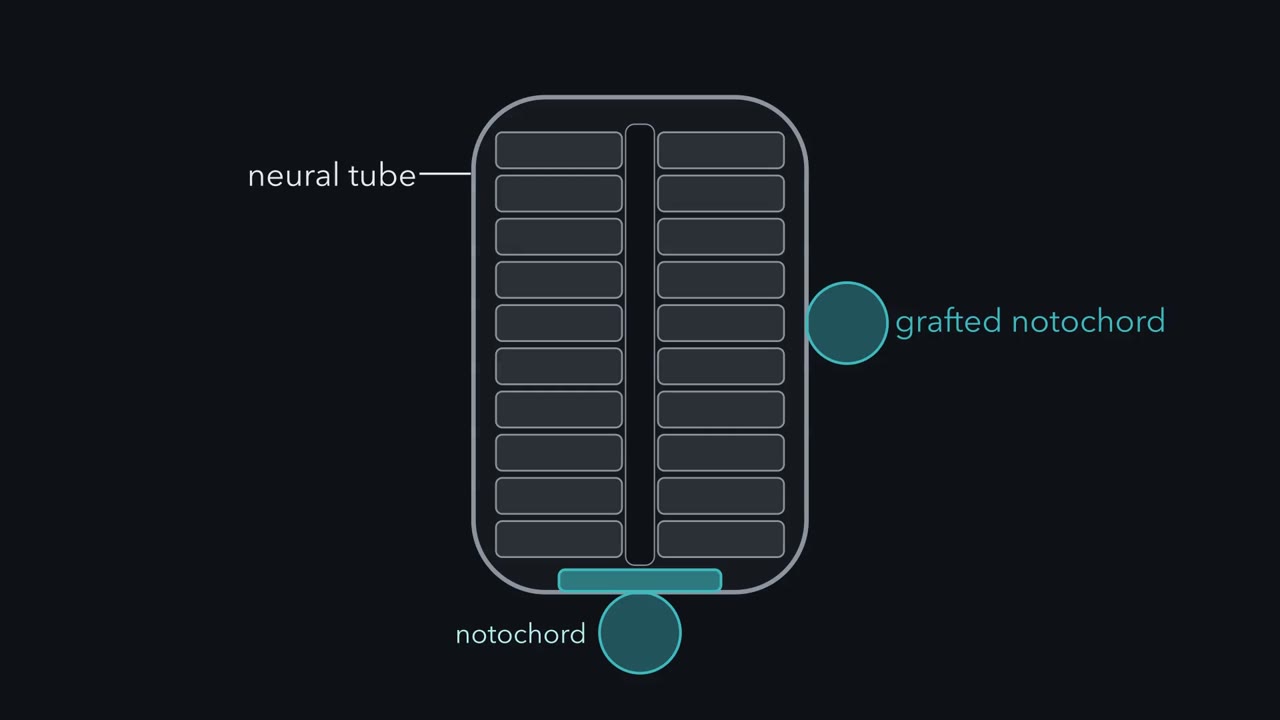
Graft a second notochord beside a chick neural tube, and the tube grows a second floor.
Pause when the reading reaches a film, and offer to play it. Saved on this device.
Watch the film now, or keep listening
Builds on:The Organizer: Spemann and MangoldNeural Induction: The Default ModelNeurulation and Neural Tube DefectsWhy these?
 ▶ Watch The Tube Gains a Second Floor · 2:55
Read the transcript of “The Tube Gains a Second Floor”
Film transcript: The Tube Gains a Second Floor
Graft a second notochord against the side of a chick neural tube. The neural cells beside it start to behave like floor plate. The tube gains a second bottom. So how does a cell in the wall know how far it sits from the bottom? Its neighbours tell it. A cell's position is written in how much signal it receives from below and above. Start with the map. The neural tube rolls up from the neural plate and becomes the brain and spinal cord. Under the tube runs the notochord, a rod of mesoderm, the embryo's middle cell layer. The tube's bottom strip is the floor plate, a narrow line of specialized cells. Its top strip is the roof plate. The epidermis, the non-neural ectoderm, ends up over the top of the tube.
In the wall between floor and roof, dividing cells called progenitors line the central canal. They will make motor neurons near the bottom, and relay neurons for body sensation near the top. The ventral half of the wall will hold five stripes of progenitors, called domains. Each makes one class of neuron. The map raises four puzzles. Who marks the bottom? What is the signal? How is a slope read? How does a slope become lines? Start with the first. Notochord and floor plate touch and share features. Is that a shared history, or an instruction? In 1990, Marysia Placzek, with Thomas Jessell, Jane Dodd and colleagues, ran the test in chick embryos.
A notochord grafted beside the side wall gave the nearby neural cells floor-plate properties. Now the reverse. Predict: what happens at the midline if you remove the normal notochord early? Floor-plate properties failed to appear at the midline. The graft shows the notochord is sufficient. It is enough to give floor-plate properties in a new place. The removal shows it is necessary. Without it, the normal floor plate does not form. Then the floor plate passes the message on. In culture, floor plate induced floor-plate markers, just as notochord did. The signals acted by contact. In this homeogenetic induction, an induced tissue recruits its neighbours to the same identity.
Keep one idea in view through every chapter. The neighbours supply a slope. The progenitors draw the lines. The takeaway: in these assays, the bottom of the tube is told, and once told, it recruits. Something passes from the notochord. What is it?
From the film
- In chick, a notochord grafted beside the neural tube gave the nearby neural cells floor-plate properties.
- Removing the notochord early stopped the normal floor plate forming, so in these assays the notochord is both sufficient and necessary.
- The floor plate then recruits its neighbours, so the bottom of the tube is told, and once told, it recruits.
Graft a second notochord against the side of a chick embryo’s neural tube, and the neural cells beside it start to behave like floor plate. The tube gains a second bottom.
So how does a dividing cell in the wall of the tube know how far it sits from the bottom, and so whether its daughters become motor neurons or relay neurons for body sensation? Its neighbours tell it. The notochord below and the skin above both send signals into the tube, and a cell’s position is written in how much of each it receives. This lesson follows those signals, Sonic hedgehog from below and bone morphogenetic proteins (BMPs) from above, and asks how the cells turn a smooth slope of signal into sharp stripes of different neurons.
The layout
Most of this lesson happens on one picture, which the first film draws. Picture the embryonic spinal cord cut crosswise, like a slice through a hollow tube, with dorsal (the back) at the top.
- The neural tube is the hollow rod that rolls up from the neural plate, a sheet of ectoderm (the embryo’s outer cell layer), and becomes the brain and spinal cord.
- The notochord is a rod of mesoderm (the embryo’s middle cell layer) running under the tube.
- The floor plate is the tube’s bottom strip, a narrow line of specialized cells just above the notochord.
- The roof plate is the tube’s top strip.
- The epidermis, the non-neural ectoderm on either side of the closing neural plate, ends up over the top of the tube.
- The ventricular zone lines the central canal, in the wall between floor and roof. Its cells are progenitors, dividing cells that will make motor neurons near the bottom and relay neurons for body sensation near the top.
The ventral half of the wall will hold five stripes of progenitors, called domains, each making one class of neuron. From dorsal to ventral they are named p0, p1, p2, pMN and p3. For now treat them as five empty slots; the code section fills them in.
The map raises four puzzles.
- Who marks the bottom? Is the floor plate born different from its neighbours, or is it told?
- What is the signal, and how does a cell receive it?
- How is a slope read? A cell can sense only the signal that reaches it. How does it turn that into “which neuron”?
- How does a slope become lines? A smooth slope should give a blur, yet the domains have sharp borders.
The top of the tube, with its own signals, bone morphogenetic proteins (BMPs) and Wnts, comes at the end.
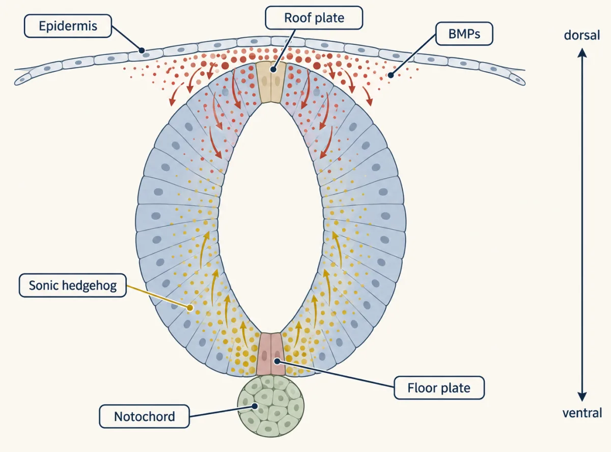
The one idea to carry through this lesson. The neighbours supply a slope; the progenitors draw the lines. The notochord and epidermis, and the floor plate and roof plate they induce, are the sources of the slope (Sonic hedgehog below, BMPs and Wnts above); they set, to a first approximation, how much signal each cell receives. The cells decide how sharp the lines are and help decide where they fall: they read the level and how long it lasts, answer with a combination of transcription factors, and pairs of neighbouring factors repress each other, which pushes each cell toward one identity and helps hold it there.
What the rule covers. Dorsal-ventral patterning of spinal cord progenitors during early neurogenesis in chick and mouse, where the evidence is strongest: the ventral Sonic hedgehog system in full, and the dorsal BMP and Wnt system in outline. It has the same shape as the rule in the netrin lesson (netrin marks a place; the growth cone decides what to do there): a signal supplies position, and the receiving cell supplies the interpretation. It has three limits, each met where it arises: sharp is not the same as accurate, the signal shapes its own slope, and not every fate is a line on this slope.
Who marks the bottom?
Notochord and floor plate lie against each other and share features. That likeness could mean a common history, or it could mean the notochord tells the neural cells what to become. A static embryo cannot tell the two apart. Moving tissue can.
In 1990 Marysia Placzek, Marc Tessier-Lavigne, Toshiya Yamada, Thomas Jessell and Jane Dodd ran the test in both directions in chick. In one direction, a notochord grafted beside the lateral wall of the tube induced floor-plate properties in the neural cells it touched: the second floor of the opening.
Predict. Now the other direction. Remove the normal notochord early, before the floor plate forms. What happens at the midline? And what does each experiment, the graft and the removal, prove on its own?
Check your prediction
Removing the normal notochord early prevented floor-plate properties at the midline. The graft shows sufficiency: the notochord is enough to give floor-plate properties in a new place. The removal shows necessity: without it, the normal floor plate does not form. Together they point to an instruction passed from notochord to neural cells, at least in these assays (Going deeper qualifies this).
The floor plate then passes the message on. An explant is a small piece of tissue cut out and grown in a dish. In a 1993 culture assay, Placzek, Jessell and Dodd found that floor plate induced floor-plate markers in neural plate explants as well as notochord did, and that the signals acted by contact. They called this homeogenetic induction: an induced tissue recruits its neighbours to the same identity. Needing contact in this assay did not settle how far the signal itself travels; the next sections treat it as a graded protein whose level falls with distance from the source.
That answers the first puzzle. The bottom is told, and once told, it recruits. Something passes from the notochord, and then from the floor plate. What is it?
Naming the signal
Read the transcript of “Naming the Signal”
Film transcript: Naming the Signal
Something passes from the notochord into the neural tube. What is it, and how does a cell receive it? The signal got a name in December 1993. Three papers appeared in one issue of the journal Cell. They described vertebrate relatives of hedgehog, a fly gene for a secreted signal. One paper was in mouse, one in zebrafish, one in chick. The gene, Sonic hedgehog, was expressed in notochord and floor plate, the tissues the grafts had pointed to. In the mouse paper, forcing the gene on where it is normally absent switched on floor-plate genes. Next, Thomas Jessell's laboratory put the protein back into a dish. An explant is a small piece of tissue grown in a dish.
In 1995, Henk Roelink and colleagues gave explants the purified signaling fragment of the protein. At the higher concentrations tested, explants made floor-plate markers. Lower ones gave motor neuron markers. One protein, at different doses, gave different ventral fates. How does a cell receive it? Think of a brake. The receptor, Patched, is the brake. On its own, it holds a second membrane protein, Smoothened, switched off. Sonic hedgehog binds Patched and lifts the brake. Smoothened is freed, and the pathway turns on. What changes is a family of transcription factors called the Gli proteins. Without the signal, Gli3 above all is cut into a short repressor.
That step depends on the primary cilium. With the signal, less repressor is made, and full-length Gli acts as an activator. In this model, a progenitor reads the balance of activator against repressor. One tool switches the pathway off. Cyclopamine, an alkaloid from a plant, blocks Smoothened. Necessity came from a knockout, a mouse with a gene deliberately disrupted. Mice lacking Sonic hedgehog have no floor plate. They lack most ventral cell types of the neural tube. Now a sharper test. Suppose Sonic hedgehog's main job is to stop Gli3 repressor from forming. Predict. In a mouse lacking Sonic hedgehog, what should removing Gli3 as well do to its ventral cells?
Removing Gli3 restores motor neurons and several ventral interneuron classes. Removing Gli3 did part of Sonic hedgehog's job. That is a double-mutant rescue. A rescue shows that two genes interact, not how. Here, the known pathway supplies the how. So much of what Sonic hedgehog does in the spinal cord is to block Gli3 repressor. The takeaway: the signal is Sonic hedgehog, read through a brake. Next, how does an amount become an address?
From the film
- The signal is Sonic hedgehog, made in the notochord and floor plate, and one protein at different doses gave different ventral fates in explants.
- Patched is a brake on Smoothened; Sonic hedgehog lifts the brake, and a progenitor reads the balance of Gli activator against repressor.
- In mice, much of what Sonic hedgehog does in the spinal cord is to block Gli3 repressor.
The signal got a name in the December 1993 issue of Cell. Three papers described vertebrate relatives of hedgehog, a fly gene for a secreted signal: Yann Echelard, Andrew McMahon and colleagues in mouse; Stefan Krauss, Jean-Paul Concordet and Philip Ingham in zebrafish; and Robert Riddle, Randy Johnson, Ed Laufer and Cliff Tabin in chick. The gene they called Sonic hedgehog (Shh) was expressed in notochord and floor plate, the tissues the grafts had pointed to, and in the mouse paper, forcing it on where it is normally absent in the central nervous system switched on floor-plate genes.
Jessell’s laboratory then put the protein back into the assay. In 1994 Henk Roelink and colleagues showed that cultured cells engineered to make a vertebrate Hedgehog induced floor-plate and motor-neuron markers in neural plate explants. In 1995 they used the purified amino-terminal fragment, the part of the protein that signals, and found that higher concentrations induced floor plate and lower ones induced motor neurons. One protein, at different doses, gave different ventral fates.
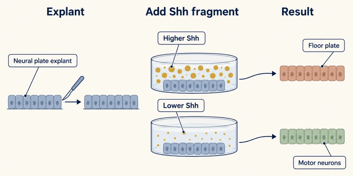
How a cell receives it: lifting a brake
Shh’s receptor, Patched, is a brake: on its own it holds a second membrane protein, Smoothened, off. Shh binds Patched, Patched stops inhibiting Smoothened, and the pathway turns on.
What the pathway changes is a family of transcription factors, the Gli proteins. Without the signal, Gli2 and Gli3 are cut into short repressors, a proteasome step that depends on the primary cilium (see that lesson). Gli3 is the main repressor; Gli2 is processed poorly and acts mainly as the activator. With the signal, less repressor is made and full-length Gli acts as an activator. What a progenitor reads, in this model, is the balance of Gli activator against Gli repressor.
Cyclopamine, an alkaloid from the plant Veratrum that causes cyclopic lambs when pregnant ewes graze on it, blocks Smoothened and so switches the pathway off.
Is Shh needed, and what does it do?
Necessity came from a knockout, a mouse with a gene deliberately disrupted. In 1996 Chin Chiang, Philip Beachy and colleagues reported that mice lacking Shh have no floor plate and lack most ventral cell types of the neural tube.
A second knockout can ask what the missing signal normally does. If removing a second gene restores what the first loss took away, the second mutation suppresses the first defect. That is a double-mutant rescue. It shows the two genes interact, not how; the mechanism needs other evidence.
Predict. Suppose Shh’s main job in the spinal cord is to stop Gli3 repressor forming. Then a mouse lacking Shh has Gli3 repressor where the signal should have stopped it. What should happen to its ventral cells if you remove Gli3 as well?
Check your prediction
Some ventral cells should come back. Chiang’s group found that also removing Gli3 restores motor neurons and several ventral interneuron classes (Litingtung and Chiang, 2000). Read with the known pathway, from Patched to Gli, this says that much of what Shh does in the spinal cord is to block Gli3 repressor.
That answers the second puzzle: the signal is Sonic hedgehog, received by lifting a brake.
Reading the slope: a code written in two classes
Read the transcript of “A Code in Two Classes”
Film transcript: A Code in Two Classes
Sonic hedgehog falls off with distance from the floor plate. How does a cell turn how much into which neuron? A signal that spreads from a source and gives different instructions at different concentrations is called a morphogen. The answer is written in transcription factors, mostly homeodomain proteins, which set cell identity across the body. In 2000, James Briscoe, Thomas Jessell and colleagues sorted them in chick. They asked one question. Does Sonic hedgehog switch the factor off, or switch it on? Class I factors are switched off by Sonic hedgehog. Each is expressed above a certain height and lost below it. Examples are Pax7, Dbx1 and Dbx2, Irx3, and Pax6.
The more sensitive the factor, the higher its border sits. Pax7 is lost at low levels, so it stays in the dorsal half. Class II factors are switched on by Sonic hedgehog. They are expressed only below a certain height. Nkx6.1 is one. Nkx2.2 needs the most signal, so it sits beside the floor plate. The names are easy to mix up. Class I proteins are the ones Sonic hedgehog represses. Each border sits at a different height, Dbx1's among them. So every stretch between two borders carries its own combination of factors: the cell's address. One more factor completes it. Olig2, a different kind of protein that Sonic hedgehog also induces, marks the motor neuron progenitors.
Predict. Compare the p3 row with the pMN row above it. Which markers differ? pMN lacks Nkx2.2, the factor that needs the most signal. It has Olig2, and a little Pax6. That combination marks the motor neuron progenitors, the domain called pMN. Now compare pMN with p2 above it. p2 lacks Olig2, and has Irx3. These rows compare neighbouring domains. They do not show a cell changing as it moves.
Notice the pattern. At each border, a pair of factors trades places. That pairing returns in chapter five. Now fill the five slots. p0, p1 and p2 make V0, V1 and V2 interneurons. pMN makes motor neurons. p3, beside the floor plate, makes V3 interneurons. You do not need every name. Three markers serve as landmarks: Pax7, Olig2, and Nkx2.2. The takeaway: a level of signal becomes an address, written as a combination of factors.
From the film
- A level of Shh signal becomes an address, written as a combination of transcription factors.
- Class I factors are switched off by Shh and class II factors are switched on, and their borders sit at different heights.
- Each domain makes one class of neuron, and Pax7, Olig2 and Nkx2.2 serve as landmarks.
A morphogen is a signal that spreads from a source and gives different instructions at different concentrations. Shh from the floor plate falls off with distance, so a progenitor near the bottom receives a lot and one halfway up receives little. The question is how a cell turns “how much” into “which neuron”.
The answer is written in transcription factors that Shh switches on or off through the Gli balance, mostly homeodomain proteins (a family of DNA-binding proteins that set cell identity across the body). In 2000 James Briscoe, Alessandra Pierani, Thomas Jessell and Johan Ericson sorted them in chick by one question: does Shh switch the factor off, or does it switch it on?
- Class I: Shh switches them off. Examples are Pax7, Dbx1, Dbx2, Irx3 and Pax6. Each is expressed above a certain height and lost below it, where Shh is stronger. The more sensitive the factor, the higher its border sits. Pax7 is switched off by low levels, so it is confined to the dorsal half.
- Class II: Shh switches them on. Nkx6.1 and Nkx2.2 need Shh and are expressed only below a certain height. Nkx2.2 needs the most signal, so it sits right next to the floor plate. Because the borders are staggered, every stretch of the wall between two borders carries a different combination of factors. That combination is the cell’s address. One more factor completes it: Olig2, a basic helix-loop-helix (bHLH) repressor rather than a homeodomain protein, which Shh also induces, marks the motor-neuron progenitors and acts with these factors (Novitch et al. 2001).
Each row of the table is a domain, and each column shows where one factor is on.
Predict. Read the table from the bottom row up. Compare the p3 row with the pMN row above it: which factors differ? Then compare pMN with p2.
| Domain (dorsal to ventral) | Dbx1 | Dbx2 | Irx3 | Pax6 | Nkx6.1 | Olig2 | Nkx2.2 | Neurons made |
|---|---|---|---|---|---|---|---|---|
| p0 | + | + | + | + | – | – | – | V0 interneurons |
| p1 | – | + | + | + | – | – | – | V1 interneurons |
| p2 | – | – | + | + | + | – | – | V2 interneurons |
| pMN | – | – | – | low | + | + | – | motor neurons |
| p3 | – | – | – | – | + | – | + | V3 interneurons |
(Simplified. Other factors, such as Nkx6.2, refine these borders.)
Check your prediction
Compared with p3, pMN lacks Nkx2.2 (the factor that needs the most Shh) and has Olig2 and a low level of Pax6: that is the motor-neuron progenitor domain. Compared with pMN, p2 lacks Olig2 and has Irx3, with Pax6 back at full level. These are neighbouring domains compared side by side, not one cell moving up: a progenitor’s identity also depends on its signaling history, as the next section shows. Notice that at each border a pair of factors trades places, Nkx2.2 for Pax6 at the bottom border of pMN and Olig2 for Irx3 at the top. That pairing is the clue to how the borders become sharp, which comes two sections on.
So the five slots are filled (the table’s last column). You do not need every name. Three markers serve as landmarks, though they do not tell every domain apart: Pax7 for the dorsal half, Olig2 for motor-neuron progenitors and Nkx2.2 for the domain beside the floor plate.
Level and time
Read the transcript of “Level and Time”
Film transcript: Level and Time
Does a cell simply measure how much Sonic hedgehog it sees? Or does time matter too? Concentration turned out to be only half of the reading. Part of the other half lies in the brake. Recall the brake. Patched holds the pathway off, and Sonic hedgehog lifts it. Here is the twist. Patched1 is also one of the genes Sonic hedgehog turns on. That sounds backwards. More receptor should mean more sensitivity. But Patched is a brake. In chick neural cells, signaling builds part of its own brake. Then it takes more signal to keep going. Picture two cells starting at once. One gets a low dose of Sonic hedgehog. The other gets a high dose. Both make more Patched as they go.
Predict. In which cell does signaling fade first? The low-dose cell. Its signaling soon fades. The high-dose cell keeps signaling for longer. In 2007, Eric Dessaud, James Briscoe and colleagues reported this in chick neural cells. The cells adapt. Signaling inside them fades, partly through rising Patched1. Higher concentrations kept signaling going longer. A level becomes a length of time. Now test it. Hold the dose fixed, and change only how long the cells are exposed. What should longer exposure do?
In 2010, the same laboratory did this in chick explants. At a fixed dose, longer exposure gave progressively more ventral identities. The time a cell spends signaling is part of its address. Roughly, a cell's identity tracks how much signaling it has had so far. It also needs the signal to continue for a while to keep that identity. In chick explants, cutting the signal off with cyclopamine made cells fall back to more dorsal fates. Duration carries information in many signaling pathways. Here, it helps set which positional identity a progenitor adopts. The simulation in this lesson lets you set both the level and the time, and watch the domain change.
The takeaway: a progenitor reads both how much signal it gets, and for how long.
From the film
- A progenitor reads both how much Shh signal it gets and for how long.
- In chick neural cells, signaling adapts partly through rising Patched1, so higher doses keep signaling going longer.
- In chick explants, longer exposure at a fixed dose gave more ventral identities, and cutting the signal off let cells fall back to more dorsal ones.
Concentration turned out to be only half of the reading, and part of the reason lies in the brake. Patched1 is also one of the genes Shh turns on. That sounds backwards, since more receptor ought to mean more sensitivity, but Patched is a brake. In chick neural cells, that extra brake contributes to adaptation: it then takes more Shh to keep signaling.
Predict. Two cells start signaling at once, one at a low dose of Shh and one at a high dose. Both cells make Patched as they go, the high-dose cell somewhat more. Which cell’s signaling falls below its working level first?
Check your prediction
The low-dose cell, because less Shh is left to overcome the rising brake. In 2007 Eric Dessaud, Briscoe and colleagues reported this adaptation in chick neural cells: signaling inside the cell fades, and higher concentrations keep it going longer. A cell can turn a level into a length of time.
If time is the variable, it should matter even at one dose. A 2010 study from the same laboratory held the dose fixed in chick explants and varied exposure time, and longer exposure gave progressively more ventral identities. Duration carries information in many pathways (see that lesson). Here, roughly, a cell’s identity tracks how much signaling it has had so far, and it needs the signal to continue for a while to keep that identity: in chick explants, ending Shh signaling with cyclopamine after a period of exposure let cells fall back to more dorsal identities than continued exposure gave (Dessaud and colleagues, 2010). The second MCAT-style question below uses simplified, illustrative numbers.
A progenitor reads both how much Sonic hedgehog it meets and for how long, because adaptation makes signaling fade and a higher dose keeps it going longer; the axes and cut-offs here are relative, a sketch of that logic and not measured values. The lesson gives floor plate only for the higher concentrations Roelink and colleagues tested, a different assay; this sketch stops at p3.
Drawing the lines: cross-repression
Read the transcript of “Drawing the Lines”
Film transcript: Drawing the Lines
A smooth slope of signal should give a blur. Yet the domains have sharp borders. Why? Picture a cell whose output simply rises with the signal, the gentle curve of simple binding. Going from 10 % to 90 percent output then takes an 81-fold change in concentration. On an illustrative slope, that change spans about eighteen cells. Eighteen cells of in-between answers is a blur. In this simple model, a steeper, switch-like response narrows the blur to one or two cells. The cells are all still there. Fewer of them give in-between answers. The domains get their switch from the factors themselves. Factors on either side of a border repress each other.
This was tested by electroporation. DNA is pushed into cells on one side of the tube. The other side is the control. James Briscoe and colleagues showed that Pax6 and Nkx2.2 repress each other. They meet at the border between pMN and p3. Novitch, Chen and Jessell found the same for Irx3 and Olig2.
Predict. Force Nkx2.2 on in the motor neuron domain. What happens to those cells? Motor neurons are suppressed on that side. The cells make V3 neurons, the fate of the domain below. One factor in the wrong place moved the line for those cells. Nothing was done to the Shh source. Two factors that repress each other can form a switch with two stable states.
In simple models, they do so only when the repression is strong and cooperative. A weak loop makes no switch. In a strongly cross-repressive circuit, a cell near a border cannot hold both factors for long. It falls to one side, and the loop helps hold it there. That gives the network some memory. In 2012, Briscoe's group tested it in mouse embryos in culture. A short pulse of a drug that activates Smoothened left the borders close to normal. Continuous treatment shifted them. There is a limit here. Sharp is not the same as accurate. A large or lasting misreading can tip a cell to the wrong side. The switch may then hold the error. Placing borders accurately is a separate problem.
In models, reading over time, and reading two opposing slopes, can help. The takeaway: the slope sets roughly where each line falls. Pairs of repressors make each line sharp, and help hold it.
From the film
- The slope sets roughly where each line falls, and pairs of repressing factors such as Pax6 and Nkx2.2 make each line sharp.
- Forcing Nkx2.2 on in the motor neuron domain suppressed motor neurons and gave V3 neurons instead.
- Sharp is not the same as accurate: a large or lasting misreading can tip a cell to the wrong side.
Why should a smooth slope give a blur? Picture a cell whose output simply rises with Shh, the gentle curve of simple binding (a Hill coefficient of 1; the Hill coefficient measures how steeply a response rises with concentration). Such a cell goes from 10 percent to 90 percent of its maximum over an 81-fold change in concentration: 9-fold to reach half, and another 9-fold to reach 90 percent. Now take an illustrative slope on which Shh falls by a factor of e (about 2.7) every 20 micrometres, the order measured for Shh protein in mouse (Cohen and colleagues, 2015), and assume progenitors 5 micrometres wide. An 81-fold change is 4.4 such steps (the natural log of 81), so it spans about 90 micrometres, roughly 18 cells of in-between answers. That is a blur.
| How steeply the cell responds | Cells spanned from 10% to 90% of the response |
|---|---|
| Hill coefficient 1 (graded) | about 18 |
| Hill coefficient 2 | about 9 |
| Hill coefficient 4 | about 4 |
| Hill coefficient 8 (switch-like) | about 2 |
(Illustrative values on that slope, not measurements for the neural tube.) In this model, where cells stay put, a border one or two cells wide needs a much steeper, switch-like response. A steep response does not remove cells; it leaves fewer of them giving in-between answers. Steepness alone does not prove a switch, and cells need not stay put: in zebrafish, progenitors are at first specified in a mixed, salt-and-pepper way and then sort into sharp domains (Xiong and colleagues, 2013). In the chick and mouse neural tube, the domains get their switch from the factors themselves: factors on either side of a border repress each other.
Briscoe and colleagues showed this in chick by electroporation, in which DNA is pushed into cells on one side of the tube with brief electric pulses and the other side serves as a control in the same embryo. Pax6 and Nkx2.2 repress each other at the border between pMN and p3. Novitch, Chen and Jessell showed in 2001 that Irx3 and Olig2 repress each other at the dorsal edge of pMN. These are the pairs you saw trading places in the table.
Predict. Force Nkx2.2 on by electroporation in the pMN domain, where cells normally carry Olig2 and a low level of Pax6. What happens to those cells, and which neurons do they make?
Check your prediction
Nkx2.2 represses Pax6 and Olig2, so the motor-neuron program goes and the cells take the p3 identity just below: motor neurons are suppressed and V3 neurons made. One factor in the wrong place moved the line for those cells, without any change to the Shh source.
Two factors that repress each other can form a switch with two stable states, the bistable circuit from that lesson. In simple models they do so only when the repression is strong and cooperative; a weak loop has a single steady state and no switch. In a strongly cross-repressive circuit a cell near a border is pushed toward one state or the other and cannot hold both for long. The loop then helps hold it there, though a lasting change in signal can still move it. That gives the network some memory.
In 2012 Nikolaos Balaskas, Vanessa Ribes, Briscoe and colleagues found in mouse embryos in culture that a short pulse of a drug that activates Smoothened left the borders close to their normal positions; continuous treatment shifted them.
Sharp is not the same as accurate. Cross-repression makes a border sharp, not necessarily accurately placed. A large or lasting misreading of the Shh level can tip a cell to the wrong side, and the switch may then hold the error; a brief fluctuation may be filtered out, as the pulse experiment suggests. Accurate placement is a separate problem; in models, reading the signal over time and reading the two opposing slopes together can help, by amounts still debated (Going deeper). This is the first limit of the rule.
The top of the tube: BMPs from the epidermis and roof plate
Read the transcript of “The Top of the Tube”
Film transcript: The Top of the Tube
Sonic hedgehog patterns the bottom of the tube. What tells the top of the tube it is the top? Recall neural induction. In frog animal caps, blocking BMP let the cells become neural. Now, inside the tube, BMP comes back. It arrives from the skin and from the roof plate. BMPs, bone morphogenetic proteins, act through receptor kinases and Smad proteins. In 1995, Karel Liem, Thomas Jessell and colleagues tested them on chick neural plate explants. BMP4 and BMP7 could mimic the dorsalizing effect of epidermis. In 1997, they proposed that BMPs from the epidermis induce the roof plate. The roof plate then becomes a second source of related signals. It also makes Wnts, including Wnt1 and Wnt3a.
The dorsal tube makes six early classes of interneurons. They are named dI1 to dI6, from the top down. Predict. Remove the roof plate early in a mouse, leaving the floor plate and notochord untouched. Which classes are lost?
In 2000, Kevin Lee, Paula Dietrich and Thomas Jessell removed the roof plate genetically. The three dorsal-most classes later failed to form. The classes further down did not depend on it, in that experiment. The two systems also oppose each other. BMP signaling alters the response to Sonic hedgehog. So the wall sits between a signal from below and one from above. Cells in the middle can be shaped by both. This lesson follows the ventral code in full and the dorsal one in outline. Now the whole rule. The neighbours supply a slope. The progenitors draw the lines. Signals from below and above set the level. The cells read it, and how long it lasts, and answer with a combination of factors.
In the ventral tube, pairs of neighbouring factors repress each other. That helps each cell settle on one identity and hold it, though a lasting change in signaling can still move it. Keep the limits too. Sharp is not the same as accurate. The signal shapes its own slope. And the floor plate follows partly different rules. One more role waits. Later, the same floor plate becomes a target for growing axons. The takeaway: two slopes, read by the cells, become the map of the spinal cord.
From the film
- BMPs from the epidermis and roof plate pattern the top of the tube, and the roof plate also makes Wnts.
- In mice, removing the roof plate early meant the three dorsal-most interneuron classes later failed to form.
- The wall sits between a signal from below and one from above, and BMP signaling alters the response to Shh.
Earlier, in neural induction, blocking BMP signaling let frog animal-cap cells become neural, though in chick BMP inhibition alone does not. Now, inside the tube, BMP comes back from the skin and the roof plate and tells the top of the tube it is the top. BMPs (bone morphogenetic proteins) are members of the TGF-beta family and act through receptor kinases and Smad proteins (see that lesson).
In 1995 Karel Liem, Gabi Tremml, Henk Roelink and Jessell found that BMP4 and BMP7 could mimic the dorsalizing effect of epidermis on chick neural plate explants. In 1997 Liem, Tremml and Jessell proposed that BMPs from the epidermis induce the roof plate, which becomes a second source of related signals. The roof plate also makes Wnts, including Wnt1 and Wnt3a.
The dorsal tube makes six early classes of interneurons, named dI1 to dI6 from the top down.
Predict. Remove the roof plate early in a mouse embryo, leaving the floor plate and notochord untouched. Which interneuron classes should be lost?
Check your prediction
In 2000 Kevin Lee, Paula Dietrich and Jessell removed the roof plate genetically in mice, and the three dorsal-most classes later failed to form. The classes nearest the roof plate needed it; dI4 to dI6, further away, did not depend on it in that experiment, and the ventral classes still had Shh from below.
The two systems also oppose each other: BMP signaling alters the response to Shh (Liem, Jessell and Briscoe, 2000), so cells in the middle of the wall can be shaped by both.
Design problem
You have intermediate neural plate explants from chick embryos, which on their own express Pax7. You also have purified Shh signaling fragment, cyclopamine and antibodies to Pax7, Olig2 and Nkx2.2. Design an experiment that tells whether progenitors read the Shh concentration, the duration of exposure, or both.
Saved in this browser only.
One way to do it
Run two series and score every explant at the same endpoint, say 48 hours, so age is the same for all.
In the dose series, give each explant a different concentration for the whole time. If concentration matters, rising dose should first switch off Pax7, then switch on Olig2, then at the highest doses switch on Nkx2.2 and lose Olig2.
In the duration series, hold one intermediate dose fixed and add cyclopamine at different times to end signaling. If duration matters, short exposures should leave cells Pax7-negative but without Olig2, longer ones should give Olig2, and with a higher fixed dose the longest exposures should also give Nkx2.2. Because progenitors can lose these markers once signaling stops, also score them at the time cyclopamine goes on, or score the neurons they made.
The expected answer from chick explant studies is that both matter. At a fixed time, higher doses push cells ventrally; at a fixed dose, longer exposure does too. Add one more test: measure a Gli reporter over time at each dose. Signaling should fade as cells adapt, faster at low dose, which is how one variable becomes two.
Putting it together
The four puzzles share one answer: the neighbours supply a slope; the progenitors draw the lines. Grafts showed that, in these chick assays, the notochord instructs the floor plate, and the floor plate recruits its neighbours. Sonic hedgehog is the signal, received by lifting a brake. Progenitors read its level and how long it lasts, and answer in class I and class II factors. Pairs of those factors repress each other, which helps make each line sharp and lets it resist brief changes. From above, bone morphogenetic proteins (BMPs) and Wnts run a matching system for the dorsal half.
The floor plate has a second job. Later the same strip of cells becomes a target for growing commissural axons, the subject of netrin and DCC.
Going deeper
Where the floor plate comes from
An earlier group had seen an extra floor plate after implanting a notochord in chick embryos in 1985; the 1990 paper added the test of necessity. Even so, the grafts did not close the “shared history or instruction?” question entirely. Later work showed that in chick part of the medial floor plate comes from cells that arise from Hensen’s node (the chick’s organizer) alongside notochord precursors, and that its induction involves early signals besides Shh (reviewed by Placzek and Briscoe, 2005). So both answers are partly true.
Nor is the floor plate simply the top step of the same dose ladder that the 1995 explants suggest. In the mouse, floor plate needs Gli2 and an early period of high signaling, while motor neurons form without Gli2 (Ribes and colleagues, 2010). That is the third limit of the rule: not every fate is a line on the slope.
Holoprosencephaly
Heterozygous mutations in human SHH can cause holoprosencephaly, in which the forebrain fails to divide into two halves. Together with the cyclopic lambs of cyclopamine, the two make the pathway’s weight clear, though both concern the head rather than the spinal cord this lesson follows.
A third pair, and a microRNA
Briscoe and colleagues also showed by electroporation in chick that Dbx2 and Nkx6.1 repress each other at the border between p1 and p2. A 2011 study found a microRNA that tunes the Irx3 and Olig2 loop by lowering Olig2.
The network moves the border
The 2012 Balaskas study also used a fluorescent reporter of Gli activity. In mice lacking both Pax6 and Olig2, Nkx2.2 spread dorsally although the measured Gli-reporter domain did not: losing the network moved the border, without a matching shift in the reporter. Whether the gradient is read directly or mainly through such feedback is still argued.
The cells shape the slope they read
Patched1, which Shh induces, also binds Shh and limits how far it spreads. So the signal partly shapes its own slope, and “the neighbours supply the slope” is a first approximation. That is the second limit of the rule.
How precise is a border?
On a steady exponential slope with fixed source level and decay length, each threshold falls at a height set by the logarithm of its concentration. On the illustrative slope used above, thresholds 2-fold apart give a domain about 3 cells wide and thresholds 10-fold apart one about 9 cells wide, so domains several cells wide need thresholds 2-fold or more apart.
A switch does not remove noise. Take an illustrative model in which each cell’s reading of Shh has a random error, independent of its neighbours, with a standard deviation of 20 percent of the concentration. The border’s position is then uncertain by about 0.8 cell (one standard deviation), so even with a sharp switch about 3 cells near it each have more than a 5 percent chance of landing on either side (a 90 percent interval 2.6 cells wide). Averaging about 10 independent readings over time cuts that to about 1; averaging does not remove an error shared by every reading. The noise level is an assumption, not a measurement. Models also suggest that reading the two opposing slopes together improves precision, though how much, and how precise one slope is alone, are debated.
BMP inhibitors from below
Bone morphogenetic protein (BMP) inhibitors such as follistatin, made by the notochord and somites, help shift cells toward ventral fates (Liem, Jessell and Briscoe, 2000).
Your notes
Select any text in the lesson to highlight it or add a note to it. Your highlights and notes are saved in this browser and collected in your notebook.
This browser is not letting the site save anything (a private window or strict privacy settings), so highlights and notes will be lost when you leave the page.
How we measure it
Tissue grafts and ablations in chick embryos
A piece of tissue, such as a notochord, is moved to a new position in a living embryo, or removed. Gain at the new site tests sufficiency; loss at the normal site tests necessity. Chick embryos suit this because they develop in an egg that can be opened and resealed.
Neural plate explants with purified signal
A small piece of neural plate is cut out and grown in a gel with a known concentration of a protein such as the Sonic hedgehog signaling fragment, for a set time. Antibodies to transcription factors then show which identity the cells took, so dose and duration can be varied one at a time.
In ovo electroporation
DNA is injected into the chick neural tube and pushed into cells on one side with brief electric pulses. The other side serves as a control in the same embryo, which is how cross-repression between pairs of transcription factors was tested.
Mouse knockouts and reporter lines
A knockout mouse carries a gene deliberately disrupted, so its embryos show what the gene is needed for. A reporter line carries a fluorescent protein driven by the target sites of a pathway, here Gli-binding sites, so signaling can be watched in the embryo.
Practice questions on this lesson (3) → MCAT-style, original, each with an explanation
Where this lesson fits
Before thisReview first
- The Organizer: Spemann and MangoldThis course · Lesson 5
The notochord's power to induce neural tissue in grafts is the same kind of experiment extended here.
- Neural Induction: The Default ModelThis course · Lesson 6
You need the BMP-inhibition default model from there to see why BMP returns at the top of the tube.
- Neurulation and Neural Tube DefectsThis course · Lesson 7
It shows how the neural plate rolls into the tube whose floor, roof and wall are mapped here.
After thisStudy next
- Transcription Factor Codes: Building a Motor NeuronThis course · Lesson 11
It takes the pMN domain and shows how Olig2 and other factors build a motor neuron from it.
- Crossing the Midline Once: Slit, Robo and CommThis course · Lesson 24
Commissural axons here grow toward the floor plate that this lesson shows is induced by Shh.
- Hedgehog and the Primary CiliumCell Signaling · Graduate school and beyond
It takes the Patched, Smoothened and Gli signalling used here further, through the primary cilium.
Elsewhere on the siteFilms, simulations and method films in other courses
- A Late Part Reads Signal LengthFilm · Cell Signaling
It shows how a cell reads both the size and the duration of a signal, as progenitors do with Shh.
Suggested by an AI model from the lessons themselves, then checked. How we use AI
Finish line
Test yourself
8 questions
Practice and MCAT-style, with explained answers.
Next · Lesson 11
Transcription Factor Codes: Building a Motor Neuron
Put Lhx3 into chick spinal cord cells that make no Isl1 and they turn on interneuron genes. Add Isl1 and the same protein turns on motor-neuron genes. If no single gene says motor neuron, how does a set of transcription factors make one, and choose the muscle its axon will reach?
Continue →← Lesson 9 · Local Organizers: The Midbrain-Hindbrain Boundary