Developmental Neuroscience · Unit 4: Making neurons
The Ventricular Zone: Radial Glia as Stem Cells
Rewritten on : new text, figures, flashcards and film. Your notes and highlights stay, but the text around them may have moved.
In rodents, radial glia, long seen as mere scaffolding, make most cortical neurons. Why does such a progenitor copy its DNA in one place and divide in another?
Pause when the reading reaches a film, and offer to play it. Saved on this device.
Watch the film now, or keep listening
Builds on:Neurulation and Neural Tube DefectsTools for Following Cells and GenesThe Wiring Problem: How a Brain Gets BuiltWhy these?
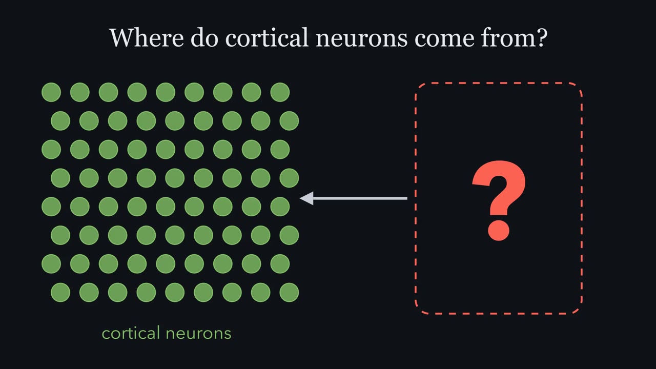 ▶ Watch One Cell Across the Wall · 3:12
Read the transcript of “One Cell Across the Wall”
Film transcript: One Cell Across the Wall
Where do the neurons of the cerebral cortex come from? In rodents, most come from one kind of cell. Here is a slice through the wall of the embryonic mouse cortex. The ventricle, a fluid-filled space, is at the bottom. The surface facing the ventricle is apical. The outer surface, under the pia, is basal. Beside the ventricle lies the ventricular zone, where progenitors live. A progenitor is a cell that still divides. Just outside it is the subventricular zone. Farther out, young neurons cross the intermediate zone to reach the cortical plate. Now light up one cell. It spans the whole wall. An apical foot sits on the ventricle. A long fibre ends in a basal foot under the pia.
Early on, cells like this are neuroepithelial cells. As neurogenesis begins, in mouse around embryonic days 10 to 12, they become radial glia. They keep the long shape. And they switch on proteins associated with glia. Look where cells divide. Most cells caught in mitosis sit against the ventricle. Cells copying their DNA sit farther out. Two words will matter. A stem cell renews itself and makes more than one kind of cell. Radial glia, as a population, meet both conditions. An asymmetric division keeps one radial glial cell. Some single lineages later make glia as well as neurons. And potential is not fate. Potential is what a cell can make alone in a dish.
Fate is what it does make in the embryo. Now the first puzzle. In fetal monkey cortex, young neurons climb radial glial fibres. Is the scaffold also the source of the neurons? The second puzzle. Why does a progenitor copy its DNA out in the ventricular zone, then come back to the ventricle to divide? Here is the idea to carry through. A radial glial cell is a stem cell anchored at the ventricle. Each time it divides, it decides how many daughters stay radial glia. Keep two, and its radial glial line doubles. Keep one, and the line holds steady while it makes neurons. Keep none, and its radial glial line ends. A daughter that leaves is spent as a stem cell.
It becomes a neuron, or an intermediate progenitor that usually makes two. Count those choices round by round, and you can see how the number of neurons depends on them. This is a counting model, for projection neurons of rodent dorsal cortex. Outer radial glia, common in primates, renew themselves without the anchor. And the rule counts. It does not explain what makes a daughter stay or leave. So hold on to one picture. One long cell across the wall, spending its divisions one choice at a time.
From the film
- In rodents, a radial glial cell is a stem cell anchored at the ventricle, and each division settles how many of its two daughters stay radial glia.
- Keep two and the radial glial line doubles; keep one and it holds steady while it makes neurons; keep none and it ends.
- A daughter that leaves is spent as a stem cell: it becomes a neuron, or an intermediate progenitor that usually makes two.
In rodents, most neurons of the cerebral cortex are made by radial glial cells, long thought to be only a scaffold. Look at a slice of embryonic mouse cortex and you can see a first clue to how they work: most cells caught in mitosis (division) sit against the ventricle, the fluid-filled space at the centre of the brain. The cells copying their DNA sit farther out.
This lesson asks three things. How can one kind of cell be both the scaffold and the source of neurons? Why does it copy its DNA in one place and divide in another? And how does the way it spends its divisions set the number of neurons the cortex gets?
The layout
The brain begins as the neural tube, an epithelium around a canal that becomes the ventricles (see the neurulation lesson). Picture a slice through the wall of the embryonic mouse cortex, with the ventricle at the bottom and the outer surface of the brain at the top.
- Apical and basal. The surface of the wall that faces the ventricle is apical. The outer surface, covered by a basement membrane and the pia, the thin membrane over the brain, is basal.
- The ventricular zone (VZ) is the layer beside the ventricle where progenitors live. A progenitor is a cell that still divides and whose daughters will become neurons or glia.
- The subventricular zone (SVZ) is the band just outside the VZ.
- The intermediate zone lies farther out. Young neurons cross it on their way to the cortical plate, the forming layer of neurons under the pia.
One cell spans all of it. Early on, the wall is made of neuroepithelial cells, each long and thin, reaching from the apical surface to the basal one. As neurogenesis begins (in mouse, around embryonic days 10 to 12), neuroepithelial cells become radial glia. They keep the shape: an apical foot on the ventricle and a long fibre that ends in a basal foot on the basement membrane under the pia. They also switch on proteins associated with glia, such as the glutamate transporter GLAST, the brain lipid-binding protein BLBP and, in primates, glial fibrillary acidic protein (GFAP).
Two more words before the puzzles.
- A stem cell renews itself and can make more than one kind of cell. Radial glia as a population meet both conditions. An asymmetric division, one that gives two different daughters, keeps one radial glial cell, and in mouse some single radial glial lineages make neurons and then glia (Going deeper).
- A cell’s potential is what it can make under set conditions, such as alone in a dish. Its fate is what it does make in the embryo. A dish tests potential; only lineage in the animal shows fate.
The map raises the first two as puzzles.
- Scaffold or source? In fetal monkey cortex, young neurons climb radial glial fibres on their way out. Is the scaffold also the source?
- Why divide at the ventricle, rather than out in the VZ where the DNA was copied?
The one idea to carry through this lesson. A radial glial cell is a stem cell anchored at the ventricle, and each division settles how many of its two daughters stay radial glia. Keep two, and its radial glial line doubles. Keep one, and the line holds steady while it makes neurons. Keep none, and its radial glial line ends. A daughter that leaves is spent as a stem cell: it becomes a neuron directly, or an intermediate progenitor that divides again, usually into two neurons. Count those choices round by round and you can see how the number of neurons depends on them.
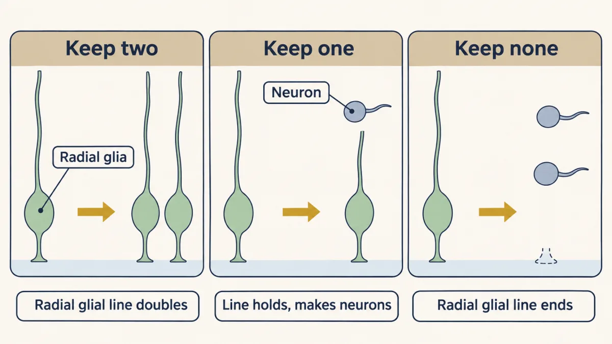
What the rule covers. It is a counting model for the excitatory projection neurons of rodent dorsal cortex, the neurons that send long axons, made during neurogenesis (in mouse, about embryonic days 11 to 17). It ignores cell death. It has four limits; three are met below, and the fourth is a later lesson.
- “Anchored at the ventricle” describes apical radial glia. Outer radial glia, rare in mouse and common in primates, renew themselves without any apical contact. So the anchor marks the stem state in this system; it does not define it.
- The rule counts; it does not explain. What makes a daughter stay or leave (Notch, unequal inheritance, G1 length) is a separate question: see the Notch lesson, proneural genes and lateral inhibition and asymmetric division.
- Radial glia and intermediate progenitors have to be counted separately. Lumping them together counts the dividing cells correctly but hides how many are stem cells, as the counting section shows.
- It does not cover the later switch to glia. Divisions can still be counted then, but daughters can enter glial lineages that divide on their own schedule, so those are counted separately (see origins of glia).
The lift: a nucleus on a timetable
Read the transcript of “The Lift: A Nucleus on a Timetable”
Film transcript: The Lift: A Nucleus on a Timetable
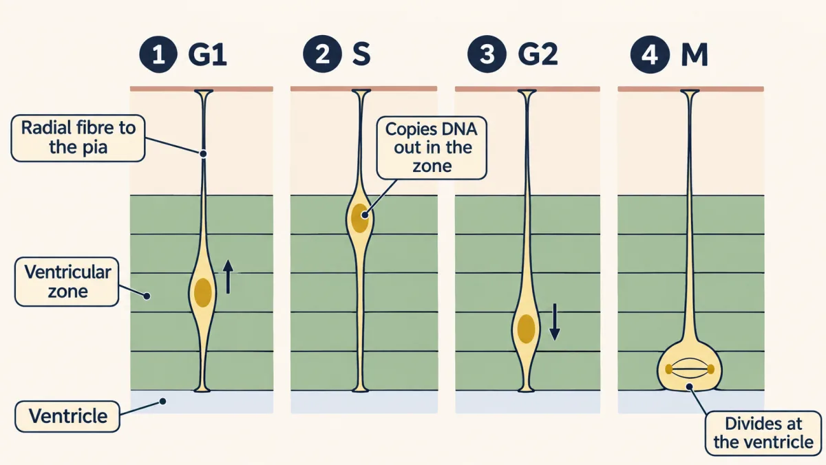
Why would a cell copy its DNA in one place, and divide in another? Nineteenth-century anatomists saw nuclei at many heights. They read the wall as several kinds of cells. In 1935, F. C. Sauer argued for a simpler picture. One kind of long cell, whose nucleus moves inside it. A single layer of cells with nuclei at many heights is called a pseudostratified epithelium. Picture a lift, with the ventricle at the bottom. The shaft is the cell. The car is the nucleus. The shaft runs all the way up to the pia. But the car only serves the lowest floors, the ventricular zone. In G1, the car rides up. It stops near the top of the ventricular zone for S phase, when DNA is copied.
In G2, it comes back down to the ground floor, the apical surface. The cell divides there. Each cell's car runs on its own timetable. That is why nuclei sit at every height. This movement is called interkinetic nuclear migration. Movement between divisions. A pulse label made Sauer's cycle a timetable. Tritiated thymidine, a radioactive DNA building block, is taken up only in S phase. Give a brief pulse, then fix the tissue at intervals. Soon after the pulse, where are the labeled nuclei? And when do labeled mitoses appear?
Soon after the pulse, labeled nuclei sit out in the ventricular zone. No cell in mitosis is labeled. Later, the labeled nuclei come down and divide at the ventricle. In 1959, two groups reported this. Labeled nuclei moved in step with the cell cycle. In eleven-day mouse embryos, no labeled mitosis appeared until an hour after the injection. That lag roughly spans G2, which includes the trip back down. So why divide at the ventricle? The answer is partly settled. The best-supported part. For much of interphase, the centrosome sits at the apical surface, at the base of the primary cilium. A dividing cell builds its spindle from its centrosomes.
On the usual account, the nucleus travels to meet them. Late in G2, the centrosome can also move to meet the nucleus. One proposal is packing. Only a cell in mitosis is round and wide. The rest can stay thin at the surface. A round cell takes about twenty times the room of a thin foot. So on average, each cell needs about a tenth of a dividing cell's room. Staggered timetables let more progenitors share the ventricle. A second proposal is signals. In zebrafish retina, Notch signalling was reported to fall from apical to basal. So how far a nucleus travels could change the signal it meets. Now the payoff. Apical radial glia divide at the surface.
So where a cell divides is a first clue to what kind it is. Most mitoses at the ventricle are apical radial glia, though some intermediate progenitors divide there too. Mitoses away from it are mostly intermediate progenitors, or outer radial glia. To name the cell, add its shape and markers. To learn what its daughters become, you need lineage, the descendants of one cell.
From the film
- The nucleus of a radial glial cell rides up in G1, copies its DNA near the top of the ventricular zone in S, comes back down in G2 and divides at the ventricle.
- Pulse labeling showed labeled nuclei out in the ventricular zone first and labeled mitoses at the ventricle later.
- Where a cell divides is a first clue to which kind of progenitor it is; the centrosome fits that, while packing and Notch signals are proposals.
The early wall looks layered, with nuclei stacked at many heights, so nineteenth-century anatomists read it as several kinds of cells. In 1935 F. C. Sauer argued for a simpler picture. Each cell spans the wall and keeps its long shape for most of the cycle while its nucleus moves inside it. The nucleus travels toward the ventricle to divide, and the daughters’ nuclei travel back out. A single layer of cells whose nuclei sit at many heights is called a pseudostratified epithelium.
Picture a lift, with the ventricle at the bottom. The shaft, the cell, runs all the way up to the pia, but the car, the nucleus, only ever serves the lowest floors, the ventricular zone (VZ). In G1 the car rides up, basally. It stops near the top of the VZ for S phase, when DNA is copied. In G2 it comes back down to the ground floor, the apical surface, and the cell divides there. Each cell’s car runs on its own timetable, which is why nuclei sit at every height. This is called interkinetic nuclear migration: movement between divisions.
Sauer’s cycle was an inference from fixed sections. A pulse label turned it into a timetable. Tritiated thymidine, a radioactive DNA building block, is taken up only by cells in S phase.
Predict. Give an embryo a brief pulse of tritiated thymidine and fix the tissue at intervals afterwards. Where are the labeled nuclei soon after the pulse? Where should labeled mitoses appear, and should they appear at once?
Check your prediction
Soon after the pulse, the labeled nuclei sit out in the VZ, where S phase happens, and no cell in mitosis is labeled. Later the labeled nuclei have come down and divide at the ventricle. In 1959 M. E. Sauer (a different Sauer) and B. E. Walker, and separately Richard Sidman, Irene Miale and Ned Feder, reported that labeled nuclei in the embryonic neural wall moved between the outer wall and the ventricle in step with the cell cycle. In eleven-day mouse embryos, Atlas and Bond (1965) saw no labeled mitotic figure until an hour after the injection. That lag is roughly G2: the cells labeled last in S phase had to pass through G2, including the trip back down to the ventricle, before any could be seen dividing.
Why divide at the ventricle?
The answer is partly settled: one part is partly known, two are proposals.
- The centrosome is there (partly known). In vertebrate neuroepithelia, for much of interphase the centrosome sits at the apical surface, at the base of the primary cilium, which pokes into the ventricle. A dividing cell builds its spindle from its centrosomes, and the usual account is that the nucleus travels to meet them. Live imaging adds a wrinkle: late in G2 the centrosome can leave the surface to meet the incoming nucleus, so mitosis can begin just short of the surface (Spear and Erickson, 2012).
- Packing (a proposal). Only a cell in mitosis is round and wide; the rest can be thin at the apical surface. Take illustrative numbers: a 12-hour cycle with half an hour of mitosis. With cells spread evenly through the cycle, about 1 in 24 is dividing at any moment. A rounded cell 10 micrometres across covers about 80 µm² of surface (π × 5²). Take an apical foot of about 2 by 2 micrometres, 4 µm², so the rounded cell takes some 20 times as much room. With 1 round cell and 23 thin ones, the average is (80 + 23 × 4) / 24, about 7 µm² per cell. That is about a tenth of what each would need if every cell took a dividing cell’s room. Staggered timetables let more progenitors share a given area of ventricle. Taverna and Huttner’s 2010 review discusses this idea.
- Signals (a proposal). Where a nucleus sits changes what it is exposed to. In zebrafish retina, Del Bene and colleagues (2008) reported a gradient of Notch signalling from apical to basal, so how far a nucleus travels could change the Notch signal it meets.
The payoff. Because apical radial glia divide at the surface, where a cell divides is a first clue to which kind of progenitor it is. Most mitoses at the ventricle are apical radial glia (or, earlier, neuroepithelial cells), though some intermediate progenitors divide there too. Most mitoses away from it are intermediate progenitors or outer radial glia. To name the cell, add its shape and markers. To learn what its daughters become, you need lineage, the descendants of one cell.
Scaffold or source?
Read the transcript of “Scaffold or Source?”
Film transcript: Scaffold or Source?
Radial glial fibres guide young neurons outward. Are those same cells also the source of the neurons? In 1972, Pasko Rakic studied fetal rhesus monkey cortex. He found young neurons pressed against radial fibres. Radial glia were seen mainly as scaffolds. Neurons, it was thought, came from some other cell in the ventricular zone. Around the year 2000, three lines of evidence in rodents, using four methods, changed the picture. Each asked the question a different way. For each one, ask: if radial glia were only a scaffold, what would we see? First, sorting, in mouse. The Götz group used mice that make GFP from a human GFAP promoter. Rodent radial glia make little GFAP protein.
But that human promoter fragment is active in them. The green cells were sorted into a dish. If they were only scaffolds, you might expect few or no neurons. Did they make any? In culture, they made neurons. But a dish shows potential, not fate. Second, fate mapping in the living mouse. An enzyme called Cre recombinase was made from a GFAP promoter. Cre cuts out a stop sequence in front of a reporter gene. The cut is a change in the DNA. So it is copied into every descendant, even after the promoter switches off in a neuron. If radial glia were only scaffolds, cortical neurons would stay dark. What did the fate map show?
Nearly all cortical projection neurons carried the reporter. Cortical interneurons, local inhibitory cells, did not. They are born in the ventral forebrain. Third, filming lineage, in rat. A retrovirus carrying GFP was injected into the ventricles. This retrovirus enters only dividing cells, and these cells divide right at the ventricle. Every descendant of a marked cell inherits the mark. A green radial glial cell divides at the ventricle. It keeps its fibre. Its daughter becomes a neuron and moves out along a parental fibre. In mouse, Miyata and colleagues saw a newborn neuron inherit the fibre itself. Last, recording, in rat. Precursors were recorded with an electrode, then filled with dye.
The filled cells had the shape of radial glia. Radial glial markers labeled nearly all cells caught in S phase or mitosis. Each method has a blind spot. A dish shows potential. An electrode samples only cells it can reach. A marker is not a family tree. But the blind spots differ. And every result went against a scaffold-only view. The fate map and the films showed neuronal ancestry directly. So in embryonic rodent cortex, the cell that guides migrating neurons is also the cell that makes most of them.
From the film
- In rodents, sorting, fate mapping, filming and recording all went against the view that radial glia are only a scaffold.
- In the mouse fate map, nearly all cortical projection neurons descended from cells that had used the GFAP promoter, and cortical interneurons did not.
- Each method has a blind spot, such as a dish showing potential and not fate, but the blind spots differ.
- In embryonic rodent cortex, the cell that guides migrating neurons also makes most of them.
Through the 1970s radial glia were known for another job. Pasko Rakic, using Golgi stains and serial electron microscopy in fetal rhesus monkey cortex, found young neurons pressed against radial fibres on their way to the cortical plate (Journal of Comparative Neurology, 1972). Radial glia were seen mainly as scaffolds, supports for neurons born from some other ventricular cell. Radial cells had been seen dividing, but few took them for the main source of neurons.
Three lines of evidence, using four methods, all in rodents and all around the year 2000, changed the picture.
- Sorting and fate mapping, in mouse. Paolo Malatesta, Eva Hartfuss and Magdalena Götz (Development, 2000) used mice that make green fluorescent protein (GFP) from a human glial fibrillary acidic protein (GFAP) promoter, and sorted the green cells. Embryonic rodent radial glia make little GFAP protein of their own, but this fragment of the human promoter is active in them. In 2003 the Götz group traced lineage in the living animal. Cre recombinase, an enzyme that recombines DNA at marked sites, was made from a GFAP promoter and cut out a stop sequence in front of a reporter gene. The cut is a change in the DNA, so it is copied into every descendant, even after the GFAP promoter has switched off in a neuron. A labeled neuron therefore means descended from a cell that once used the promoter.
- Filming lineage, in rat. Stephen Noctor, Arnold Kriegstein and colleagues (Nature, 2001) injected a GFP retrovirus into the ventricles of embryonic rats, so that every descendant of a cell carrying the virus inherits the mark, and filmed living slices of cortex. This kind of retrovirus can enter the DNA only of a dividing cell, and these cells divide right at the ventricle, where it was injected. Miyata and colleagues (Neuron, 2001) filmed mouse slices.
- Recording and filling, in rat. The same group (Journal of Neuroscience, 2002) recorded from precursors with an electrode, then filled each recorded cell with dye.
Predict. Suppose radial glia were only a scaffold, and neurons came from some other ventricular cell. What would each of these methods have shown?
Check your prediction
| Method | What it tests | If only a scaffold | What was seen | Blind spot |
|---|---|---|---|---|
| Sorting green cells into a dish (mouse, 2000) | potential | few or no neurons expected, though a dish can unmask unused potential | in culture they made neurons | a dish shows potential, not fate |
| Cre fate map (mouse, 2003) | fate in the animal | cortical neurons unlabeled | nearly all cortical projection neurons labeled; cortical interneurons, local inhibitory neurons born in the ventral forebrain, not | rests on when the promoter switches on (Going deeper) |
| Filming labeled cells in slices (rat and mouse, 2001) | one cell’s daughters | radial glia never make a neuron | rat radial glia divided at the ventricle, kept their fibre, and made daughters that became neurons and moved out along a parental fibre; in mouse, Miyata and colleagues saw a neuronal daughter inherit the parent’s fibre and pull itself up along it | a slice, followed for hours to days |
| Recording and dye filling (rat, 2002) | who is dividing | dividing cells likely to look different | filled cells had radial glial shape; radial glial markers labeled nearly all cells caught in S phase or mitosis | an electrode samples only the cells it can reach, and a marker is not a family tree |
Every result went against a scaffold-only view. The dish and the recordings made it unlikely; the fate map and the films showed neuronal ancestry directly. Each has a blind spot, but the blind spots differ, so together they persuaded. In embryonic rodent cortex, the cell that guides migrating neurons also makes most of them.
Two routes to a neuron
Read the transcript of “Two Routes to a Neuron”
Film transcript: Two Routes to a Neuron
In an asymmetric division, a radial glial cell keeps one daughter. What happens to the other one? It can take one of two routes. In direct neurogenesis, the radial glial cell divides into another radial glial cell and a neuron. In indirect neurogenesis, the second daughter is an intermediate progenitor. It lets go of the ventricle and moves into the subventricular zone. There it divides once more, in rodent cortex usually into two neurons. Filming rat slices, Noctor and colleagues tracked both routes. Most radial glial divisions were asymmetric. About nine in ten intermediate progenitor divisions gave two neurons. But only 19 divisions away from the ventricle were seen, so read it as rat.
In mouse, intermediate progenitors may make most cortical projection neurons. Transcription factors, proteins that switch genes on or off, mark the steps. Pax6 marks radial glia. Tbr2 marks intermediate progenitors. Tbr1 marks young neurons. They appear in that order, as cells move outward. But are the markers only labels? In 2008, two groups removed Tbr2 from the developing mouse brain. Count mitoses at the ventricle, and away from it. If Tbr2 is a step in the indirect route, which count should fall?
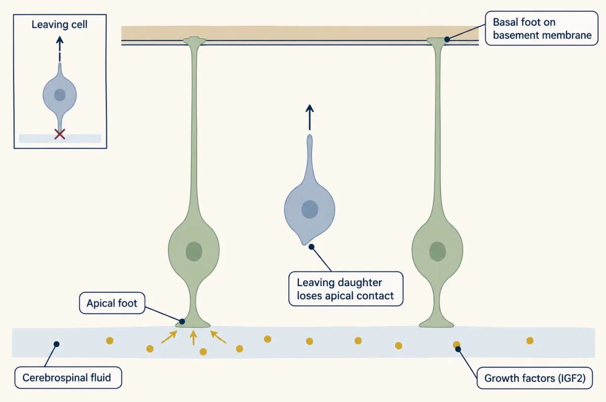
Division at the ventricle went on. Division away from it fell. And fewer cortical neurons were made. The position of mitosis, read with the markers, pointed to the lost progenitor. In those mice, Tbr2 was needed for the indirect route. Primates have a third progenitor in far greater numbers. Outer radial glia divide far from the ventricle. Mouse has a few. Humans have far more. Now, what does a leaving daughter give up? A niche is the local setting that keeps a stem cell a stem cell. The apical foot faces the cerebrospinal fluid. In mouse, that fluid carries growth factors, among them IGF2, that support proliferation. The basal foot rests on the basement membrane under the pia.
Neighbouring cells, through Notch, are part of the setting too. In an asymmetric division, the daughter that leaves lets go of the apical surface. It gives up the fluid side of the niche, and its growth signals. The daughter that stays keeps it. Losing apical contact goes with leaving, but it is not the same thing. An intermediate progenitor has let go, and still divides once more. So a spent daughter makes one neuron directly, or usually two through an intermediate progenitor. Follow its markers and divisions to tell which.
From the film
- In direct neurogenesis a radial glial cell divides into a radial glial cell and a neuron; in indirect neurogenesis the second daughter is an intermediate progenitor that, in rodent cortex, usually divides into two neurons.
- Pax6, Tbr2 and Tbr1 mark radial glia, intermediate progenitors and neurons in that order, and removing Tbr2 in mice reduced divisions away from the ventricle and made fewer cortical neurons.
- A daughter that leaves gives up apical contact with the cerebrospinal fluid niche, though losing that contact is not the same as leaving.
A radial glial daughter can take one of two routes. In direct neurogenesis, a radial glial cell divides into another radial glial cell and a neuron. In indirect neurogenesis, the second daughter is an intermediate progenitor. It lets go of the ventricle, moves into the subventricular zone (SVZ) and divides there once more, in rodent cortex usually into two neurons.
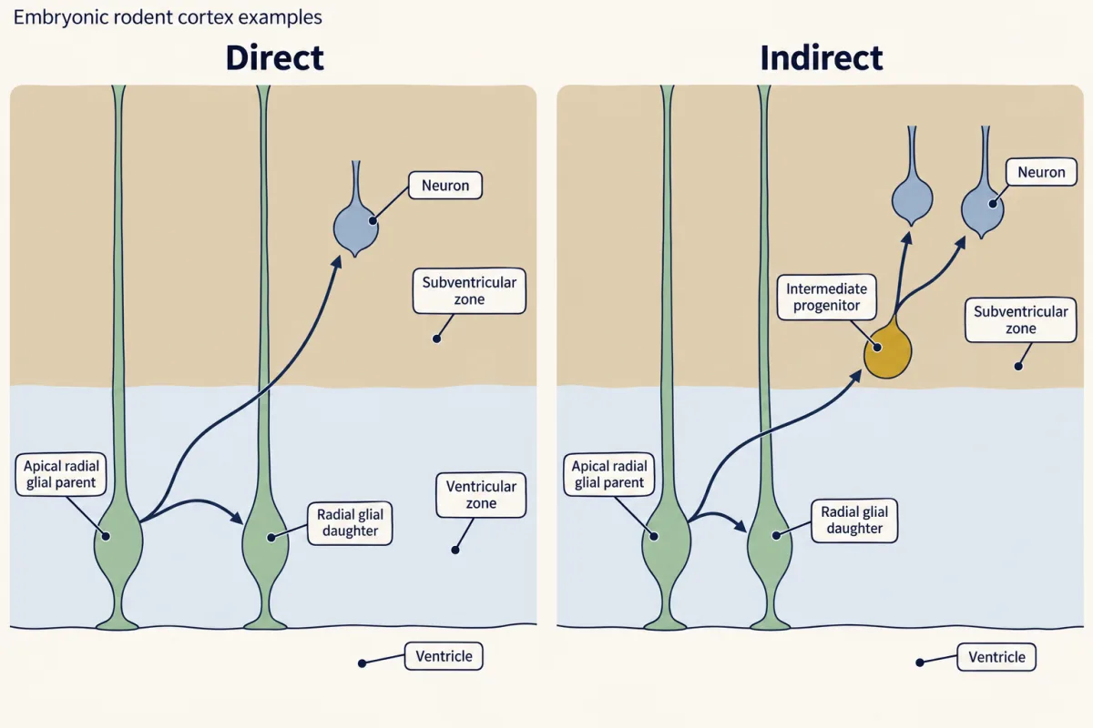
Noctor and colleagues (Nature Neuroscience, 2004) tracked both routes in rat cortical slices labeled with a GFP retrovirus at embryonic day 16 and followed to day 19. About 79 percent of radial glial divisions were asymmetric, giving a radial glial cell plus a neuron or an intermediate progenitor, and about 90 percent of intermediate-progenitor divisions gave two neurons. The sample was 146 daughter cells, with only 19 divisions seen away from the ventricle, so read the result as rat, at those stages, in that study. In mouse, Tbr2 intermediate progenitors may make most cortical projection neurons (Kowalczyk and colleagues, 2009).
Transcription factors, proteins that switch genes on or off, mark the steps. In mouse cortex, Chris Englund, Robert Hevner and colleagues (2005) found three in order as cells move outward (Tbr2 is also called Eomes):
radial glia (Pax6) → intermediate progenitor (Tbr2) → neuron (Tbr1)
Are the markers only labels? In 2008 two groups removed Tbr2 from the developing mouse brain (Arnold and colleagues; Sessa and colleagues).
Predict. Count mitoses in two places, at the ventricle and away from it. If Tbr2 is a step the indirect route needs, which count should fall? What should happen to the number of cortical neurons?
Check your prediction
Division at the ventricle went on, division away from it fell, and fewer cortical neurons were made. This is the lift paying off: the position of mitosis, read with the markers, pointed to the progenitor that had been lost. In those mutants Tbr2 was needed for the indirect route, not for all neurogenesis. Results differ between deletion models: in a later one, divisions away from the ventricle rose and the intermediate progenitors failed to become neurons (Mihalas and colleagues, 2016).
A third progenitor, the outer radial glial cell, divides far from the ventricle and feeds further progenitors. Mouse cortex has a few (Shitamukai and colleagues, 2011); human cortex has them in far greater numbers (Hansen and colleagues, 2010). They are taken up in human brain expansion.
What a leaving daughter gives up
A niche is the local setting that keeps a stem cell a stem cell. An apical radial glial cell touches two parts of one. Its apical foot faces the cerebrospinal fluid (CSF) in the ventricle; in mouse, that fluid carries growth factors, among them IGF2, that support progenitor proliferation (Lehtinen and colleagues, 2011). Its basal foot rests on the basement membrane under the pia. Neighbouring cells are part of the setting too: Notch signalling between neighbours helps keep radial glia as progenitors, and in mouse forebrain an activated form of Notch1 pushed cells toward radial glial identity (Gaiano, Nye and Fishell, 2000).
When a division is asymmetric, the daughter that leaves lets go of the apical surface and gives up the CSF side of the niche. The daughter that stays keeps it, and with it the signals that support dividing. Losing apical contact goes with leaving in this system, but it is not the same thing: an intermediate progenitor has let go and still divides once more, and outer radial glia renew themselves without it.
Counting what stays
Read the transcript of “Counting What Stays”
Film transcript: Counting What Stays
If every division keeps some daughters and spends others, how many neurons does the cortex get? Start with a simple model. A pool of dividing cells, each dividing once per round, with none dying. Every cell makes two daughters. Call P the fraction of daughters that stay progenitors. Call Q the fraction that quit as neurons. Together they make one. The next pool comes from the daughters that stay. The neurons come from the daughters that quit. This pool is every dividing cell, not just stem cells. Suppose Q is 0.5. Half the daughters quit. Does the pool shrink, hold or grow? And does it make neurons?
It holds, and it makes neurons. It is as if every division keeps one daughter and spends one. A steady pool is not an idle one. Below 0.5, the pool grows. Above it, the pool shrinks. In mouse dorsal cortex, Takahashi and colleagues estimated about eleven cycles of neurogenesis. Q starts near zero. It passes 0.5 in the eighth cycle, and reaches about 0.8 by the tenth. Takahashi measured Q for the ventricular zone proper, apart from cells dividing farther out. Those old compartments are not today's cell classes. Follow one founder through that schedule. The numbers are illustrative. Early on, almost nothing is made, and the pool multiplies. Late, the pool is spent.
Output peaks at cycle 9, just after Q passes 0.5, while the pool is still near its largest. The schedule is a lever. In this model, moving the crossing one cycle later nearly doubles the total, over the same eleven cycles. That is why a few extra proliferative cycles are a candidate for how bigger cortices are built. Now a trap. Suppose every radial glial division makes a radial glial cell and an intermediate progenitor. In that round, no daughter quits. Counted as one pool, it seems to double. Follow one radial glial cell for four cycles. Each intermediate progenitor divides into two neurons. How many radial glia, and how many neurons?
Still one radial glial cell, and six neurons. One more intermediate progenitor is about to make two more. That first doubling was not a gain in stem cells. So count two pools. In this model, radial glia are the source that keeps neurogenesis going. Intermediate progenitors multiply, and delay, what each spent daughter yields. The cycle itself slows. In mouse dorsal cortex, it lengthens from about 8 hours to about 18. Nearly all the added time is in G1. One hypothesis: a longer G1 gives fate determinants more time to act. In cultured mouse embryos, a drug that lengthened G1 led to early neurogenesis. Whether a longer G1 causes the switch is still open.
The first puzzle has an answer: the scaffold is the source. The second has a partial answer: the trip to the ventricle fits the centrosome and may let more cells share the surface. What the cortex gets is a count: how many radial glia each division keeps, and what each spent daughter yields.
From the film
- In the counting model, a steady pool at Q of one half still makes neurons; below one half the pool grows and above it the pool shrinks.
- In the model of mouse dorsal cortex, output peaks just after Q passes one half, and moving the crossing one cycle later nearly doubles the total.
- Count radial glia and intermediate progenitors separately: radial glia keep neurogenesis going, and intermediate progenitors multiply what each spent daughter yields.
- The cycle lengthens, mostly in G1, as Q rises, but whether a longer G1 causes the switch is still open.
Now count, with a deliberately simple model. Every cell in the pool divides once per round (one cell cycle for the whole pool), none dies or arrives from elsewhere, and each daughter either keeps dividing or becomes a neuron. Start with N dividing cells. One round makes 2N daughters. Call P the fraction of daughters that stay progenitors and Q the fraction that quit the cycle as neurons, so P + Q = 1.
next pool = 2N × P; neurons made this round = 2N × Q
The pool is every dividing cell, so 2N × P is not a count of stem cells (the trap below).
Predict. Q is 0.5: half of all daughters quit. Does the pool shrink, hold or grow? Does it make neurons?
Check your prediction
It holds, and it makes neurons. 2N daughters, half staying, gives N progenitors again, plus N neurons. That is what you get if every division keeps one daughter and spends one. Below Q = 0.5 the pool grows; above it, the pool shrinks.
Run the clock forward. In mouse dorsal cortex, Takahashi, Nowakowski and Caviness estimated that neurogenesis lasts about eleven cycles, with Q near zero at the start, passing 0.5 in the eighth cycle and reaching about 0.8 by the tenth. Their Q is for the ventricular zone proper, which they separated from cells dividing farther out. Those older compartments do not map one to one onto today’s radial glia and intermediate progenitors, so their Q is not a direct count of stem cells kept.
Follow one founder through a smooth schedule of that shape, Q = 1 / (1 + e^−(cycle − 7.5)/1.8). The numbers are illustrative expected counts, so fractions are averages, and each daughter that quits counts as one neuron, a simplification, since a cell leaving the ventricular zone proper may still divide outside it.
| Cycle | Q | Dividing pool entering | Neurons made | Pool leaving |
|---|---|---|---|---|
| 1 | 0.03 | 1 | 0.1 | 1.9 |
| 2 | 0.05 | 1.9 | 0.2 | 3.7 |
| 3 | 0.08 | 3.7 | 0.6 | 6.9 |
| 4 | 0.13 | 6.9 | 1.7 | 12 |
| 5 | 0.20 | 12 | 4.8 | 19 |
| 6 | 0.30 | 19 | 12 | 27 |
| 7 | 0.43 | 27 | 23 | 31 |
| 8 | 0.57 | 31 | 35 | 26 |
| 9 | 0.70 | 26 | 37 | 16 |
| 10 | 0.80 | 16 | 26 | 6.4 |
| 11 | 0.87 | 6.4 | 11 | 1.6 |
In the first five cycles almost nothing is made and the pool multiplies. In cycles 6 to 8 the pool is still growing, or has just peaked, while output climbs steeply. Late, the pool is spent. Output peaks at cycle 9, just after Q passes 0.5, while the pool is still near its largest. Over the eleven cycles this founder makes about 150 neurons. The schedule is a lever: in this model, moving the Q = 0.5 crossing one cycle later nearly doubles the total (about 1.8 times over the same eleven cycles, with about a dozen cells still dividing at the end). That is why a few extra proliferative cycles are a candidate for how bigger cortices are built (human brain expansion).
One trap: intermediate progenitors. An intermediate progenitor divides, so it does not count as having quit, though it will divide only once more, usually into two neurons.
Predict. One radial glial cell makes a radial glial cell and an intermediate progenitor every cycle, and each intermediate progenitor divides into two neurons. After four cycles, how many radial glia and how many neurons are there?
Check your prediction
There is still one radial glial cell, and there are 6 neurons: none in the first cycle, then 2 in each of the next three. One more intermediate progenitor, made in cycle 4, is about to divide into 2 more. Counted as a single pool, Q went 0, then 0.5, 0.5, 0.5, so the formula reads cycle 1 as a doubling of the pool, yet no stem cell was gained. Count the two pools separately. In this model, radial glia are the source that keeps neurogenesis going; intermediate progenitors multiply, and delay, what each spent daughter yields. In real cortex, the timing and survival of both pools shape the total.
The clock
The same group timed the cycle (1995). In mouse dorsal cortex it lengthens from about 8 hours at embryonic day 11 to about 18 hours near the end of neurogenesis, and nearly all the added time is in G1. Eleven cycles ramping across that range add up to about six days, which fits neurogenesis from about embryonic day 11 to 17. One hypothesis links the two trends, the longer G1 and the rising Q: a longer G1 gives fate determinants, the proteins that steer a daughter to stay or leave, more time to act. In cultured mouse embryos, a drug that lengthened G1 without stopping the cycle led to early neurogenesis in the forebrain neuroepithelium (Calegari and Huttner, 2003). Whether a longer G1 causes the switch, follows from a fate already chosen, or both, is still open.
Design problem
One radial glial cell divides six times, each time keeping one radial glial daughter. How many neurons does it make if every division is direct (radial glia plus neuron)? How many if every division is indirect (radial glia plus an intermediate progenitor that divides once into two neurons)? Which change in division mode would most enlarge the cortex?
Saved in this browser only.
One way to do it
Direct: six divisions make 6 neurons, and one radial glial cell remains.
Indirect: each division adds one intermediate progenitor, which later makes two neurons. Once all six have divided, that is 12 neurons, and one radial glial cell still remains. (Right after the sixth division, counted as in the trap, it is 10 neurons and one intermediate progenitor still to divide.) Routing output through intermediate progenitors doubles the neurons per radial glial division without spending the radial glial cell.
The bigger lever is earlier. A symmetric proliferative division, radial glia into two radial glia, makes no neurons now but doubles every later output. Once every intermediate progenitor has divided, one such division before the six indirect ones gives two radial glial cells and 24 neurons; two such divisions give four and 48.
Putting it together
The first puzzle has an answer: in rodent cortex, the scaffold is also the source. The second has a partial one. Apical mitosis is where the centrosome and cilium base sit for much of interphase, and staggered timetables may let more progenitors share the ventricular surface; the signalling idea is still a proposal. That trip is also why the place of a mitosis is a first clue to the progenitor.
What the cortex gets is a count. Each radial glial division keeps two, one or no radial glia, and each spent daughter yields one neuron or, through an intermediate progenitor, usually two. Once Q passes 0.5, the pool starts to be spent; a lengthening G1 goes with that rise and may help cause it.
The same radial fibre has one more job elsewhere. In mouse spinal cord, ventricular-zone progenitors supply netrin-1 that collects on their radial fibres and at the pial surface, where it is thought to act as a local path for commissural axons (netrin and DCC). Next: how newborn neurons climb the fibre (radial migration), how birth date sets layer (birth order and layers), and how the same lineage turns to glia (origins of glia).
Going deeper
Motors behind interkinetic nuclear migration
The movement has motors. In embryonic rat cortex, Jin-Wu Tsai, Richard Vallee, Arnold Kriegstein and colleagues (2010) knocked down motor proteins with RNA interference and found that the kinesin Kif1a carries the nucleus basally in G1 and cytoplasmic dynein pulls it apically in G2. In mouse, Kosodo and colleagues (2011) argued that the basal movement is largely passive, with G1 nuclei pushed outward by G2 nuclei crowding toward the ventricle, so whether the basal step is driven by a motor is still debated. In zebrafish retina the apical movement has been reported to depend on actomyosin instead, so it is not a rule for every vertebrate.
The clonal study: potential, not fate
Sally Temple did clonal analysis, the study of the family of a single founder, in the most literal way (Nature, 1989). She dissociated the forebrain of rat embryos at embryonic days 13.5 to 14.5, isolated cells able to divide, and grew them one per tiny culture, with live conditioning cells present because the founders would not divide without them. Most of the clones held only neurons or only glia, while about a fifth (22 percent) were mixed: neurons together with non-neuronal cells. Such cells are called multipotent progenitors and, where a cell can also renew itself, neural stem cells. A well shows potential, not fate, and her cells came from the forebrain as a whole, so they need not have been cortical progenitors.
When the GFAP promoter switches on
The Cre fate map gave a different answer outside the cortex. In the basal ganglia few neurons carried the reporter. A 2004 fate-mapping study (Anthony and colleagues) found that radial glia made the neurons of every region, and a 2008 comparison traced the gap to the glial fibrillary acidic protein (GFAP) promoter switching on in ventral radial glia only after most of their neurons were made. A fate map is only as early as the promoter that drives it.
How many neurons per radial glial cell?
Single-cell lineage tracing in mouse cortex (MADM; Gao and colleagues, 2014) reported that a neurogenic radial glial cell makes a fairly fixed number of neurons, roughly eight or nine, and that a minority of these cells then go on to make glia. The table above follows a founder that first expands its pool, and lumps intermediate progenitors with radial glia; the MADM figure is per radial glial cell once it has turned to making neurons. The table shows the shape of a schedule and is not a fit: its 12 cells entering cycle 5 go on to make about 148 neurons, some 12 each. If the MADM figure holds, it constrains any count of this kind, which would need tuning (its Q schedule, its intermediate-progenitor yield) to match it. It is also a direct case of one cell making more than one kind of cell.
Your notes
Select any text in the lesson to highlight it or add a note to it. Your highlights and notes are saved in this browser and collected in your notebook.
This browser is not letting the site save anything (a private window or strict privacy settings), so highlights and notes will be lost when you leave the page.
How we measure it
Pulse labeling of DNA synthesis
A labeled DNA building block, tritiated thymidine or the analogue BrdU, is given briefly and is taken up only by cells copying DNA. Fixing tissue at later times shows where the labeled nuclei go and when they divide, which gives the position of S phase, the movement of the nucleus and the length of the cell cycle.
Retroviral lineage labeling with time-lapse imaging
A retrovirus carrying a marker gene such as GFP is injected into the ventricle, where it enters dividing progenitors and inserts into their DNA, so all descendants of the cell that carries it are marked. Filming living slices then shows which cell divided and what each daughter became.
Cell sorting and Cre-based fate mapping
Cells that make a fluorescent protein from a cell-type promoter can be sorted and grown alone to test what they can make. Cre recombinase made from the same promoter switches on a reporter permanently, so every descendant stays marked and lineage can be read in the animal.
Whole-cell recording with dye filling
An electrode records a single cell's electrical properties and then fills it with a dye that does not pass easily to neighbours. The cell's full shape can then be matched to its physiology, which tied precursor-like recordings to radial glial shape.
Practice questions on this lesson (3) → MCAT-style, original, each with an explanation
Where this lesson fits
Before thisReview first
- Neurulation and Neural Tube DefectsThis course · Lesson 7
The neural tube and its apical and basal surfaces are the layout this lesson builds on.
- Tools for Following Cells and GenesThis course · Lesson 4
Fate mapping and lineage tracing from there are the tools used to show radial glia make neurons.
- The Wiring Problem: How a Brain Gets BuiltThis course · Lesson 1
It sets up the question of how neuron number and types are generated from few genes.
After thisStudy next
- Proneural Genes and Lateral InhibitionThis course · Lesson 14
It explains how Notch and proneural genes decide which daughter stays a progenitor and which leaves.
- Asymmetric Division: Numb and the NeuroblastThis course · Lesson 15
It explains how one division gives two different daughters, the stay-or-leave choice that is only counted here.
- Radial Migration: Climbing the Glial LadderThis course · Lesson 17
Newborn neurons climb the same radial glial fibre that this lesson shows is also the source.
Elsewhere on the siteFilms, simulations and method films in other courses
- Copy First, Then ShareFilm · Life Science
It shows the cell cycle phases (interphase and mitosis) whose order sets where the nucleus sits.
- Mitosis stepper and phase counterSimulation · Biology
It lets you step through the phases of the cell cycle that the moving nucleus follows.
Suggested by an AI model from the lessons themselves, then checked. How we use AI
Finish line
Test yourself
8 questions
Practice and MCAT-style, with explained answers.
Next · Lesson 14
Proneural Genes and Lateral Inhibition
Every cell in the cluster could become the neuron. Why does only one?
Continue →